Equine hepatocytes: isolation, cryopreservation, and
applications to in vitro drug metabolism studies
Khaled A. Shibany, Sabine Tötemeyer, Stefanie L. Pratt & Stuart W. PaineSchool of Veterinary Medicine and Sciences, University of Nottingham, College Road, Sutton Bonington, Leicestershire LE12 5RD, United Kingdom
Keywords
Cell viability, cryopreservation, drug metabolism, equine, hepatocyte isolation
Correspondence
Stuart W. Paine, School of Veterinary Medicine and Sciences, University of Nottingham, College Road, Sutton Bonington, Leicestershire LE12 5RD, United Kingdom. Tel: +44 (0)115 9516615; Fax: +44 (0)115 951 6440;
E-mail: stuart.paine@nottingham.ac.uk
Funding Information
Funding was provided by the Ministry of Higher Education and Scientific Research-Libya and in part by The Wellcome Trust (Grant number 109186/Z/15/Z). The content is solely the responsibility of the authors and does not necessarily represent the views of Ministry of Higher Education and Scientific Research-Libya and The Wellcome Trust.
Received: 22 July 2016; Accepted: 27 August 2016
Pharma Res Per, 4 (5), 2016, e00268, doi: 10.1002/prp2.268
doi: 10.1002/prp2.268
Abstract
Despite reports of the successful isolation of primary equine hepatocytes, there are no published data regarding the successful cryopreservation of these isolated cells. In this study, a detailed description of the procedures for isolation, cryop-reservation, and recovery of equine hepatocytes are presented. Furthermore, the intrinsic clearance (Clint) and production of metabolites for three drugs were
compared between freshly isolated and recovered cryopreserved hepatocytes. Pri-mary equine hepatocytes were isolated using a two-step collagenase perfusion method, with an average cell yield of 2.472.629106cells/g of perfused liver tissue and viability of 84.12.62%. These cells were cryopreserved with Wil-liam’s medium E containing 10% fetal bovine serum with 10% DMSO. The via-bility of recovered cells, after a 30% Percoll gradient, was 7711% and estimated recovery rate was approximately 27%. These purified cells were used to determine the in vitro Clintof three drugs used in equine medicine;
omepra-zole, flunixin, and phenylbutazone, via the substrate depletion method. Cryopre-served suspensions gave a comparable estimation of Clintcompared to fresh cells
for these three drugs as well as producing the same metabolites. This work paves the way for establishing a bank of cryopreserved equine hepatocytes that can be used for estimating pharmacokinetic parameters such as the hepatic metabolic in vivo clearance of a drug as well as producing horse-specific drug metabolites.
Abbreviations
BSA, bovine serum albumin; CLint, intrinsic clearance; DMSO, dimethyl sulfoxide; EGTA, ethylene glycol tetraacetic acid; EPC, effective plasma drug concentration; ESI, electrospray ionization; FBS, fetal bovine serum; HBSS, Hanks balanced salt solution; HPLC, high-performance liquid chromatography; IPC, irrelevant plasma concentration; MS, mass spectrometer; PD, pharmacodynamics; PK, pharmacoki-netics; TB, Trypan Blue; WEM, Williams medium E.
Introduction
Horses have both a commercial and a domestic role within society, ranging from companion animals to the equine sports and food industries. In all cases, the welfare of the horses must be considered, including appropriate medica-tion when necessary. The optimal dosing regimen for a med-ication is dependent on a thorough understanding of the drug pharmacokinetics (PK). The clearance of a drug is par-ticularly important, especially when determining the average concentration of a drug once steady-state has been reached.
upon the above definition that also takes into account variation in both PK and pharmacodynamic (PD) param-eters for a population of horses. An estimate of the irrele-vant plasma concentration (IPC) is based upon a pharmacologically effective plasma drug concentration (EPC) divided by an appropriate safety factor. The EPC is the average concentration at steady-state and is estimated from the therapeutic dose, dosing interval, and clearance of the drug. The IPC can be used for deriving a drug screening limit, which in turn can be used in conjunction with drug in vivo PK profiles to obtain appropriate drug withdrawal times.
In vivo PK studies including bioanalysis are expensive as well as involving invasive sampling from the animal. It is now standard practice within the pharmaceutical industry to use in vitro tissue models to estimate the clearance of a drug in human and reduce the number of preclinical ani-mals used. The use of primary hepatocytes has been prac-ticed for more than five decades (Berry and Edwards 2001). Primary hepatocytes are considered as a “gold standard” for evaluating hepatic metabolism and toxicity of drugs and other xenobiotics in vitro and potential drug-drug interactions (Dambach et al. 2005; Hewitt et al. 2007; Li 2007). Although freshly isolated hepatocytes have a limited viability in suspension of up to 4 h, this period is long enough to determine the metabolic stability of a drug and to allow identification of major metabolites (Jouin et al. 2006; Soldatow et al. 2013). The limited availability of fresh cells restrains their use, however, cryopreservation offers an option for long-term storage of hepatocytes providing a continuous and sufficient “off the shelf” supply (Bachmann et al. 2003; Griffin and Houston 2004; Terry et al. 2010).
Fresh equine primary hepatocytes have successfully been isolated and cultured (Bakala et al. 2003; Stefanski et al. 2013). However, there is no literature evidence describing the cryopreservation and recovery of equine hepatocytes and the resulting cell viability and activity of xenobiotic metabolizing enzymes. A better understanding of equine drug metabolism is important for optimal administration regimens of drugs and the resulting effects of the applied treatment. As a consequence, equine veteri-nary surgeons will be better informed with regard to the outcomes of administered drugs and as a result potential drug-drug interactions may be avoided. This will ulti-mately lead to improved welfare of the horse population. Furthermore, a greater understanding of how drugs are metabolized in the horse, including rates of metabolism and metabolites generated using in vitro systems, will aid the equine sports industry regulate for the abuse of medi-cations and doping substances with a reduction in animal usage and overall costs.
The aim of this work was to develop and optimize a pro-tocol for the cryopreservation of equine hepatocytes that can
be recovered with good cell viability and metabolic activity for use in metabolism studies of the commonly used equine drugs omeprazole, flunixin, and phenylbutazone.
Materials and Methods
Materials
Collagenase and Calcium-free Hanks Balanced Salt Solu-tion (HBSS) 910, HEPES buffer, William’s medium E, and fetal bovine serum (FBS) were purchased from Thermo Fisher Scientific (Paisley, UK). Dimethyl sulfox-ide, Percoll, flunixin, ketoprofen, phenylbutazone, diclofe-nac, and trypan blue were purchased from Sigma (Dorset, UK). Omeprazole and lanzaprozole were purchased from Tocris Bioscience (Bristol, UK). All other chemicals were of reagent grade.
Liver samples
Livers were obtained from seven horses post slaughter (2– 22 years old, of both sexes). The horses were slaughtered in accordance with the Welfare of Animals at the Time of Killing (UK) Regulations 2015. Liver lobes (preferably the left lateral, caudal, or quadrats segment) were excised by a single transverse cut and immediately after excising immersed in HBSS (0–2°C) prior to being transported to the laboratory.
Hepatocyte preparation
Purification of the initial cell suspension
The digested liver tissue was dispersed in 100 mL HBSS buffer containing 0.1% w/v albumin at 4°C. The resulting cell suspension was twice filtered, first through a 250lm nylon mesh followed by a 100lm nylon mesh. Next, HBSS buffer containing albumin was added to the filtered suspension to reach a final volume of 300 mL. Once resuspended, the cell suspension was centrifuged for 5 min at 75g followed by 3 min at 50g. Cell pellets were resuspended in 45 mL HBSS buffer containing albumin and placed on ice.
Determination of hepatocytes yield and viability
The yield and viability of freshly isolated hepatocytes were assessed with Trypan Blue (TB) dye exclusion test (Ten-nant 1964). A quantity of 15lL TB was mixed with the same volume of cell suspension and stained (nonviable) and unstained (viable) cells were counted in a hemocy-tometer. Viability % was determined from the percentage of live cells to total cell count.
Cryopreservation of freshly isolated hepatocytes
Hepatocytes were cryopreserved according to a modified protocol by Adams et al. (1995): The main modification was use of 50% v/v Williams E medium, 40% v/v Fetal calf serum, and 10% v/v DMSO as cryopreservation media. Cells were pelleted at 50g for 3 min at 4°C, the supernatant was removed by aspiration, and the pellet was resuspended in cryopreservation media prepared fresh at the time of cryopreservation under sterile conditions. The cryopreservation medium was added dropwise while the suspension was gently shaken. After resuspension, the cells were placed on ice and aliquoted (1 mL of cell sus-pension/cryovial at a concentration of 19107cells/mL). The vials were then transferred into Mr Frosty iso-propanol Containers (Fisher, UK) and cooled at a rate of 1°C/min in a 80°C Freezer. After 18 h, the vials were transferred to liquid nitrogen for long-term storage at
196°C.
Cell recovery
Cryopreserved equine hepatocytes were recovered by thawing the vials in a 37°C water bath under gentle agita-tion. The cells were diluted 1:10 by drop wise addition of prewarmed WEM plus 10% fetal bovine serum as previ-ously described by Terry et al. 2010. Cells were pelleted at 50g for 5 min at room temperature and suspended in
prewarmed WEM. Hepatocyte yield and viability was determined immediately post-thaw by TB exclusion.
Recovery of cryopreserved hepatocytes for drug clearance assays
After thawing, viable cells were purified using two differ-ent Percoll gradidiffer-ents (25 and 30%). A quantity of 15 mL of the Percoll solution was overlaid with the content of one vial and centrifuged at 110gat 4°C for 20 min (Krea-mer et al. 1986; SciKon Innovation 2010). After centrifu-gation, the dead cells and the cell debris were located at the interface while viable cells were pelleted. The pelleted cells were resuspended in WEM and washed by centrifu-gation for 3 min at 50g.
Determination of intrinsic clearance
Freshly isolated or recovered cryopreserved hepatocytes were diluted to 1 million cells/mL, using WEM with 0.125% v/v bovine serum albumin (BSA), and were prein-cubated for 30 min at 37°C with 5% CO2 as previously
described by Jouin et al. (2006). All incubations were per-formed in 1.2 mL cluster tubes (Sigma, Dorset, UK) in a 96-well heater block (BioShake iQ), set at 750 rpm and 37°C with a total incubation volume of 1 mL. Drug stocks were prepared in DMSO at 1009 final incubation concentration (5lmol/L). The reaction was initiated by adding 10lL of each drug stock to cell suspension. Then, 100lL aliquots were removed at 0, 5, 10, 20, and 30 min and quenched in 200lL of ice-cold methanol containing internal standard (10lmol/L). Samples were subsequently frozen at 20°C until further analysis. Samples were cen-trifuged at 31gfor 10 min at 4°C prior to HPLC analysis. The supernatant was removed and transferred into HPLC vials and analyzed.
LC/MS analysis
monitoring mode using predetermined parent MH/Z and oxidative metabolites MH+16/Z. The intrinsic clearance was determined using the drug substrate depletion approach where the slope of the logarithm of MS parent drug response versus time was determined and converted into appropriate units.
Statistical analysis
The data are presented as mean of seven cell preparations
standard deviations. Statistical significance was esti-mated by the paired, two-tailed Student’s test. AP<0.05 was considered significant.
Results and Discussion
Yield and viability of fresh and cryopreserved equine hepatocytes
The average of total yielded viable fresh hepatocytes was 249 198 9106 cells per isolation, which equated to 4.36 2.479106 cells per gram of perfused tissue. Hepatocyte viability was on average 84.12.62% (Table 1) and is comparable to that reported by Bakala et al. (2003) of 82.7 10.2% and Stefanski et al. (2013) of 84.6 6.4%. However, the number of hepatocytes iso-lated per gram of perfused tissue was fivefold higher than that reported by Stefanski et al. (2013) (n =9) but five-fold lower than in the study by Bakala et al. (2003). Although protocols were similar, Bakala et al. (2003) used a University of Wisconsin proprietary solution for trans-portation, which may have superior organ preservation capacities, enabling a larger viable cell yield to be obtained. Another potential factor is the number of liver samples, which was greater (n=264) .
Cryopreserved equine hepatocytes had a post-thaw via-bility of 60.3 12% (Table 2). The results obtained in
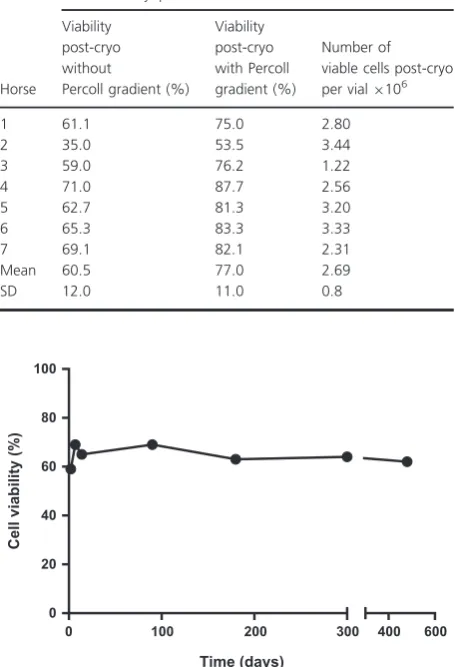
[image:4.595.310.538.93.259.2]this study were consistent with data obtained from other species such as rats and human. For rat cryopreserved hepatocytes, the immediate post-thaw viability of 60% was achieved by Sosef et al. (2005). Meanwhile, Terry et al. (2010) obtained post-thaw viability of 529% using cryopreserved human hepatocytes. A batch of cry-opreserved equine hepatocytes were stored for up to 16 months in liquid nitrogen and no effect on post-thaw viability was observed in that time frame (Fig. 1). Two concentrations of Percoll gradient (25% and 30%) were used in this study to examine their effect on the percent-age viability and recovery. A 25% Percoll treatment increased hepatocyte viability from 5214 to 62.3 11% with 31.66% recovery (n= 3). However, the average viability after 30% Percoll treatment was 7711% with an average recovery of 277%
Table 1. Summary of equine liver cell viability and yield pre-cryopre-servation.
Age at slaughter (years)
Weight of liver (g)
Number of viable cells isolated per gram perfused tissue9106
Average cell viability (%)
1 19 100 4.10 87.7
2 18 90 9.33 83.0
3 3 100 3.83 81.0
4 6 90 4.88 83.7
5 18 80 1.6 82.6
6 14 80 2.4 87.7
7 25 80 4.4 82.7
Mean 4.36 84.1
[image:4.595.311.539.105.439.2]SD 2.47 2.62
Table 2. Post-thaw viability with and without 30% Percoll gradient and number of viable cells after Percoll gradient.
Horse
Recovered cryopreserved cells
Viability post-cryo without
Percoll gradient (%)
Viability post-cryo with Percoll gradient (%)
Number of viable cells post-cryo per vial9106
1 61.1 75.0 2.80
2 35.0 53.5 3.44
3 59.0 76.2 1.22
4 71.0 87.7 2.56
5 62.7 81.3 3.20
6 65.3 83.3 3.33
7 69.1 82.1 2.31
Mean 60.5 77.0 2.69
SD 12.0 11.0 0.8
0 100 200 300 0
20 40 60 80 100
400 600
Time (days)
Cell viability (%)
[image:4.595.66.292.558.702.2](Table 2). The recovery percentage was higher with 25% Percoll and is consistent with results reported by McGin-nity et al. (2004). However, the post-thaw viability after 25% Percoll reported herein was lower than the accept-able limit for enzyme kinetic studies. In their study, McGinnity et al. (2004), found that a 25% Percoll treat-ment increased dog and human hepatocyte viability from 521 to 78 8% and 662 to 883%, respec-tively. Therefore, a 30% Percoll concentration was used for kinetic studies since it gave a higher post-thaw viabil-ity with an acceptable recovery. The Percoll densviabil-ity gradi-ent has often been used for cell separation and to increase viability by separating live from dead cells (Kreamer et al. 1986; Innes et al. 1988; Diener et al. 1993). Moreover, several studies have found that the use of this gradient has an advantage for cell function, including cytochrome P450 function and cell attachment (Dou et al. 1992; Utesch et al. 1992; McGinnity et al. 2004).
The present work uses simple conventional commer-cially available solutions and tissue culture materials and methods for isolation and cryopreservation of equine hep-atocytes with no need for specialized equipment or tech-niques and appears very effective. However, further optimization of the described methods may lead to an increased number of hepatocytes isolated per gram of perfused tissue and improvement of post-thaw cell viabil-ity and function.
Comparison of intrinsic clearance and metabolite formation between fresh and cryopreserved equine hepatocytes
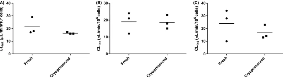
This work also investigated the effect of cryopreservation on xenobiotic metabolizing enzymes. Intrinsic clearance is used as an indicator for the efficiency of metabolism and can be scaled to in vivo plasma clearance with knowledge of physiological parameters such as microsomal protein
content, hepatocellularity, liver weight, and a model of hepatic extraction such as the well-stirred model (Hous-ton 1994; Naritomi et al. 2001). Intrinsic clearances for omeprazole, flunixin, and phenylbutazone were deter-mined by the substrate depletion method. These drugs were chosen because they are known to produce metabo-lites from in vivo studies (Neto et al. 1996; Kanazawa et al. 2002; Jedziniak and Szprengier-juszkiewicz 2005; Jedziniak et al. 2007). The results of this study show that there is no significant difference (P>0.5) in intrinsic clearance between fresh and cryopreserved hepatocytes for the drugs investigated (Fig. 2). The meanSD (n=3) of Clint for omeprazole, flunixin, and phenylbutazone
were 21.36.7, 16.30.5, 19 6.2, and 18.74, 2412.49 and 16.75.5 in fresh and cryopreserved hepatocytes, respectively.
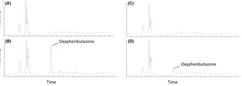
[image:5.595.60.523.533.663.2]Moreover, Figure 3 shows the mass chromatograms of oxidation metabolites (MH+16/Z) generated for omepra-zole in fresh (A and B) and cryopreserved (C and D) equine hepatocytes. Metabolites of omeprazole were characterized by comparison of the negative control chromatogram (0 time point, Fig. 3A and C) with the chromatogram of the 30 min time point (Fig. 3B and D). Oxidation metabolite peaks are presented by M1, M2, and M3 with retention times (RT) 4.22, 4.96, and 5.57 min. This suggests that M1 and M2 are more polar than omeprazole, whereas M3 is less. Kanazawa et al. (2002) determined a RT order of 5-hydroxyomeprazole, omeprazole, and then omeprazole sul-fone suggesting either M1 or M2 is 5-hydroxyomeprazole and M3 is omeprazole sulfone. This study also identified 3-hydroxyomeprazole as a minor metabolite, and although no RT has been published, it is a reasonable assumption that either M1 or M2 is 3-hydroxyomeprazole. The ratio between peak areas of M1, M2, and M3 was 2:1:1, respec-tively, in both fresh and cryopreserved hepatocytes (Fig. 3A and B). A phenylbutazone oxidation metabolite was char-acterized by comparison of the negative control
chromatogram (0 time point, Fig. 4A and C) with the chromatogram of the 30 min time point (Fig. 4B and D). This metabolite was identified as oxyphenbutazone since it showed an identical retention time to an authentic oxyphenbutazone standard. Oxyphenbutazone was detected for both fresh and cryopreserved hepatocytes, however, the intensity, after normalizing for internal stan-dard, was 50% lower for the latter which may suggest some degradation in the metabolizing enzyme. No significant oxidation metabolites were observed for flunixin which is known to mainly undergo phase II metabolism in camel (Wasfi et al. 1998). Selected ion monitoring for glu-curonide metabolites (MH+176/Z) showed no detectable metabolites. The observations observed in this study with
equine hepatocytes are consistent with the literature for other species (Diener et al. 1993; McGinnity et al. 2004). Moreover, Griffin and Houston (2004) found that cryopre-served human hepatocytes gave similar high clearances for propranolol and diazepam compared to fresh hepatocytes.
[image:6.595.67.539.68.266.2]In conclusion, this study has shown that equine hepa-tocytes can be successfully cryopreserved using a conven-tional freezing protocol. The use of a Percoll centrifugation after thawing yielded equine hepatocytes with high viability, which appear to possess an enzymatic metabolic capability similar to that of freshly isolated hepatocytes. A continuous availability of cryopreserved equine hepatocytes represents a major advantage as a tool for drug screening in the horse.
Figure 3. High-performance liquid chromatography-mass spectrometer (HPLC-MS) chromatograms of oxidation metabolites of Omeprazole in suspension cultures of fresh (A=0 min and B=30 min) and recovered cryopreserved (C=0 min and D=30 min) equine hepatocytes. The ratio between the peak areas of M1, M2, and M3 was 2:1:1, respectively, in both fresh and cryopreserved hepatocytes.
[image:6.595.71.536.331.496.2]Acknowledgements
The authors acknowledge the support from the biobank within the School of Veterinary Medicine and Science at the University of Nottingham and especially Wayne Sanders.
Author Contributions
Participated in research design: K. A. Shibany, S. T€ ote-meyer, S. L. Pratt, S. W. Paine; Conducted experiments: K. A. Shibany and S. L. Pratt; Performed data analysis: K. A. Shibany and S. L. Pratt; Contributed to writing manuscript: K. A. Shibany, S. T€otemeyer, S. L. Pratt, S. W. Paine.
Disclosures
None declared.
References
Adams RM, Wang M, Crane AM (1995). Effective cryopreservation and long-term storage of primary human hepatocytes with recovery of viability, differentiation, and replicative potential. Cell Transplant 4: 579–586.
Bachmann K, Byers J, Ghosh R (2003). Prediction of in vitro hepatic clearance from in vitro data using cryopreserved human hepatocytes. Xenobiotica 33: 475–483.
Bakala A, Karlik W, Wiechetek M (2003). Preparation of equine isolated hepatocytes. Toxicol In Vitro 17: 615–621.
Berry MN, Edwards AM (2001). The hepatocyte review. Kluwer Academic Publisher, Dordrechet, The Netherlands.
British Horseracing Authority (2013) Economic Impact of British Racing 2013 Forward Executive Summary Introduction Section 1 : Overall Economic Impact Racing’s Position in the Leisure Industry Feature Articles. Available at: http://www. britishhorseracing.presscentre.com/imagelibrary/downloadMe dia.ashx? MediaDetailsID=235. (accessed 15 May 2014).
Dambach DM, Andrews BA, Moulin F (2005). New
technologies and screening strategies for hepatotoxicity: use of in vitro models. Toxicol Pathol 33: 17–26.
Diener B, Dietmar U, Beer N, Heike D, Oesch F (1993). A method for the cryopresrvation of liver parenchymal cells for studies of xenobiotics. Cryobiology 30: 116–127.
Dou M, Lacarelle B, Placidi M, Lechene P (1992). Human hepatocytes in primary culture. Cryobiology 469: 454–469.
Griffin JS, Houston JB (2004). Comparison of fresh and cryopreserved rat hepatocyte suspensions for the prediction of in vitro intrinsic clearance. Drug Metab Dispos 32: 552–558.
Hewitt NJ, de Kanter R, LeCluyse E (2007). Induction of drug metabolizing enzymes: a survey of in vitro methodologies and
interpretations used in the pharmaceutical industry–do they comply with FDA recommendations? Chem Biol Interact 168: 51–65.
Houston JB (1994). Utility of in vitro drug metabolism data in predicting in vivo metabolic clearance. Biochem Pharmacol 47: 1469–1479.
Innes GK, Fuller BJ, Hobbs KEF (1988). Functional testing of hepatocytes following their recovery from cryopreservation. Cryobiology 30: 23–30.
Jedziniak P, Szprengier-juszkiewicz T (2005). Determination of phenylbutazone and oxyphenbutazone in bovine plasma using high performance liquid chromatography with UV detection. Bull Vet Inst Pulawy 49: 223–226.
Jedziniak P, Szprengier-Juszkiewicz T, Olejnik M, Jaroszewski J. (2007). Determination of flunixin and 5-hydroxyflunixin in bovine plasma with HPLC-UV method development, validation and verification. Bull Vet Inst Pulawy 51: 261– 266.
Jouin D, Blanchard N, Alexandre E, Delobel F, David-Pierson P, Lave T, et al. (2006). Cryopreserved human hepatocytes in suspension are a convenient high throughput tool for the prediction of metabolic clearance. Eur J Pharm Biopharm 63: 347–355.
Kanazawa H, Okada A, Matsushima Y, Yokota H, Okubo S, Mashige F, et al. (2002). Determination of omeprazole and its metabolites in human plasma by liquid chromatography-mass spectrometry. J Chromatogr 949: 1–9.
Kreamer B, Staecker J, Sawada N, Salter G, Hsia M, Pitot H (1986). Use of a low-speed, iso-density percoll
centrifugation method to increase the viability of isolated rat hepatocyte preparations. In Vitro Cell Dev Biol 22: 201–211.
Li AP (2007). Human hepatocytes: isolation, cryopreservation and applications in drug development. Chem Biol Interact 168: 16–29.
McGinnity DF, Soars MG, Urbanowicz RA, Riley RJ (2004). Evaluation of fresh and cryopreserved hepatocytes as in vitro drug metabolism tools for the prediction of metabolic clearance. Drug Metab Dispos 32: 1247–1253.
Naritomi Y, Terashita S, Kimura S, Suzuki A, Kagayama A, Sugiyama Y (2001). Prediction of human hepatic clearance from in vivo animal experiments and in vitro metabolic studies with liver microsomes from animals and humans. Drug Metab Dispos 29: 1316–1324.
Neto L, Andraus M, Salvadori M (1996). Determination of phenylbutazone and oxyphenbutazone in plasma and urine samples of horses by high-performance liquid chromatography and gas chromatography-mass spectrometry. J Chromatogr B Biomed Appl 678: 211–218.
com/wp-content/uploads/2011/10/Mouse-Hepatocyte-Care-Ma nual.pdf. (accessed 5 July 2015).
Selgen PO (1976). Preparation of isolated rat liver cells. Methods Cell Biol 13: 29–83.
Soldatow VY, Lecluyse EL, Rusyn I (2013). In vitro models for liver toxicity testing. Toxicol Res 2: 23–39.
Sosef MN, Baust JN, Sugimachi K, Fowler A, Tompkins RG, Toner M (2005). Cryopreservation of isolated primary rat hepatocytes enhanced survival and long-term hepatospecific function. Ann Surg 241: 125–132.
Stefanski A, Mevissen M, Moller A-M, Kuehni-Boghenbor K,€ Schmitz A (2013). Induction of cytochrome P450 enzymes in primary equine hepatocyte culture. Toxicol In Vitro 27: 2023– 2030.
Tennant JR (1964). Evaluation of the trypan blue technique for determination of cell viability. Transplantation 2: 685–693.
Terry C, Dhawan A, Mitry RR, Lehec SC, Hughes RD (2010). Optimization of the cryopreservation and thawing protocol for human hepatocytes for use in cell transplantation. Liver Transpl 16: 229–237.
Toutain PL, Lassourd V (2002). Pharmacokinetic/ pharmacodynamic approach to assess irrelevant plasma or urine drug concentrations in postcompetition samples for drug control in the horse. Equine Vet J 34: 242–249.
Utesch D, Diener B, Molitor E, Oesch F, Platt KL (1992). Characterization of cryopreserved rat liver parenchymal cells by metabolism of diagnostic substrates and activities of related enzymes. Biochem Pharmacol 44: 309–315.