DEVELOPMENT OF A PREDICTIVE DNA DOUBLE STRAND
BREAK ASSAY FOR THE IDENTIFICATION OF INDIVIDUALS
WITH HIGH NORMAL TISSUE RADIOSENSITIVITY
Emma Jane Hay Brown
A Thesis Submitted for the Degree of MD at the
University of St Andrews
2008
Full metadata for this item is available in Research@StAndrews:FullText
at:
http://research-repository.st-andrews.ac.uk/
Please use this identifier to cite or link to this item:
http://hdl.handle.net/10023/855
Development of a predictive DNA double strand break assay for the
identification of individuals with high normal tissue radiosensitivity
A thesis submitted in fulfilment of the
requirements for the degree of
Doctor of Medicine
University of St Andrews
II
Table of contents
Table of contents ________________________________________________ II Table of Figures ________________________________________________ VII Acknowledgements ______________________________________________ IX Thesis Declaration _______________________________________________ X Abbreviations __________________________________________________ XII Summary _____________________________________________________ XIII 1. Introduction ________________________________________________________ 1
1.1 Radiotherapy - clinical importance. _________________________________ 1
1.2 Principles of radiotherapy planning and delivery ______________________ 2
1.3 Normal Tissue Toxicity ___________________________________________ 5 1.3.1 Clinical features of radiotherapy toxicity. ____________________________ 6 1.3.2 Grading systems ________________________________________________ 9 1.4 What determines the radiation dose prescribed in today’s clinical practice?
______________________________________________________________ 10 1.4.1 Factors influencing the risk of and severity of radiation toxicity __________ 12
1.4.1.1 Treatment-related factors ____________________________________ 12 1.4.1.2 Patient-related factors _______________________________________ 13 1.5 Individual intrinsic normal tissue radiosensitivity ____________________ 14
1.6 Predictive assays for normal tissue radiosensitivity ___________________ 15 1.6.1 Clinical value of a predictive assay of normal tissue radiosensitivity ______ 15 1.6.2 Development of a predictive assay of normal tissue radiosensitivity ______ 20 1.6.2.1 Functional cell-based assays __________________________________ 21 1.6.2.2 Other strategies in development of predictive assays of normal tissue
radiosensitivity: ___________________________________________ 24 1.6.3 Have functional cell-based assays been proven to be of no clinical utility? _ 25 1.7 H2AX phosphorlyation in human peripheral blood lymphocytes – a
III 2. Materials and methods _______________________________________________ 30
2.1 Materials ______________________________________________________ 30
2.2 Methods _______________________________________________________ 30 2.2.1 Collection of peripheral blood samples from healthy volunteers __________ 30 2.2.2 Gamma source operation and dosimetry ____________________________ 31 2.2.3 Immunomagnetic isolation of CD4 and CD8 positive peripheral blood
lymphocytes __________________________________________________ 32 2.2.4 Immunostaining procedure for focus analysis by microscopy ____________ 33 2.2.5 Fluorescence microscopy, digital image capture and image analysis ______ 34 2.2.6 Cell separation procedure using CPT tubes __________________________ 34 2.2.7 Immunofluorescent staining of PBLs for flow cytometric analysis ________ 35 2.2.8 Flow cytometric analysis of γH2AX staining_________________________ 36 2.2.9 Cell phenotyping ______________________________________________ 37 2.2.10 Flow cytometer performance monitoring with CaliBrite beads _________ 38 2.2.11 Measurement of DNA double-strand break re-joining in human peripheral
blood lymphocytes ___________________________________________ 39 2.2.12 Detection of apoptosis in human peripheral blood lymphocytes after
irradiation __________________________________________________ 40 2.2.13 Statistical analysis ___________________________________________ 41 3. Systematic review of the current evidence base relating to functional cell-based predictive assays of normal tissue radiosensitivity. ____________________________ 42
3.1 Methodology of diagnostic test development and assessment of clinical utility 42
3.1.1 Technical efficacy______________________________________________ 43 3.1.2 Diagnostic accuracy ____________________________________________ 44 3.1.3 Ideal study design in assessment of diagnostic accuracy of predictive assays of normal tissue radiosensitivity _____________________________________ 44 3.1.4 Statistical methods employed in assessment of diagnostic accuracy. ______ 46 3.1.5 Hypothesis generating and validation data sets _______________________ 49 3.1.6 Rationale for a systematic review of the current literature pertaining to
functional cell-based assays in the predictive testing of normal tissue
IV 3.2 Method ________________________________________________________ 50
3.2.1 Identification of relevant studies __________________________________ 50 3.2.2 Data Extraction ________________________________________________ 51 3.2.3 Development of a scoring system for study “quality” __________________ 51 3.2.4 Extraction of individual patient data, construction of ROC curves and
calculation of diagnostic odds ratio. ________________________________ 52 3.3 Results ________________________________________________________ 53
3.3.1 Assay under investigation and cell types used. _______________________ 53 3.3.2 Study design. _________________________________________________ 53 3.3.3 Reporting of tumour and patient characteristics. ______________________ 55 3.3.4 Reporting of radiotherapy details. _________________________________ 56 3.3.5 Recording and reporting of radiotherapy toxicity. _____________________ 56 3.3.6 Details of the laboratory assay under investigation. ____________________ 58 3.3.7 Treatment of confounding factors and statistical analysis _______________ 59 3.3.8 Hypothesis generating and validation data sets _______________________ 61 3.3.9 Summary of the results from identified studies _______________________ 62 3.3.10 Extraction of individual patient data, ROC curve construction and
calculation of diagnostic odds ratios _____________________________ 74 3.4 Conclusion _____________________________________________________ 79
4.γH2AX induction and loss as a potential assay of normal tissue radiosensitivity - quantification of γH2AX foci by microscopy _______________________________ 80
4.1 Quantification of γH2AX foci by microscopy in irradiated mammalian
fibroblasts _______________________________________________________ 81 4.1.1 Immunofluorescent detection of γH2AX in human peripheral blood
lymphocytes. __________________________________________________ 82 4.2 Techniques for quantification of γH2AX foci in human PBLs by microscopy _ ______________________________________________________________ 85 4.2.1 Analysis of digital images _______________________________________ 86 4.3 Determination of the most precise technique for focus quantification. ____ 90
V 4.3.3 Determination of the most precise method for focus quantification by
microscopy - intra-sample precision________________________________ 95 4.3.4 Inter-sample precision. __________________________________________ 98 4.3.5 Assay failure rate _____________________________________________ 102 4.3.6 Time taken to obtain assay results. ________________________________ 102 4.3.7 Inter - individual variation. ______________________________________ 102 4.4 Discussion ____________________________________________________ 102
5.Quantification of γH2AX in human peripheral blood lymphocytes by flow
cytometry. __________________________________________________________ 108
5.1 Development of basic technique for flow cytometric analysis of γH2AX
staining in human PBLs. __________________________________________ 110 5.1.1 Lymphocyte isolation from whole blood ___________________________ 110 5.1.2 Immunostaining procedure and flow cytometric analysis ______________ 111 5.1.3 Quantification of the dose response relationship between γH2AX induction
and increasing radiation dose by flow cytometry. ____________________ 112 5.1.4 Kinetics of γH2AX induction and loss after 2Gy _____________________ 114 5.1.5 Potential assay end-points for measuring γH2AX kinetics _____________ 115 5.1.6 Assay practicality: ____________________________________________ 123 5.1.7 Kinetics of γH2AX induction and loss in human PBLs after different radiation
doses _______________________________________________________ 124 5.1.8 Comparison of the kinetics of radiation-induced DNA double strand break
repair and γH2AX induction and loss in human peripheral blood lymphocytes. ___________________________________________________________ 127 5.1.9 Discussion ___________________________________________________ 132 6. Inter-individual comparison of kinetics of γH2AX induction and loss in irradiated
human peripheral blood lymphocytes. ___________________________________ 139
6.1 Introduction __________________________________________________ 139
6.2 Methods: a study of 8 volunteers. _________________________________ 139
6.3 Results: Individual results for 8 volunteers and intra- and inter-individual variation for each assay end-point. ________________________________ 140
VI 7. Effect of blood sample storage duration and conditions on γH2AX induction in vitro. _________________________________________________________________ 147
7.1 Rationale for investigation of effects of sample storage on assay results. _ 147
7.2 Method _______________________________________________________ 147
7.3 Results _______________________________________________________ 148
7.4 Discussion. ____________________________________________________ 151
8. Final discussion ____________________________________________________ 154
8.1 Future work __________________________________________________ 161
9. References ________________________________________________________ 163
VII Table of Figures
Figure 1.1 Basic principles of radiotherapy planning (adapted from Dobbs, 1999) ... 3
Figure 1.2 Definitions of target volumes for radiotherapy planning ... 4
Figure 1.3 Frequency distribution of normal tissue response amongst patients treated with an identical radiotherapy schedule ... 16
Figure 2.1 Flow cytometric analysis of γH2AX stained PBLs ... 37
Figure 3.1 ROC curve for % reduction in Binucleated index after radiation in human fibroblasts and risk of wound healing complications after post-operative radiotherapy for soft-tissue sarcoma. AUC is 0.805 (95% CI 0.432-1) (Data for analysis extracted from (Akudugu, Bell et al. 2006) ... 49
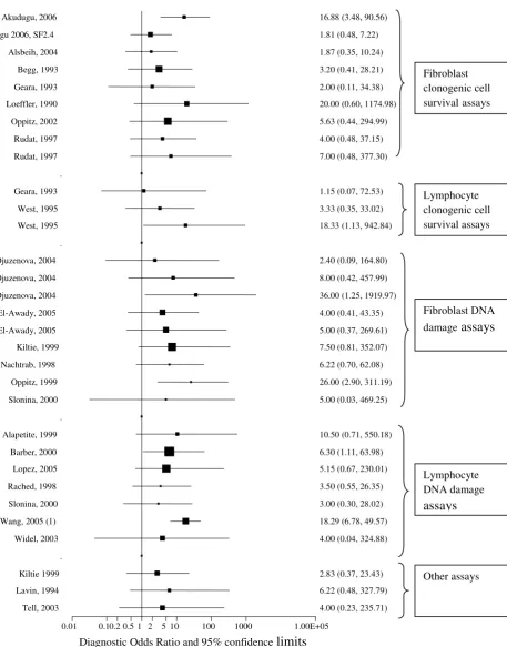
Figure 3.2 Forest plot of diagnostic odds ratio +/- 95% confidence intervals for individual comparisons of assay result and acute radiation toxicity end-points... 75
Figure 3.3 Forest plot of diagnostic odds ratio +/- 95% confidence intervals for individual comparisons of assay result and late radiation toxicity end-points. ... 76
Figure 3.4 Funnel plot of effect (log DOR) vs. quality index (A) and sample size (B) to test for presence of bias in systematic review. ... 77
Figure 4.1 Irradiated Muntjac fibroblast after 0.5Gy γ-rays showing discrete, easily visualised and quantifiable foci of γH2AX. ... 82
Figure 4.2 Cytospin preparations of human peripheral blood CD4 and CD8 T-lymphocytes fixed and stained for γH2AX 30 minutes after irradiation and viewed at x100 magnification. ... 84
Figure 4.3 “SPOT” image analysis ... 87
Figure 4.4 “SPOT” image analysis ... 87
Figure 4.5 “SPOT” image analysis ... 88
Figure 4.6 “SPOT” image analysis ... 88
Figure 4.7 Semi-automated focus quantification ... 89
Figure 4.8 Dose-response of γH2AX focus induction after irradiation (microscopy). ... 91
Figure 4.9 Kinetics of γH2AX focus induction and loss in human PBLs following 0.4Gy γ-irradiation in vitro (microscopy). ... 94
Figure 4.10 Modelling of the kinetics of γH2AX loss ... 95
VIII Figure 4.12 Inter-sample variability - assay results plotted against time for 5 assay repeats over 4 months using fresh blood samples from the same individual to assess for systematic drift in results over the study period ... 101 Figure 5.1 Dose response of γH2AX induction in human PBLs 30 minutes after
γ-irradiation in vitro. ... 113 Figure 5.2 Visual confirmation H2AX focus induction in samples analysed by flow
cytometry ... 113 Figure 5.3 Kinetics of γH2AX induction and loss in human peripheral blood lymphocytes after 2Gy ... 115 Figure 5.4 Intra-sample/inter run precision – mean and standard deviation for flow
cytometric quantification of γH2AX in human PBLs at 1 hour post 2Gy from a single blood sample. ... 118 Figure 5.5 Change in γH2AX assay results in a single individual over the study period. 120 Figure 5.6 Relationship between duration of storage of fixed samples and assay results . 122 Figure 5.7 Kinetics of γH2AX induction and loss in isolated PBLs from a single volunteer following different test doses of in vitro irradiation ... 124 Figure 5.8 Correlation of end-points of γH2AX kinetics experiments with in vitro test dose of radiation ... 126 Figure 5.9 Kinetics of DNA double strand break formation and repair and γH2AX
induction and loss in PBLs after 20Gy ... 129 Figure 5.10 Kinetics of DSB induction and repair and γH2AX induction and loss for the 6 hours immediately post irradiation (20Gy) in isolated human peripheral blood
IX
Acknowledgements
I would like to express my gratitude to Professor Hugh MacDougall, and Dr Peter Bryant from the University of St Andrews for their invaluable advice, and continuous support and encouragement throughout this project.
I would also like to express my sincere appreciation to Professor Alistair Munro, University of Dundee for all his time, advice, help with statistics, encouragement and reassurance at all stages of this project.
Many thanks to the eight volunteers who kindly donated blood samples and without whose generosity this project would not have been possible.
Dr Michael Boylen, University of Dundee kindly provided advice regarding flow cytometry.
X
Thesis Declaration
I, Emma Jane Hay Brown, hereby certify that this thesis, which is approximately 50,000 words in length, has been written by me, that it is the record of work carried out by me and that it has not been submitted in any previous application for a higher degree.
I was admitted as a research student in [month, year] and as a candidate for the degree of Doctor of Medicine in November 2003, the higher study for which this is a record was carried out in the University of St Andrews between 2003 and 2006.
Signature of candidate ………. ………Date………...
I hereby certify that the candidate has fulfilled the conditions of the Resolution and Regulations appropriate for the degree of Doctor of Medicine in the University of St Andrews and that the candidate is qualified to submit this thesis in application for that degree.
Signature of supervisor………. ………Date………...
Signature of supervisor………. ………Date………...
XI right to migrate my thesis into new electronic forms as required to ensure continued access to the thesis. We have obtained any third-party copyright permissions that may be required in order to allow such access and migration, or have requested the appropriate embargo below.
The following is an agreed request by candidate and supervisors regarding the electronic publication of this thesis:
Access to all or part of printed copy but embargo of all electronic publication of thesis for a period of 2 years on the following grounds:
Publication would preclude future publication
Signature of candidate ………. ………Date………
Signature of supervisor………. ………Date………
XII
Abbreviations
AUC Area Under the Curve BMI Body Mass Index BNI Binucleated Index
CFGE Constant Field Gel Electrophoresis CPT Cell Preparation Tube
CTV Clinical Target Volume CV Coefficient of Variation DOR Diagnostic Odds Ratio DSB Double strand break DVH Dose Volume Histogram FCS Foetal Calf Serum
FITC Fluorescein Isothiocyanate GTV Gross Tumour Volume
Gy Gray
LDR Low Dose Rate
LMP low melting point (agarose) MPC Magnetic Particle Concentrator MTD Maximum Tolerated Dose
MTT 3-(4,5-dimethylthiazol-2-yl)-2,5-diphenyl tetrazolium bromide NFR Normalised Fluorescence Ratio
NTCP Normal Tissue Complication Probability PBL Peripheral blood lymphocyte
PBS Phosphate Buffered Saline PE Phycoerytherin
PFA Paraformaldehyde
PFGE Pulsed Field Gel Electrophoresis PI Propidium iodide
PST PBS/donkey serum/Triton X-100 PTV Planning Target Volume
ROC Receiver Operator Characteristic ROS Reactive Oxygen Species
SF2 Surviving Fraction at 2Gy
SNP Single Nucleotide Polymorphism TAE Tris/acetic acid/EDTA
XIII
Summary
A genetically determined high level of intrinsic normal tissue radiosensitivity may account for the 5% of patients who experience unexpectedly severe normal tissue side effects following radiotherapy. The pre-treatment identification of these individuals by a diagnostic test or “predictive assay “ may allow appropriate modification of treatment plans and improve the therapeutic index of radiotherapy.
Results from studies of cell-based assays measuring the response of a single cell type taken from patients to in vitro irradiation have been inconsistent, leading to the opinion of many that they are of no value in the prediction of normal tissue radiosensitivity.
A systematic review of the literature presented here, however, suggests that poor methodology of study design often with inadequate control for those factors other than normal tissue radiosensitivity which influence radiotherapy toxicity and lack of reporting of assay precision means that it is difficult to form any conclusions, positive or negative about the diagnostic accuracy of the cell-based assays studied so far. Analysis of individual patient data extracted from these studies suggests that at least some of these assays may possess some discriminatory value.
1
1.
Introduction
The medical use of ionising radiation to treat disease (radiotherapy) is most often used as part of cancer treatment. Approximately half of all patients diagnosed with cancer will receive radiotherapy during the course of their illness.
It has been estimated that approximately 5% of patients treated with radiotherapy experience unexpectedly severe side effects that could not have been predicted from the patient-related and treatment-related factors known to influence radiation toxicity. It is thought that the normal tissues of these individuals possess an intrinsically high level of sensitivity to radiation damage and that this may be genetically determined. If it were possible to measure an individual’s normal tissue radiosensitivity before treatment it may be possible to predict their likely radiation toxicity and modify their treatment accordingly. At present no such predictive assay of normal tissue radiosensitivity exists in clinical practice despite over a decade of research interest. The majority of studies published so far have examined the role of functional cell-based assays - giving a test dose of radiation in vitro to a tissue sample from the individual in question and examining the response. As results have been perceived to be disappointing interest has moved away from these cell-based assays to assays cell-based on genotyping – or “radiogenomics”.
Inadequate study design may have contributed to the apparent failure of cell-based assays to deliver a clinically useful diagnostic test. The aims of this project were therefore to evaluate the current evidence regarding functional cell-based assays in the predictive testing of normal tissue radiosensitivity to determine if the perception that they are of no clinical utility is justified and, if then deemed appropriate, to develop a novel functional cell-based assay in a methodical and systematic fashion which might have a potential role as a predictive assay for normal tissue radiosensitivity in the clinic
1.1 Radiotherapy - clinical importance.
2 patients were treated with radiotherapy in Scotland in 1999 and it is projected that by 2011 this will increase to 16 500 (Scottish Executive 2004).
Radical radiotherapy may be used in some cancer types as an alternative to surgery to achieve long term tumour control and cure. Long-term tumour control can often be achieved with acceptable cosmetic and functional results, which may be superior to those following radical surgical resection. Increasingly chemotherapy or other biological targeted therapies are being administered in combination with radiotherapy in an attempt improve the chances of local tumour control and survival over those achievable with radiotherapy alone.
Adjuvant radiotherapy is used in combination with surgery to improve the chance of long term tumour control. Radiation is delivered pre- or post-operatively to eradicate microscopic residual disease in or around the tumour bed that may be left behind by the surgeon.
Palliative radiotherapy also has a key role in the relief of distressing symptoms such as pain, breathlessness and bleeding when the cancer cannot be cured. Palliation is the most common indication for radiotherapy and can be very effective – for example, 80% of patients with pain secondary to metastatic tumour deposit in bone will experience relief after palliative radiation treatment (Hoskin, Yarnold, 2001).
1.2 Principles of radiotherapy planning and delivery
The ultimate aim of radiotherapy is to deliver a clinically effective dose of ionising radiation to a tumour to kill or limit the proliferation of tumour cells that would normally multiply causing the cancer to survive and grow.
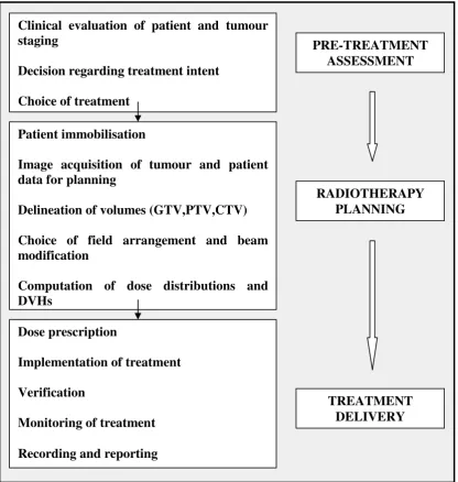
3 Figure 1.1 Basic principles of radiotherapy planning (adapted from Dobbs, 1999)
Clinical evaluation of patient and tumour staging
Decision regarding treatment intent
Choice of treatment
Patient immobilisation
Image acquisition of tumour and patient data for planning
Delineation of volumes (GTV,PTV,CTV)
Choice of field arrangement and beam modification
Computation of dose distributions and DVHs
Dose prescription
Implementation of treatment
Verification
Monitoring of treatment
Recording and reporting
TREATMENT DELIVERY RADIOTHERAPY
PLANNING PRE-TREATMENT
4 The tumour volume to be irradiated is defined according to international definitions for tumour and normal tissue volumes ( Figure 1.2) (ICRU 1993; ICRU 1999)
Figure 1.2 Definitions of target volumes for radiotherapy planning
Organs at risk are defined as those normal tissues likely to be irradiated during the course of treatment.
Radiotherapy planners must then determine the beam energy, arrangement of radiation beams, and required beam modification, required to achieve the aim of delivering the prescribed dose in a uniform distribution to encompass the entire PTV, whilst minimising the dose to the organs at risk. This part of the planning process has been greatly assisted by the development of sophisticated radiotherapy planning software.
The radiation dose to be delivered to the PTV will vary according to the tumour type, and aim of treatment. Treatment is usually fractionated with fractions delivered on a daily basis over a number of weeks. The radiotherapy prescription must specify the total dose, number and size of each fraction, overall time over which the treatment is to be delivered and the point to which the prescribed dose is to be delivered.
Gross tumour volume
(demonstrable macroscopic extent of tumour)
Clinical target Volume
(accounts for potential microscopic spread of tumour outside GTV)
Planning Target Volume
5 For different tumour types there are generally accepted standard radiotherapy schedules usually defined by clinical experience or evidence from randomised controlled trials. Radiation dose is defined as the amount of energy absorbed per unit mass and is measured in Gray, where 1 Gray is equal to 1 Joule of energy absorbed per kilogram of tissue. As an example, radical radiotherapy to the lung may be delivered as 55Gy in 20 fractions over 4 weeks. Palliative radiotherapy is usually a lower dose delivered more quickly e.g. 20Gy in 5 fractions over 5-7 days.
The technological systems for planning and delivery of radiation therapy are becoming increasingly more sophisticated. 3-D conformal radiotherapy allows precise shaping of the radiation beam around the target. Intensity Modulated radiotherapy (IMRT) allow shaping of the radiation dose around critical normal structures, and Image-guided radiotherapy (IGRT) improves precision of radiation delivery to the PTV by allowing for patient and/or organ motion during the course of treatment delivery.
1.3 Normal Tissue Toxicity
Despite technological improvements in radiotherapy delivery it will always be impossible to treat a cancer without simultaneously irradiating surrounding normal tissue. Often the volume of normal tissue within the planning target volume will exceed the gross tumour volume. Radiation beams must traverse normal tissue below the patient’s surface to reach deep-seated tumours. The unavoidable irradiation of normal tissues causes the side effects associated with radiotherapy.
The pathological processes that lead to radiation injury begin immediately after radiation exposure but may not become clinically apparent for days, weeks, months or even years. By convention, radiation effects on normal tissues are usually divided into acute and late reactions.
6 Acute effects typically involve rapidly renewing tissues with a hierarchical cell lineage such as the skin, lining of the GI tract and haematopoietic system. These tissues are composed of a stem cell compartment in which the cells divide and give rise to differentiating daughter cells. Acute radiation reactions occur when damage to the stem cell compartment caused by radiation means that differentiated cells in the tissue lost during normal tissue turnover are not replaced at a sufficient rate. The time of onset of acute reactions therefore is determined by the lifespan of the differentiated cells. On completion of radiotherapy or even sometimes during it, compensatory proliferation of the remaining stem cells is followed by replacement of the functional cells and recovery.
If acute toxicity is very severe a course of radical or adjuvant radiotherapy treatment may have to be abandoned or interrupted impacting detrimentally on the long term probability of tumour control and cure (Hendry, Bentzen et al. 1996). Acute severely symptomatic toxicity following palliative radiotherapy is obviously undesirable given that the aim of treatment is to improve symptoms and quality of life.
Late normal tissue effects are those which appear more than 90 days after completion of radiotherapy and may not appear for months or years after radiation exposure. Once established, late reactions tend to be irreversible and their severity can progress with time resulting in increasing functional loss and cosmetic changes. Functional loss can be severe and have a major impact on an individual’s quality or even duration of life. Examples include hemi or quadriplegia in the case of spinal cord damage, loss of upper limb function with brachial plexus damage, focal neurological deficit due to brain necrosis, severe dyspnoea due to radiation induced pulmonary fibrosis and renal failure due to radiation nephropathy. Cosmetic disfigurement such as visible skin changes on the hands or face, or retardation of bone or muscle growth in the case of children can result in significant psychological or social morbidity. The clinical importance of late normal tissue toxicity is growing as long-term survival after cancer therapy continues to improve.
1.3.1 Clinical features of radiotherapy toxicity.
7 a) Skin and submucosa
Acute skin toxicity is a well-recognised complication of radiotherapy and before the advent of modern megavoltage linear accelerators was frequently the dose-limiting toxicity encountered in clinical practice. It is still a frequent complication of treatment of breast, head and neck and ano-genital malignancies.
Erythema develops in the 2nd or 3rd week of a fractionated course of radiotherapy followed by dry, then moist desquamation due to depletion of the basal stem cell population and failure to replace functional cells (Archambeau, Pezner et al. 1995). Moist desquamation may lead to ulceration. Acute skin toxicity is accompanied by pruritis, hypersensitivity and pain, is distressing for the patient, may require intensive nursing input and can also lead to breaks in or curtailment of treatment. It may begin to heal by the end of treatment or may not resolve for several weeks after the completion of therapy.
Late changes in the skin and submucosa are characterised by atrophy, fibrosis and telangiectasia, which are thought to be a result of vascular injury with endothelial cell loss, vessel dilation and increased blood flow in remaining vessels. Marked fibrosis has obvious cosmetic implications and depending on the site can result in impairment of function.
b) Oral Mucosa
Acute radiation toxicity in the oral and pharyngeal mucosa is a significant complication in treatment for head and neck cancer. As with radiation dermatitis, mucositis also results from loss of functional cells from the mucous membrane lining of the oral cavity and pharynx. Severe confluent mucositis is painful and can lead to diminished oral intake often requiring hospital admission for enteral feeding. Temporary treatment interruption may be required.
c) GI tract
8 rectum, nausea and gastritis. If severe, treatment may have to be temporarily interrupted and can occasionally be life-threatening.
The resulting breakdown in the mucosal barrier in the GI tract results in inflammation which may subside rapidly once treatment is completed, or give rise to waves of ongoing inflammation with induction of necrosis, vascular sclerosis and fibrosis (Hauer-Jensen, Richter et al. 1998). Late effects may be consequential to this on-going inflammation and vascular damage and include fibrosis and ischaemia in the submucosa and muscle wall of the bowel, along with development of telangiectasia and other vascular abnormalities. Clinical symptoms of late bowel toxicity include increased stool frequency, urgency, spotting of blood and faecal leakage. Occasionally mucosal ulceration, severe bleeding, pain, fistulation, stricture formation and severe incontinence can occur (O'Brien 2001)
d) Brain
Cerebral oedema with increased intracranial pressure and accompanying headache and nausea can occur during radiotherapy. The most important and potentially devastating consequences of normal tissue damage in the brain tend to occur a few months to several years after radiotherapy. Transient demyelination in the CNS can occur in the first 6 months causing “somnolence syndrome” characterised by drowsiness, lethargy and anorexia (Faithfull and Brada 1998). Transient memory impairment has also been reported as an delayed acute effect of cranial irradiation (Armstrong, Ruffer et al. 1995; Vigliani, Sichez et al. 1996). Features of late radiation damage to the brain occurring 6 months to several years following treatment are demyelination and necrosis leading to permanent and sometimes progressive neurological and cognitive deficit. In the first year after radiotherapy histological changes are mostly limited to the white matter, with increasing grey matter changes and pronounced vascular lesions developing later. Histological changes include necrosis, glial atrophy and vasculopathies with telangiectasia and haemorrhage (Van der Kogel 1991).
e) Spinal cord
9 several months following treatment and can last several months. Clinical features are of shock-like sensations radiating to the hands and feet when the neck is flexed.
Late radiation damage includes a permanent demyelination and necrosis of white matter which can begin 6-18 months post-treatment. A later manifestation with a latent period of 1-4 years is progressive vascular damage with telangiectasia, haemorrhages and on-going necrosis. The clinical features of both of these processes are neural dysfunction with severe functional loss and permanent paraplegia
f) Lung
The lung is very sensitive to radiation damage. It is frequently irradiated as part of treatment for lung, breast and oesophageal cancers and lymphoma.
Acute radiation toxicity becomes apparent 1-3 months post-treatment as “pneumonitis” manifested by cough, breathlessness, fever and occasionally chest pain. Histological changes are of inflammation with oedema and inflammatory cell infiltrate including alveolar macrophages (McDonald, Rubin et al. 1995). Type II pneumocytes are increased and there are a reduced number of parenchymal cells. The alveoli fill with fibrinous exudates and gas exchange is impaired. Radiological changes are of pulmonary infiltrates within the irradiated volume.
Pneumonitis resolves but may then be followed by late radiation toxicity consisting of chronic inflammation and fibrosis that may continue to develop for many years after radiotherapy. Histologically there is evidence of vascular damage and collagen deposition (McDonald, Rubin et al. 1995). The patient may experience no symptoms if the irradiated volume was small, but if a large lung volume has been damaged they may be permanently and severely breathless due to diminished gas exchange.
1.3.2 Grading systems
10 radiotherapy regimens. Several attempts have been made to devise systems for accurate reporting and grading of radiation normal tissue toxicity, none of which has gained general acceptance as the “gold standard”.
The ideal toxicity scoring system should be
• comprehensive – it should be possible to score any relevant adverse effect
• reproducible – intra- and inter- observer variation in scoring should be low
• sensitive – the system should be able to detect small increases or decreases in rates of adverse effects
In addition the ideal system should be easy to use, clinically relevant and ensure that information is of sufficient quality to be of use to both clinicians and radiobiologists wishing to assess treatment outcomes. A number of systems have been devised and are in clinical use. These include:
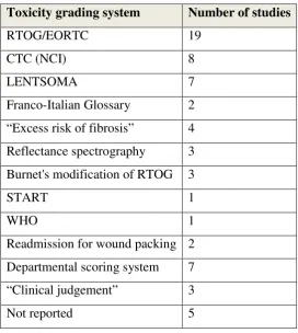
• RTOG/EORTC Acute Radiation Morbidity Scoring criteria and Late Morbidity scoring criteria (Cox, Stetz et al. 1995).
• LENT/SOMA (Late Effects on Normal Tissues/ Subjective Objective Management Analytic) scales (Rubin, Constine et al. 1995; Rubin, Constine et al. 1995; Denekamp, Bartelink et al. 1996; Denekamp, Bartelink et al. 1996; Dorr and Hendry 2001)
• NCI CTC (Common Toxicity Criteria) system (Trotti, Byhardt et al. 2000; Trotti 2002; Trotti, Colevas et al. 2003; Trotti and Bentzen 2004)
• UCLA index
• Franco-Italian Glossary.
Many centres have devised their own scoring systems or modified those above.
1.4 What determines the radiation dose prescribed in today’s clinical practice?
11 animal tumour systems and in clinical practice (Fletcher 1972; Suit 1982; Steel and Peacock 1989; Withers 1992; Okunieff, Morgan et al. 1995; Zelefsky, Leibel et al. 1998; Kuban, Pollack et al. 2003; Bradley, Graham et al. 2005; Belderbos, Heemsbergen et al. 2006). Equally increasing radiation dose also increases the probability of normal tissue complications (Bentzen 1994; Bentzen 2002). The radiotherapy schedules in clinical practice have been developed to balance tumour control probability with normal tissue complication probability to try to maximise tumour control whilst keeping the risk of severe normal tissue toxicity in the treated population at an acceptable level. What constitutes an “acceptable” level of risk depends on the specific toxicity and its effect on the functioning of the patient. Spinal cord damage has a potentially devastating effect on the patient and so even a small risk is unacceptable, whilst a slightly higher risk of damage that is primarily cosmetic may be tolerated.
Few prospective dose-escalation studies have been performed to determine the maximum tolerated radiation dose (MTD) in any given tissue and there is little quantitative data on normal tissue tolerance, particularly for those tissues where damage can lead to a catastrophic functional outcome, such as the spinal cord. MTD-finding studies are difficult to conduct as it is the late irreversible and severe effects on normal tissues rather than acute reversible toxicities that are generally dose limiting. A number of dose escalation studies aiming to define MTD in the modern radiotherapy era do exist, but in general the dose- effect relationships for toxicity of individual normal tissues have been derived empirically from clinical observation, retrospective data and consensus opinion. (Emami, Lyman et al. 1991). This has led to the development of parameters attempting to define the risk of normal tissue toxicity for a given radiotherapy schedule such as the TD5/5 i.e. the
12 1.4.1 Factors influencing the risk of and severity of radiation toxicity
The risk of normal tissue toxicity is not only defined by the prescribed radiation dose. There are other features of the treatment itself and patient-related factors that are known influence the risk of developing normal tissue complications due to radiotherapy.
1.4.1.1Treatment-related factors
Dose per fraction
It is recognised that delivering the total radiation dose in multiple small fractions rather than fewer larger fractions results in a reduction in severity of late effects. Late effects are more sensitive to changes in fraction size than acute effects (Thames, Withers et al. 1982).
Overall treatment time
Acute effects are sensitive to changes in the overall treatment time but late effects less so – it has been demonstrated that a reduction in the overall treatment time in head and neck cancer by treating with two or three fractions of radiotherapy per day (accelerated fractionation) increases the risk and severity of acute effects, with a decrease in some late toxicity end-points(Dische, Saunders et al. 1997; Bourhis, Calais et al. 2004).
Volume of irradiated tissue
13 Dose homogeneity
Despite careful attention to beam modification and arrangement patient outline and tumour position may mean that there are unavoidable “hot-spots” within the irradiated volume which will receive a higher dose and dose per fraction than other areas. As a consequence this tissue will be at a higher risk of complications.
Concurrent chemotherapy
The concurrent administration of chemotherapy with radiotherapy has been shown in randomised controlled trials and large meta-analyses to improve local control and survival in some tumour types, particularly in cervical and head and neck cancer (Pignon, Bourhis et al. 2000; Green, Kirwan et al. 2005). There is also clear randomised controlled evidence that concurrent chemotherapy increases the risk and severity of acute radiation reactions although the effect on late toxicity is less clear (Bourhis, Calais et al. 2004; Denis, Garaud et al. 2004; Green, Kirwan et al. 2005).
1.4.1.2Patient-related factors
Co-morbidity
Patient comorbidities can influence the development of radiation normal tissue toxicity. Co-morbidities that affect the vascular system especially diabetes and uncontrolled hypertension appear to increase the risk of radiation toxicity (Turesson, Nyman et al. 1996; Herold, Hanlon et al. 1999). A systematic review has identified that connective tissue disease is associated with an increased risk of late radiation toxicity (Holscher, Bentzen et al. 2006)
Smoking
Smoking during therapy can increase the risk and severity of both acute and late normal tissue radiation toxicity (Johansson, Bjermer et al. 1998; van der Voet, Keus et al. 1998; Eifel, Jhingran et al. 2002; Twardella, Popanda et al. 2003; Wells, Macmillan et al. 2004)
Body Mass Index
14 Haemoglobin
Haemoglobin level during radiotherapy may influence the development of normal tissue toxicity. There is some evidence suggesting that a low haemoglobin during treatment is associated with a lower risk of normal tissue toxicity but this has not been a consistent finding by all groups (Bentzen and Overgaard 1994; Henke, Bechtold et al. 2000; Daly, Poulsen et al. 2003).
Genetic syndromes
Some groups of patients may have a genetic susceptibility to radiation normal tissue damage – rare but recognised genetic syndromes associated with increased normal tissue radiosensitivity include Ataxia-telangiectasia, Blooms’, Fanconi’s anaemia and Nijmegen Breakage syndrome. (Rogers, Plowman et al. 2000; Gatti 2001; Alter 2002; McMillan 2002)
1.5 Individual intrinsic normal tissue radiosensitivity
15 syndromes mentioned above, a component of which is enhanced radiation sensitivity of normal tissue
If the biological determinants of intrinsic normal tissue radiosensitivity could be identified they could potentially form the basis of a diagnostic test or “predictive assay” to identify individuals at risk of severe normal tissue damage.
1.6 Predictive assays for normal tissue radiosensitivity
1.6.1 Clinical value of a predictive assay of normal tissue radiosensitivity
If a high precision assay of normal tissue radiosensitivity existed which could accurately and reliably predict an individual’s risk of developing severe normal tissue radiation toxicity, how could its results be incorporated into clinical practice and used to improve the therapeutic index of radiotherapy?
16
0 200 400 600
0 5 10 15 20
+/- 2 SDs from mean
A
A = highly radiosensitive minority of normal population Dose required for normal tissue isoeffect
F
r
e
q
u
e
n
c
y
Figure 1.3 Frequency distribution of normal tissue response amongst patients treated with an identical radiotherapy schedule
If it were possible to measure an individual’s propensity to develop normal tissue damage prior to starting radiotherapy then theoretically their radiation treatment could be modified with the aim of preventing serious toxicity in those who are radiosensitive. A strategy using normal tissue radiosensitivity testing and prospective prediction of normal tissue response might also permit safe dose escalation in appropriate “non-sensitive” individuals which should result in improved rates of tumour control. In this way treatment could be “biologically” individualised and the therapeutic ratio of radiotherapy could be improved. The results from a predictive assay of normal tissue radiosensitivity could therefore potentially be used to:
a) Screen for the minority of individuals with very high normal tissue radiosensitivity and treat these with a reduced radiation dose or offer an alternative to radiotherapy such as surgical resection.
b) Screen out the radiosensitive minority and escalate the dose in the remainder.
17 A number of authors have modelled the potential impact of normal tissue radiosensitivity testing on outcome after radiotherapy (Norman, Kagan et al. 1988; West and Hendry 1992; Tucker, Geara et al. 1996; Bentzen 1997; MacKay, Niemierko et al. 1998; Mackay and Hendry 1999) . In most cases a clinically useful improvement in tumour control probability without a corresponding increase in normal tissue radiation toxicity has been predicted.
Tucker et al (Tucker, Geara et al. 1996) have estimated the potential for individualising dose prescription in order to attain a uniform 15% risk of severe late damage to mucous membrane and bone, based on results from a study correlating fibroblast radiosensitivity in vitro and late normal tissue complications. They estimate that for the 8 patients in the study who were sensitive to radiation, an average dose reduction of 13.1Gy would have been necessary to reduce the Normal Tissue Complication Probability (NTCP) to 15%. In 20 of the 21 remaining cases, a dose escalation averaging 7.7Gy could be tolerated whilst still maintaining the risk of severe late damage at 15% for each individual. The resulting effect on tumour control probability is not modelled but the authors assume that these dose modifications would result in higher Tumour Control Probability (TCP) than observed in reality as there would have been twice as many dose increases as decreases.
18 In the context of an ideal predictive assay, the predicted gain in population TCP resulting from tailoring individual patient dose to attain a NTCP of 5% is 30% and is predicted to be highest when individuals have a higher sensitivity and the distribution of radiosensitivity in the population is greatest. When the distribution of radiosensitivity in the population is high, the spread of doses required to achieve the isoeffect of 5% NTCP is wide, with a small tail of very high doses achievable of up to 200Gy. In reality, the inherent inaccuracies in the fibroblast radiosensitivity data used to generate the model have probably resulted in an overestimate of the spread of intrinsic radiosensitivity in the population. If radiosensitivity is modelled with a narrower distribution within the population, possibly more reflective of reality, the range of doses predicted to give a NTCP of 5% is narrower and the high dose tail much smaller.
In reality results of predictive assays measuring a biological endpoint such as normal tissue radiosensitivity are likely to possess an inherent variability or “noise” – i.e. the same test performed on repeated occasions on the same individual may give differing results. MacKay and Hendry demonstrated that when assay noise is fitted into their model no gain in TCP could be achieved if the assay coefficient of variation was greater than 50% of the inter-individual biological variation. They conclude that a crucial factor in gaining a therapeutic advantage by individualisation of radiotherapy dose is the development of an accurate and reliable assay.
19 control probability. In the simulation the individualisation of dose increased the average dose received by the population as a whole by 1.4Gy, but tumour control dropped by 3%. Because of the non-linearity of the dose response curve for TCP, a positive dose increment of a given size will increase the TCP by a smaller amount than the same dose decrement would decrease TCP. These conclusions have been criticised by other authors who feel that some of the assumptions used in Bentzen’s model are based on too small a data set and are therefore inaccurate (MacKay, Niemierko et al. 1998; Mackay and Hendry 1999).
An alternative to complete individualisation of dose prescription is to use predictive testing of normal tissue radiosensitivity to split the population into three groups – high, average and low radiosensitivity - and to treat each group differently (Mackay and Hendry 1999) – (Table 1). This would avoid the potential problems likely to be encountered whilst trying to individualise dose prescriptions using an assay that is less than completely reliable.
Table 1.1 Theoretical division of population into 3 groups according to normal tissue radiosensitivity and the dose to which each group could be treated to maintain NTCP<5%
CV = 0.2 CV = 0.1
Radiosensitivity High Average Low High Average Low
% Population 12 43 45 18 42 40
Prescribed dose (Gy) 56 66 78 64 70 78
Results are shown for 2 different populations with log normal distribution of SF2=0.36 and CV (coefficient of variation) of assay results across the population of 0.2 and 0.1. Before splitting into groups the whole population would have been treated with 60Gy (CV = 0.2) or 66Gy (CV = 0.1) in order to keep the population NTCP <5%. (Mackay and Hendry 1999)
20 When the assay CV is 50% of the biological variation within the population it is still possible, however, to have a potential gain in TCP of 27%. This contrasts to the effect of assay unreliability on complete individualisation of dose prescription where no gain in TCP was expected with this level of assay unreliability. In fact, by dividing the population into groups in this way it is predicted that an increase in TCP of 11% is still possible even if the assay CV is the same as the biological CV. Tripartite separation of the population would therefore seem to be less sensitive to assay uncertainty than complete individualisation of radiotherapy dosing.
If tumour radiosensitivity is correlated with normal tissue radiosensitivity then the potential clinical gains resulting from normal tissue radiosensitivity testing are even greater (MacKay, Niemierko et al. 1998). However, whether such a relationship exists is debatable and if present is likely to be weak. (West and Hendry 1992; Geara, Peters et al. 1996; Bernier, Thames et al. 1998; West, Davidson et al. 1998).
Mathematical models are provisional and require appropriate caveats (Jones and Dale 1999) – their output is dependent on the quality of the data entered into the model and so results cannot be assumed to be generalisable, and they must be re-tested when new clinical data is available. They do however provide a useful tool for estimating possible outcomes of changes in therapy without conducting clinical trials which, in the case of normal tissue radiosensitivity testing, would not only be costly and time-consuming, but potentially dangerous with a concomitant risk of loss of tumour control and increased radiation toxicity. Mathematical modeling so far would seem to support the concept that normal tissue radiosensitivity testing could potentially improve the therapeutic index of radiation therapy if incorporated into clinical practice.
1.6.2 Development of a predictive assay of normal tissue radiosensitivity
21 accuracy is the ability of an assay to correctly diagnose the condition in question and is measured by assay sensitivity and specificity. Ideally an assay should be generalisable to a large population and could be performed in different laboratories with reproducible results. The results of a diagnostic assay should influence clinical decisions - to do this it must not only be reliable, precise and accurate, it must be able to produce results quickly in a clinically relevant time-frame. It must also be affordable and acceptable to patient and clinician without the need for unpleasant or dangerous invasive procedures otherwise it will not be adopted in routine clinical practice no matter how efficiently it performs.
A fundamental issue in the development of any diagnostic assay is determining which parameter should be measured and how to measure it reliably. One of the main problems in the development of a biological assay to predicate normal tissue toxicity has been the relative lack of knowledge of the molecular, cellular and tissue pathophysiology underlying acute and late radiation toxicity. Strategies tested so far have had mixed outcomes.
1.6.2.1Functional cell-based assays
Assays that are based on sampling of living cells from an individual and examining their response to ex-vivo irradiation have until recently been the main focus of most work on predictive testing. The fundamental principle underlying these assays is that there is a relationship between in vitro cellular response to irradiation and normal tissue toxicity and that it is possible to test for this relationship using a single cell type sampled from an individual as a surrogate for the normal tissue in question. Assay end-points examined have included clonogenic cell survival, assays of radiation-induced DNA damage, apoptosis, and differentiation. Fibroblasts derived from skin biopsy and peripheral blood lymphocytes have been the most frequently tested surrogate tissue
Clonogenic cell survival.
22 between fibroblast radiosensitivity as measured by a clonogenic cell survival assay and late skin toxicity in 6 patients treated with post-mastectomy chest wall radiation(Burnet, Nyman et al. 1992; Burnet, Nyman et al. 1994) . Subsequent studies confirmed this relationship (Geara, Peters et al. 1993; Brock, Tucker et al. 1995; Johansen, Bentzen et al. 1996) whilst others did not (Russell, Grummels et al. 1998; Peacock, Ashton et al. 2000; Oppitz, Baier et al. 2001). Fibroblast radiosensitivity correlated with the development of late central nervous system complications after stereotactic radiosurgery for arterio-venous malformations in a small group of Canadian patients (Raaphorst, Malone et al. 2002) Late radiation effects after head and neck radiotherapy, however, did not correlate with fibroblast SF2 in a prospective study of 25 patients (Rudat, Dietz et al. 1999)
Similarly studies examining the relationship between acute radiotherapy toxicity and the clonogenic survival of skin fibroblasts have revealed conflicting results (Begg, Russell et al. 1993; Rudat, Dietz et al. 1997; Oppitz, Baier et al. 2001; El-Awady, Mahmoud et al. 2005)
Lymphocyte radiosensitivity measured by clonogenic survival has been correlated with the development of late effects after pelvic radiotherapy (West, Davidson et al. 2001)
Assays of radiation-induced DNA damage
23 level of DNA double-strand breaks (DSBs) rather than with other types of DNA damage. Cells are very sensitive to formation of DNA DSBs and even a single DSB can trigger the damage sensing process (Huang, Clarkin et al. 1996) and can lead to cell death if unrepaired (Bennett, Lewis et al. 1993). Maintaining the integrity of DNA therefore seems to be very important biologically, and the ability to detect and repair DNA damage appears to determine whether a cell will survive following radiation.
It follows then that an individual’s ability to detect, process and repair DNA damage may in part determine their intrinsic normal tissue radiosensitivity and radiation tolerance and several studies have studied the relationship between in vitro DNA damage and repair and clinical radiosensitivity using assays of DNA damage such as the comet assay or pulsed-field gel electrophoresis (PFGE) or cytogenetic endpoints of DNA double-strand breaks such as the micronucleus assay, G2 chromatid radiosensitivity or studies of chromosome re-arrangements
A positive correlation has been found between cellular radiosensitivity as measured by the micronucleus assay and both early (Widel, Jedrus et al. 2003) and late radiotherapy toxicity (Nachtrab, Oppitz et al. 1998; Barber, Burrill et al. 2000; Lee, Allison et al. 2003; Widel, Jedrus et al. 2003). Rached et al however found no such relationship between micronucleus yield and acute radiotherapy toxicity (Rached, Schindler et al. 1998)
Chromosome aberrations or G2 chromatid damage induced by in vitro irradiation have been found to correlate with acute (Kearsley, Fang et al. 1998) and late radiation toxicity (Borgmann, Roper et al. 2002)( Borgmann, Roper et al. 2000; (Neubauer, Dunst et al. 1997; Barber, Burrill et al. 2000)
24 al. 2000; Dickson, Magee et al. 2002; El-Awady, Mahmoud et al. 2005; Lopez, Guerrero et al. 2005; Wang, Chen et al. 2005; Pinar, Lara et al. 2007)
Radiation-induced apoptosis
Crompton et al have demonstrated that the lymphocytes in blood samples from patients with high levels of both acute and late radiation toxicity undergo less radiation-induced apoptosis in vitro as measured by flow cytometry than those from patients with average levels of normal tissue toxicity (Crompton, Miralbell et al. 1999). The same group confirmed this finding in a prospective study demonstrating a relationship between increased late radiation toxicity and lower levels lymphocyte apoptosis after in vitro irradiation of peripheral blood samples (Ozsahin, Crompton et al. 2005). Another group however, could not correlate apoptotic rate in human peripheral blood lymphocytes after in vitro irradiation with rates of breast fibrosis, telangiectasia or breast retraction after radiotherapy for breast cancer (Barber, West et al. 2000)
1.6.2.2 Other strategies in development of predictive assays of normal tissue radiosensitivity:
As shown above, the results from functional cell-based assays of normal tissue radiosensitivity have been inconsistent. Authors have raised concerns that the assays lack reliability due to confounding external influences (West, Davidson et al. 2001). Even when a statistically significant relationship between an assay result and normal tissue toxicity has been demonstrated considerable overlap in assay results between patients with high and low normal tissue toxicity has led authors to conclude that cell based assays are not sufficiently discriminatory to be of any use in the clinic (West, McKay et al. 2005; Burnet, Elliott et al. 2006).
25 those involved in DNA repair, free radical scavenging, cell cycle control and cytokine release, it may be possible to predict radiation sensitivity(Andreassen, Alsner et al. 2002) A tissue bank with linked data regarding patients demographics, comorbidity, tumour, and precise radiotherapy details and dosimetry has been set up in the EU to enable this process (GENEPI)(West, McKay et al. 2005) . Initial results from genotyping assays have been promising but positive results have so far not been confirmed in appropriate validation studies (Andreassen, Alsner et al. 2003; Andreassen, Alsner et al. 2005; Andreassen, Alsner et al. 2006).
Other assays based on serum cytokine measurements during radiotherapy have shown a correlation between increased TGF-β at the end of radiotherapy and interleukin 6 levels during radiotherapy for lung cancer and the development of subsequent radiation pneumonitis (Anscher, Kong et al. 1997; Chen, Hyrien et al. 2005). These assays have already been tested to see if radiation dose can safely be escalated in those patients identified as having a low susceptibility to pneumonitis (Anscher, Marks et al. 2003). This study confirmed the principle that TGF-β could predict those at risk of pneumonitis – unfortunately whilst patients who were dose escalated had acceptable rates of pneumonitis they did suffer severe complications in other normal tissues which had not been predicted by the assay.
26 all is not yet known (Rodningen, Overgaard et al. 2005). It remains to be seen whether SNPs in a few of these candidate genes can reliably predict a highly radiosensitive phenotype.
In the meantime, functional assays have the advantage of not relying on an in depth knowledge of the molecular machinery governing radiation response. In effect the cell is a “black box” – we can put in a signal (the test dose of radiation) and measure the output signal (the assay result) without knowledge of the circuitry and wiring within. Is there sufficient evidence to be sure that function cell based assays have no role in predictive testing of normal tissue radiosensitivity?
As described above many groups have tried to develop functional cell-based assays of intrinsic normal tissue sensitivity based on study designs which correlate in vitro cellular radiosensitivity with severity of normal tissue reaction. These studies must collect standardised information on radiotherapy toxicity as well as radiation exposure and factors other than intrinsic normal tissue radiosensitivity known to modify normal tissue response so allowing potential confounding factors to be taken into account during data analysis. Twardella et al performed a systematic review of studies examining the relationship between cell based assays and normal tissue toxicity in patients receiving radiotherapy for breast cancer (Twardella and Chang-Claude 2002) . They found that of the 25 studies identified limitations in study design were frequently found, with potential sources of bias arising from misclassification of patients due to non-standardisation of assessment of treatment related side effects, selection bias by studying convenient patient groups rather than truly representative groups and confounding due to failure to adjust analysis for important factors influencing normal tissue reaction. An estimate of assay sensitivity and specificity was performed in only one of the studies identified. Given the methodological problems in assay design and testing reported by Twardella et al it is possible that the assumption that functional cell based assays are unhelpful in predictive testing of normal tissue sensitivity may be premature.
27 identified – as with predictive testing of normal tissue sensitivity studies looking at similar assay end points have yielded inconsistent results with methodological differences, poor study design, small sample sizes, non-standardised or non-reproducible assays, and inappropriate statistical analyses being thought to account for these inconsistencies (Fielding, Fenoglio-Preiser et al. 1992; Simon and Altman 1994; Hayes, Bast et al. 1996; Hall and Going 1999; Schilsky and Taube 2002).
A joint initiative between the National Cancer Institute (NCI) and European Organisation for the Research and Treatment of Cancer (EORTC) has identified poor study design and analysis, assay variability and inadequate reporting of studies as some of the major barriers to progress in this field. Following this the reporting recommendations for tumour marker prognostic studies have been published (REMARK) (McShane, Altman et al. 2005). These recommendations suggest the systematic reporting of key study features including quality control assessment procedures for the prevention of bias in assay and clinical measurement, statistical analysis and appropriate reporting of findings which should allow transparent and complete reporting with relevant information so that the usefulness of the data can be judged clearly by others. The same reporting standards are transferable to predictive assays of normal tissue radiosensitivity.
1.7 H2AX phosphorlyation in human peripheral blood lymphocytes – a potential predictive assay of normal tissue sensitivity.
28 1.7.1 DNA DSBs and γH2AX induction
When a DSB is introduced into DNA the histone protein H2AX becomes rapidly phosphorylated at serine 139 within its COOH-terminal region involving hundreds to thousands of H2AX molecules in a megabase region on either side of the DSB (Rogakou, Pilch et al. 1998; Stiff, O'Driscoll et al. 2004) . A commercially available monoclonal antibody to the phosphorylated form of H2AX (γH2AX) has been developed and using immunocytochemistry it is possible to visualise these DNA DSBs as large foci within the cell nucleus (Rogakou, Boon et al. 1999). A single DSB is sufficient for the formation of a γH2AX focus and there appears to be a 1:1 correspondence between the number of DSBs
and γH2AX foci after DNA damage induction (Sedelnikova, Rogakou et al. 2002). Immunofluorescent staining for γH2AX foci can detect DSB induction at much lower doses than established DNA DSB assays and has been reported to be sensitive enough to detect DSBs in cells after doses as low as 0.001Gy (Rothkamm and Lobrich 2003).
The phosphorylation of H2AX is thought to recruit DNA repair factors to the site of the DNA DSB (Paull, Rogakou et al. 2000) and may also be involved in the amplification of DNA damage signals that activate the G2/M checkpoint to prevent damaged cells from entering mitosis (Fernandez-Capetillo, Chen et al. 2002). Inability to form γH2AX foci has been correlated to radiosensitivity, genomic instability and other repair defects (Bassing, Chua et al. 2002; Celeste, Petersen et al. 2002; Kuhne, Riballo et al. 2004; Taneja, Davis et al. 2004). The mechanism by which γH2AX is removed following DNA repair is incompletely understood. In some studies the kinetics of γH2AX loss mirrors the kinetics of DNA DSB rejoining (Rothkamm and Lobrich 2003) whilst others have found that although the number of γH2AX foci formed after irradiation correlates with the number of double strand breaks formed the kinetics of foci development and loss differ from those characteristic of double-strand break re-joining, and loss of γH2AX may therefore be indicative of more than simple DSB re-joining..(MacPhail, Banath et al. 2003).
29 culture correlates with clonogenic survival at 2Gy (MacPhail, Banath et al. 2003) and the rate of γH2AX disappearance is slower in radiosensitive tumour cells and radiosensitive murine normal tissue than radioresistant cell lines or normal tissue (Olive and Banath 2004). The techniques for immunofluorescent staining and γH2AX quantification in cultured cells are quick and yield results in a number of days rather than many weeks. Quantification of γH2AX induction and the kinetics of γH2AX loss in normal human cells or tissues after a test dose of radiation could therefore potentially form the basis of a predictive assay of human normal tissue radiosensitivity.
Normal cells sampled from patients and utilised for predictive testing should be plentiful and easily and rapidly accessible by non-invasive means. For γH2AX quantification the cells would ideally be non-cycling as γH2AX foci are also induced at collision of replication forks during DNA replication (Furuta, Takemura et al. 2003). Human peripheral blood lymphocytes (PBLs) obtained by venepuncture in the clinic fulfil both these criteria and could be used as a surrogate tissue to test if there is a relationship between cellular γH2AX induction and loss in vitro after irradiation and normal tissue radiosensitivity.
1.8 Aims
The aims of this project were therefore:
• To conduct a systematic review of the literature regarding functional cell-based assays in the predictive testing of normal tissue radiosensitivity to ascertain whether studies so far have been performed with sufficiently rigorous approach to assay quality control, avoidance of bias in study design, and statistical analysis of results, and to determine if the perception that cell-based assays are not helpful in predicting normal tissue radiosensitivity is really valid based on current literature.