Vol. 66, published by Elsevier, and the attached copy is provided by Elsevier for the author's benefit and for the benefit of the author's institution, for non-commercial research and educational use including without limitation use in instruction at your institution, sending it to specific colleagues who know you, and providing a copy to your institution’s administrator.
All other uses, reproduction and distribution, including without limitation commercial reprints, selling or licensing copies or access, or posting on open internet sites, your personal or institution’s website or repository, are prohibited. For exceptions, permission may be sought for such use through Elsevier's permissions site at:
http://www.elsevier.com/locate/permissionusematerial
From: Joanne Carney, Sammy A. Mason, Cedric Viero, and Alan J. Williams, The Ryanodine Receptor Pore: Is There a Consensus View? In Irina Serysheva, editor: Current Topics in Membranes, Vol. 66, Burlington: Academic Press, 2010, pp. 49-67.
ISBN: 978-0-12-381037-3 © Copyright 2010 Elsevier Inc.
CHAPTER 3
The Ryanodine Receptor Pore:
Is There a Consensus View?
Joanne Carney, Sammy A. Mason, Cedric Viero, and Alan J. Williams
Department of Cardiology, Wales Heart Research Institute, School of Medicine, Cardiff University, Heath Park, Cardiff, United Kingdom
I. Overview II. Introduction III. Ion Handling in RyR
A. Basic Properties of Ion Handling in the RyR Isoforms
B. What is Responsible for the High Rate of Ion Translocation in RyR? IV. Where is the PFR in the RyR Channel?
A. Evidence from Cryo-Electron Microscopy
B. Analogies with KþChannels and Evidence from Functional Studies of Mutant RyR Channels
C. Further Analogies Between the PFR of KþChannels and RyR V. Attempts to Identify the Structure of the RyR PFR
A. A Model of the RyR Pore Using KcsA as a Template B. High-Resolution Images of the PFR of RyR
C. A RyR PFR Model Based on a High-Resolution Cryo-EM Structure VI. Theoretical Approaches to Understanding the Mechanisms Underlying Ion
Translocation and Discrimination in RyR
VII. Testing Physical and Theoretical Models of the RyR PFR by Residue Substitution A. RyR1
B. RyR2
VIII. Concluding Remarks References
I. OVERVIEW
The intracellular Ca2þ-release channel referred to as the ryanodine
recep-tor (RyR) is a key facrecep-tor in a plethora of biological and pathophysiological processes and is therefore the focus of interest of many scientists and
Current Topics in Membranes, Volume 66 1063-5823/10 $35.00
clinicians. In recent years, an ever-growing body of evidence has emerged detailing the ligands and mechanisms involved in the regulation of RyR activity, and how this might be altered in disease. This information is reviewed in other articles in this publication. Here we review a fundamental aspect of RyR function, namely the structures and mechanisms that control which, and how many, ions can flow through the open channel and underpin the unusual ion handling characteristics of RyR and its great efficiency as a
Ca2þ-release channel. Therefore, we will limit our discussion to a relatively
small region of the channel that provides the pathway for ion movement across the membrane: the pore-forming region (PFR). Our present under-standing of these processes has been informed by a wide range of approaches. Information on the likely structure of the PFR has been provided by high-resolution cryo-electron microscopy (cryo-EM) and by molecular modeling, following the identification of structural analogies between RyR and other ion channels. The mechanisms involved in discrimination and translocation have emerged from detailed characterization of channel function, theoretical models, and the interpretation of the consequences of residue mutation in the PFR. Together, these approaches have been used to identify regions of RyR that are critical for ion movement and may contribute to the binding site for ryanodine.
II. INTRODUCTION
Endo/sarcoplasmic reticulum (ER/SR) intracellular membrane systems
contain two species of Ca2þ-release channel, the inositol trisphosphate
receptor (InsP3R) and the RyR, so named because this channel possesses a
high-affinity binding site for the plant alkaloid ryanodine (Sutko, Airey,
Welch, & Ruest, 1997). Three mammalian isoforms of RyR have been identified: RyR1 found predominantly in skeletal muscle, RyR2 found in both cardiac muscle and the brain, and RyR3 which is expressed at low levels
in a wide range of tissues (Fleischer, 2008). Functional RyR channels are
homotetramers with each monomer containing approximately 5000 amino acids. Detailed information on the functional properties of RyR channels has been obtained following the incorporation of individual channels into artificial phospholipid bilayers which permits the measurement of single
channel gating and ion handling (Williams, West, & Sitsapesan, 2001).
RyR channels are extremely efficient Ca2þ-release channels and this
III. ION HANDLING IN RYR
A. Basic Properties of Ion Handling in the RyR Isoforms
The first characterization of ion handling in RyR was carried out by
Meissner and colleagues using purified rabbit RyR1 (Smith et al., 1988).
The studies demonstrated that RyR1 (a) is cation selective, (b) is divalent selective, (c) has a higher unitary conductance for monovalent than divalent cations, and (d) is essentially nonselective between group 1a monovalent cations. These basic features were confirmed in detailed characterizations
of monovalent (Lindsay, Manning, & Williams, 1991) and divalent ion
handling (Tinker & Williams, 1992) in purified sheep RyR2 (seeTable I).
In addition to the above, RyR2 was found to be relatively nonselective between individual mono- or divalent cations, somewhat selective for divalents
ðPX2þ=PKþ6Þ, while exhibiting a high affinity (mM) for divalents (Tinker,
Lindsay, & Williams, 1992b) compared to a relatively low affinity (mM) for monovalent cations. The different experimental conditions that have been used to examine ion handling in the RyR isoforms makes a direct comparison of
properties difficult; however, basic features are compared inTable II.
TABLE I
Ion Handling in RyR2
Xþ pXþ/pKþ g(pS) KD(mM) gmax(pS)
Kþ 1.00 723 19.9 900
Naþ 1.15 446 17.8 516
Csþ 0.61 440 34.0 588
Rbþ 0.87 621 n.d. n.d.
Liþ 0.99 215 9.1 248
X2þ pX2þ/pKþ g(pS) *K
D(mM) pX2þ/pBa2þ
Ba2þ 5.8 2025 0.165 1.0
Sr2þ 6.7 1664 0.123 1.1
Ca2þ 6.5 1355 0.116 1.1
Mg2þ 5.9 894 0.116 1.1
pXþ/pKþ: bi-ionic 210 mM.
g(pS): determined at symmetrical 210 mM [Xþ] or bi-ionic with [Kþ/X2þ].
KDandgmaxfrom Michaelis–Menten conductance–activity plots. [Xþ] or *predicted from RyR Eyring
rate theory model (Tinker et al., 1992b). pX2þ/pKþ: bi-ionic 210 mM.
pX2þ/pBa2þ: bi-ionic with 210 mM [Ba2þ/X2þ].
n.d.: not determined.
Differences between the RyR isoforms are minimal; however, there are some limited differences in the relative conductance of monovalent cations and permeability sequences of RyR1 and RyR2 that may suggest some minor differences in the mechanisms governing ion handling in these channels. The sequence for monovalent and divalent cation binding in RyR2
corres-pond toEisenman (1962) and Sherry (1969)sequences XI and VII,
respec-tively, indicating the involvement of high field strength sites in the process of
discrimination (Tinker et al., 1992b). Based on the structure of Ca2þbinding
proteins, it was predicted that cation binding at such sites could be coordi-nated by carboxyl oxygens. To account for the higher relative permeability of divalents over monovalents, it was proposed that the RyR PFR was likely to
contain a high density of such sites (Tinker et al., 1992b).
B. What is Responsible for the High Rate of Ion Translocation in RyR?
The maximum rate of cation translocation in RyR is phenomenal, with saturating unitary conductance for monovalent cations approaching 1 nS (ap-proximately twice the theoretical limit for a selective channel based on the laws
of diffusion;Hille, 1991). Clearly an enormous unitary conductance is a useful
property for a Ca2þ-release channel, but how is this achieved? A range of
potential contributing mechanisms have been proposed, for example, rates of delivery of cations could be maximized by a high density of acidic residues at the
entrance of the PFR (Tinker et al., 1992b) or the RyR PFR could be occupied
TABLE II
Comparison of Ion Handling in RyR Subtypes
RyR1 RyR2 RyR3
[Kþ]g(pS) 772 723 777
[Ca2þ]g(pS) 123 135 137
pCa2þ/pKþ 6.6 6.5 6.3
Permeability sequence Liþ>Naþ>Kþ>Rbþ>Csþ Naþ>Kþ>Liþ>Rbþ>Csþ n.d. Conductance sequence Kþ>Csþ>Rbþ>Naþ>Liþ Kþ>Rbþ>Naþ>Csþ>Liþ n.d.
RyR1, RyR2 and RyR3 K+ slope conductances were examined in symmetrical [K+] 250 mM/ 210 mM/250 mM, respectively. Divalent permeability ratios were examined in bi-ionic [K+
/Ca2+
] at 50/ 50 mM, 210/210 mM and 250/250 mM, respectively. Monovalent permeability ratios were examined in bi-ionic [K+
/X+
] 210/200–210 mM, 210/210 mM and 250/250 mM, respectively. Conductance sequences were compared in symmetrical [X+
] 200–215 mM (RyR1) and in symmetrical 210 mM (RyR2). n.d. : not deter-mined. Readers may refer to the following publications for further information: RyR1 (Shomer et al., 1994; Smith et al., 1988; Xu et al., 1993), RyR2 (Lindsay et al., 1991; Tinker & Williams, 1992) and RyR3
by more than one ion at a time leading to increased rates of ion exit by ion–ion
repulsion (Smith et al., 1988). Another suggestion, based on earlier predictions
for large conductance Kþchannels (Latorre & Miller, 1983), was that the PFR
of RyR, in comparison with other ion channels, was both wide and short so
maximizing both rates of ion entry and exit (Williams, 1992).
The dimensions of some aspects of the RyR PFR have been investigated.
The minimum radius of the RyR PFR has been estimated as 3.5 A˚ based on
the relative permeability of organic monovalent cations of known
dimen-sions (Tinker & Williams, 1993). However, subsequent experiments have
shown that this region of the PFR is likely to be flexible. With a high driving
force neomycin (minimum radius 5 A˚ ), a normally impermeant blocking
polycation, can move through the PFR (Mead & Williams, 2002a). The
binding of ryanodine to RyR prevents this translocation presumably by
decreasing the flexibility of the PFR (Mead & Williams, 2002b). Information
on aspects of the length of the PFR is also available.
The length of the voltage drop across the channel has been measured at
approximately 10 A˚ by monitoring blocking parameters of bis-quaternary
ammonium ions of varying length (Tinker & Williams, 1995) and the region
of RyR in which single-file diffusion occurs has been estimated as 9 A˚ from
measurements of streaming potentials (Tu, Velez, Brodwick, & Fill, 1994).
Taken together these parameters are consistent with the proposal that the PFR of RyR is, in comparison with more selective, lower conductance,
channels such as the bacterial Kþchannel KcsA, relatively wide and short
(Williams et al., 2001).
IV. WHERE IS THE PFR IN THE RYR CHANNEL?
A. Evidence from Cryo-Electron Microscopy
Three-dimensional structures of RyR tetramers have been obtained by reconstruction of images gathered from electron micrographs of frozen-hydrated single isolated channels. Of the three RyR isoforms, the most extensive structure–function analysis has been carried out with RyR1 due to its high abundance in skeletal muscle and the relative ease with which it can be purified. Until recently, the most detailed structures available were
at resolutions of only 25–30 A˚ (for a review seeSerysheva, Chiu, & Ludtke,
2007). Although no secondary structure elements can be identified at this
resolution, it is clear that RyRs have an overall mushroom shape with a large cytoplasmic foot assembly connected to a transmembrane (TM)
stalk-like structure (Radermacher et al., 1994; Serysheva et al., 1995). This
technique has been used to identify domains in the cytoplasmic foot which
are binding sites for various effectors (Liu, Zhang, Wang, Wayne Chen, & Wagenknecht, 2004; Samso & Wagenknecht, 2002; Wagenknecht et al.,
1997). By analogy with other ion channels, the PFR of RyR is likely to be
located in the TM portion of the structure and an apparent opening running axially into the TM domain has been identified in what is believed to be an
open conformation of RyR1 (Orlova, Serysheva, van Heel, Hamilton, &
Chiu, 1996). This opening is not visible in the closed conformation of the channel and these studies have also highlighted other structural alterations in RyR that may occur during the transition from a closed state to an open state of the channel. Recent technical advances have yielded structural informa-tion on the TM domain of RyR1 at considerably improved resoluinforma-tion and
this will be discussed inSection V.B.
B. Analogies with KþChannels and Evidence from Functional Studies
of Mutant RyR Channels
In 1999,Balshaw, Gao, and Meissner (1999)identified a motif located in a
luminal loop between the last two membrane spanning helices in the primary
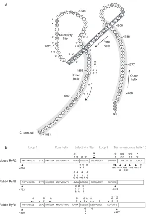
structure of RyR (GGGIGD) (seeFig. 1) as being analogous to the signature
selectivity sequence of Kþchannels and suggested that this region was likely
to be a component of a PFR in RyR. Consistent with this proposal, mutation of various residues within this motif, and of residues flanking this motif, were
shown to alter ion handing properties of both RyR1 and RyR2 (Fig. 1).
1. RyR1
Meissner’s laboratory expressed rabbit RyR1 in HEK cells and screened
amino acids at the end of the predicted ‘‘loop 2’’ of the PFR (seeFig. 1).
R4913E RyR1 channels released Ca2þafter caffeine stimulationin situ, but
[3H]-ryanodine binding was lost and unitary Kþconductance was decreased
by 60% while Ca2þcurrent was also reduced. In contrast, D4917A mutants
showed no caffeine-induced Ca2þtransients, no [3H]-ryanodine binding, no
detectable Ca2þ current, but retained 60% of the unitary Kþconductance
(Gao et al., 2000). Of particular interest are mutations of two residues conserved in all RyR isoforms: D4903A and D4907A. Both mutants retained
Ca2þ-dependent [3H]-ryanodine binding and had unitary conductance
indis-tinguishable from wild-type (WT) channels. Caffeine-evoked Ca2þtransients
were seen in cells expressing both mutants, but were increased by the D4907A
substitution (Gao et al., 2000). The mutant G4894A induced dramatic
changes in ion conductance. Maximum unitary Kþconductance was severely
decreased and Ca2þcurrent was abolished, while responses to caffeine and
* P D D D M K C D 4808 R F F F A A A V K V V 4777 4768 V V N NH 2 V V N 4788 Pore helix Outer helix E E E 4838 A B I E D Selectivity filter G G G A G G G AR V G V Y Y Y Y C Y M M H F F F F F V V I I I I I D A F G E L R D Q Q Q V 4881 C-term. tail Loop 1 Mouse RyR2 Rabbit RyR2 Rabbit RyR1 4790 4792 4860
RKFYNKSEDG DTPD MKCDDM LTCYMFHMYV GVRA GGGIGD EIEDPAGDEY EIYRIIFD
RKFYNKSEDG DTPD RKFYNKSEDE DEPD MKCDDM LTCYMFHMYV MKCDDM MTCYLFHMYV GVRA GVRA GGGIGD GGGIGD EIEDPAGDEY EIYRIIFD EIEDPAGDEY ELYRVVFD ..TFF....IV...LL...IQGLII & * & & & * * * * * * * * * & * & * * & * * * * Loop 2
Pore helix Selectivity filter Transmembrane helix 10
K E Q G L I T T T T L L L L L L L L Q G G M I D D D DT
P K K S E F Y N F F I I I R Y I * * @ 4828 4858 Inner helix 4868 # # # # # # # # # @ @ @ @ @ @ @ @ @ @ @ @ @ @ @ & * @ & * @ & * @ & * *@ & * @ @ # @ # @ # @ # # * @ # @ # @ @ # # @ # # # @ # @ # @ # # @ @ @ @ @ @ @ 4847 4849 4917
[image:8.432.68.358.44.475.2]4849 4855 4858 4862 @ @@ @ @ @ @ @
Recently, MacLennan’s laboratory characterized the I4898T substitution in the human RyR1 sequence (corresponding to I4897T in rabbits and I4895T in mice), a mutation in the putative selectivity filter which occurs in
central-core disease (CCD) patients (Zvaritch et al., 2007). Transient expression of
the rabbit ortholog in HEK-293 cells led to the identification of leaky
channels (Lynch et al., 1999). However, homozygous I4895T mice displayed
altered developmental features with a suppressed RyR1-mediated Ca2þ
release while the morphology of in situ RyR1 clusters was preserved
(Zvaritch et al., 2007).
In addition, previous investigations from the same group provided evi-dence that R4892W, I4897T, and G4898E (three CCD mutations) resulted in
reduced Ca2þ sensitivity and amplitude of Ca2þ-dependent Ca2þ release,
together with a decrease of [3H]-ryanodine binding, when rabbit channels
were coexpressed with SERCA1a in HEK-293 cells (Du, Khanna, Guo, &
MacLennan, 2004).
2. RyR2
The involvement of residues in and around the putative selectivity filter in RyR2 has also been investigated. Residue substitutions in the rabbit motif,
GVRAGGGIGD (Fig. 1), were studied by MacLennan and colleagues (Du,
Guo, Khanna, & MacLennan, 2001). With the exception of I4829A and
I4829T (corresponding to the CCD mutant I4897T in RyR1), in situ
caf-feine-evoked Ca2þtransients were observed in all mutants (either Ala
substi-tution or Ala into Val). Moreover, there was a complete loss of [3
H]-ryanodine binding in all mutants except G4826A, I4829V, and G4830A.
A high concentration of ryanodine (10M) inducedin situCa2þrelease in
all Ala mutants from 4823 to 4827. Ryanodine also raised the amplitude of
caffeine-evoked Ca2þtransients in G4828A and restored caffeine sensitivity/
responsiveness in I4829A and I4829T. Single-channel recording was possible
for all mutants except G4822A and A4825V; however, the unitary Kþ
conductance of all mutants within this group except I4829V and G4830A was reduced; in G4824A (corresponding to G4894 in RyR1) conductance
was reduced by 97%. The ability to discriminate between Ca2þand Kþwas
lost in all of these mutants, except G4826A, I4829V, and G4830A (Du et al.,
2001). These findings confirmed the results of earlier investigations reported
by Chen and colleagues (Zhao et al., 1999) who characterized residues in the
equivalent motif (GVRAGGGIGD) in mouse RyR2 channels. These studies established that residue substitutions at R4822, G4825, G4828, and D4829
reduced or abolished high-affinity [3H]-ryanodine binding, while in situ
caffeine-induced Ca2þ release in cells expressing these mutant RyR2s was
comparable with that of WT channels. In contrast, substitution at G4824
decreased caffeine-induced Ca2þ release. The Kþ unitary conductance of these channels was reduced drastically (22 pS as compared with 798 pS for
the WT). Mutations at G4820 and G4826 reduced caffeine-induced Ca2þ
release and abolished [3H]-ryanodine binding.
C. Further Analogies Between the PFR of KþChannels and RyR
A breakthrough in our understanding of the mechanisms involved in the function of ion channels came about when the crystal structure of the
bacterial Kþ channel, KcsA, was solved by MacKinnon and coworkers
(Doyle et al., 1998). This prototypical ion channel structure revolutionized our understanding of the structures involved in ion discrimination, ion
translocation, and the gating process of Kþchannels, allowing direct
com-parisons with other cation channels. The pore of KcsA is formed by four identical subunits arranged around a fourfold symmetry axis to form a
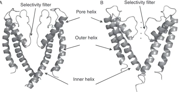
functional tetramer, each consisting of two TM helices (Fig. 2). An
extracel-lular loop, which folds back into the membrane, connects the helices and is composed of a short pore helix and a sequence of amino acids that contain a
selectivity filter incorporating the Kþchannel signature selectivity sequence.
The pore forms an inverted tepee with a gate at the cytosolic entrance. The transition from a closed to an open state occurs by the flexing of a
A Selectivity filter B Selectivity filter
Pore helix
Outer helix
[image:10.432.57.368.373.534.2]Inner helix
FIGURE 2 (A) RyR pore (analogy model fromWelch et al., 2004) and (B) KcsA with four potassium ions shown in the four binding sites of the selectivity filter (1BL8.pdb,Doyle et al., 1998). Two opposing monomers of each channel are shown for clarity.
glycine hinge located approximately halfway along the inner helix (Jiang et al., 2002b). The relatively rigid structure of the selectivity filter of
KcsA allows precise coordination of Kþions with backbone carbonyl atoms
of residues. The hydration state of the permeating ion is also a crucial factor
in selecting Kþover Naþ(Dudev & Lim, 2009).
V. ATTEMPTS TO IDENTIFY THE STRUCTURE OF THE RYR PFR
A. A Model of the RyR Pore Using KcsA as a Template
Following the identification of a presumed selectivity filter, secondary structure predictions for the loop linking the last two TM helices in RyR indicated that this region was likely to contain structural elements equivalent
to those found in the PFR of Kþchannels (Williams et al., 2001). To further
test possible structural similarities between the PFRs of RyR and Kþ
chan-nelsWelch, Rheault, West, and Williams (2004) constructed of a model of the putative RyR PFR using the recently solved crystal structure of KcsA as
a template (seeFig. 2).
The model comprises just 2.4% of the total RyR2 monomer incorporating the last two TM helices and the luminal loop of each monomer. By combin-ing information from secondary structure prediction programs and compar-ing sequence analogies with KcsA, correspondcompar-ing structural elements in RyR were identified and, using molecular modeling software, a stable tetrameric model for RyR was constructed.
By making quantitative comparisons of the physicochemical properties of
KcsA and the RyR homology model, Welch et al. (2004) were able to
KcsA, and the higher density of negative charge around the mouths are likely to be key determinants in the observed differences in ion handling between RyRs and KcsA. Molecular dynamics (MD) simulations performed in the RyR model correlate extremely well with previous experimental data with
regards to selective ion permeation (Williams et al., 2001) and block of Kþ
translocation by tetraethyl ammonium ions (Tinker, Lindsay, & Williams,
1992a). The reader is referred to the original paper for more detailed and quantitative comparisons and assessments of structural components, physicochemical comparisons, and ion handling capabilities of the model (Welch et al., 2004). The RyR pore analogy model contains a glycine residue
in the putative inner helix (GLIIDA) (Fig. 1) that could act as a hinge in a
gating motif (GXXXXA), as originally identified byJiang et al. (2002b)in
Kþ channels. This raises the possibility that the conformational changes
underlying gating in RyRs could be similar to those of Kþchannels.
B. High-Resolution Images of the PFR of RyR
Two reports in 2005 detailed the structures of presumed closed states
of RyR1 obtained using cryo-EM at resolutions of around 10 A˚ . The
first structure was compared directly to the atomic model of KcsA, docking
this structure into the 3D map of RyR1 (Samso, Wagenknecht, &
Allen, 2005).
Apart from the outer helices, the overall orientation and arrangement of structural elements display a high degree of architectural compatibility, with the selectivity filter orientated toward the lumen and the constriction of the inner helices forming a gate at the cytoplasmic face of the structure. The
second structure, published by Ludtke, Serysheva, Hamilton, and Chiu
(2005)is modeled independently of any known structures and identifies five
a-helices in the RyR1 TM domain of which two are identified as the inner
helix and pore helix. In contrast to the cryo-EM structure obtained bySamso
et al. (2005) the inner helix is modeled with a kink at the putative glycine gating hinge and is therefore compared to the open structure of the
Kþchannel MthK (Jiang et al., 2002a; Samso et al., 2005).
The different conformations of the inner helices in these two structures mean that disagreement exists regarding the closed structure of RyR and the theory that kinking of the inner helices is required to open the channel. This may be due to the relatively low level of resolution and the ambiguity as to which conformational state the protein is in during the imaging process (Hamilton & Serysheva, 2009). What is clear from these cryo-EM structures is that the RyR PFR orientates around a central fourfold axis of symmetry and that a funnel like cavity is formed with a wide entrance at the luminal
side tapering in towards the cytoplasmic side. These observations strongly
correlate with the earlier analogy model (Welch et al., 2004) and indicate that
the PFR of RyRs are indeed similar in architecture to Kþchannels and could
gate using similar elements.
C. A RyR PFR Model Based on a High-Resolution Cryo-EM Structure
In an attempt to bridge the gap between the low resolution limits of
cryo-EM and the previously published RyR PFR model, Ramachandran,
Serohijos, Xu, Meissner, and Dokholyan (2009)recently built a molecular model of the putative PFR of RyR1 based on the cryo-EM structure of
Ludtke et al. (2005). The unambiguous assignment of several helix-like
densities gave a model with remarkable similarities to the MthK Kþchannel,
crystallized in the open state. The two main helices, inner helix and pore-forming helix, were found to face each other and line the channel pore, as evidenced in all previous cryo-EM structures. As in previous assignments of the pore structure, acidic residues are located at the mouth of the pore with residues D4899 and E4900, postulated to be important for channel function
in RyR1, positioned within the selectivity filter (Wang, Xu, Pasek, Gillespie,
& Meissner, 2005; Xu, Wang, Gillespie, & Meissner, 2006).
The low resolution limits of cryo-EM do not allow for modeling of amino acid side chains. However, by replacing the missing side chains and adding
the luminal loop, Ramachandran et al. (2009) were able to perform MD
simulations and, by taking into consideration data from previously published single channel experiments, were able to examine interactions of the PFR
with mono- and divalent ions known to permeate the channel (Lindsay et al.,
1991; Tinker & Williams, 1992). MD simulations of selectivity filter mutants known to alter ion handling correlate well with experimental data with ion occupancies observed in the model agreeing well with the charge space competition (CSC) theory, as discussed below. Specific residues located
near the selectivity filter show preferential affinity for Ca2þ over Kþ,
sup-porting the accuracy of the model as a Ca2þ-selective channel. The proposed
glycine gating hinge occurs at residue 4934 in RyR1, analogous to G4864
modeled in RyR2 byWelch et al. (2004). That recent structures resemble the
VI. THEORETICAL APPROACHES TO UNDERSTANDING THE MECHANISMS UNDERLYING ION TRANSLOCATION AND DISCRIMINATION IN RYR
In the absence of detailed information on the structure of the RyR PFR, potential mechanisms underlying ion handling have been explored using theoretical models. A major area of debate that has arisen from these studies is whether the high unitary conductance of RyR is achieved as the result of ion–ion repulsion in a multi-ion pore. It is frequently acknowledged within the field that the interpretation of experimental ion channel permeation phenomena and ion channel occupancy are model dependent. For example, the saturation of conductance in RyR suggests that ions do not move independently of one another (as in solution) with a fit consistent with a
single occupancy pore (Lindsay et al., 1991; Tinker & Williams, 1992);
however, multi-ion models can show saturation (Gillespie & Boda, 2008;
Gillespie & Eisenberg, 2002).
For this reason the ion occupancy state of RyR has been a matter of strong debate with no clear conclusion made due to the presence of two opposing theories; rate theories such as that of Eyring and continuum models such as
Poisson–Nernst–Planck (PNP) (Levitt, 1986; Miller, 1999; Nonner, Chen &
Eisenberg, 1999) which both describe the patterns of ion permeation in RyR (Chen, Xu, Tripathy, Meissner, & Eisenberg, 1997, 1999; Tinker et al., 1992b). Although there are multiple lines of ‘‘model-dependent’’
experimen-tal evidence supporting multi- (Smith et al., 1988) or single-ion occupancy
(Lindsay et al., 1991; Tinker & Williams, 1992; Tinker et al., 1992b) one phenomenon has become central to the argument of ion occupancy in RyR, the anomalous mole fraction effect (AMFE). AMFE is observed as a minima in current, conductance or Erev when the concentration of ions within a bi-ionic mixture varies to produce different ratios of ions (mole fractions). AMFE can be interpreted in terms of Eyring rate theory with a single filing
multi-ion pore (Lindsay et al., 1991; Tinker & Williams, 1992) or in terms of
resistors in series (PNP—continuum model) which can be shown when (on
average)<1 ion is in the pore (Nonner, Chen, & Eisenberg, 1998). It has long
been established that multi-ion channels may not necessarily display AMFE (Hille & Schwarz, 1978) and that single filing is not necessary to observe this
phenomenon (Gillespie, Boda, He, Apel, & Siwy, 2008). In RyR, the
pres-ence of AMFE is highly dependent on the choice of cation, ion activities, and
the method of detecting AMFE (Tomaskova & Gaburjakova, 2008).
There-fore, advocates of rate theory models of RyR will be satisfied that single
occupancy exists in some ion mixtures (Lindsay et al., 1991; Tinker &
Williams, 1992) whereas in others it does not (Gillespie, Giri, & Fill, 2009;
Tomaskova & Gaburjakova, 2008). Recent PNP theories adopt density function theories (DFT) to improve on the previous flawed models which do not describe ions of a finite size and do not consider interactions between ions and channel structure. New models describe selectivity in RyR by CSC where ions can interact with flexible, time-averaged, charged residues where ‘‘charge space competition created by ions in a confined geometry results in
significant selectivity’’ (Gillespie & Eisenberg, 2002). In this model, RyR, on
average, is multiply occupied in various mole fraction mixtures of monova-lent and divamonova-lent cations while one, approaching two, divamonova-lent cation(s) are
present in the pore in pure divalent solutions (Gillespie, Xu, Wang, &
Meissner, 2005). However, this model assumes that ions are dehydrated once reaching the pore, whereas an alternative model of RyR demonstrates
that ions are partially hydrated within the pore (Welch et al., 2004). In
summary, there is agreement within the literature that there is multi-ion occupancy in RyR and therefore this could serve as a mechanism, along
with pore dimensions (Section III.B) and negative charge (Sections V.A and
VII.B) to maximize ion conduction in the channel.
VII. TESTING PHYSICAL AND THEORETICAL MODELS OF THE RYR PFR BY RESIDUE SUBSTITUTION
A. RyR1
Meissner’s group identified potentially important residues by using muta-genesis in conjunction with a theoretical PNP–DFT model and MD simula-tions in their model of the PFR of RyR1. D4899 and E4900 were shown to be critical for both conductance (ion permeation) and selectivity (high ion binding) (Wang et al., 2005). Simulations and electrophysiological experiments revealed
that the mutant D4899Q exhibited a loss of Ca2þpreference in the selectivity
filter (Ramachandran et al., 2009). Furthermore, analysis of G4898R, a CCD
mutant showed a decrease of Ca2þconductance experimentally, whereas Kþ
conductance was constitutively high. These data were confirmed by simulations
with a decrease in the preference of Ca2þ over Kþ in the selectivity filter
(Ramachandran et al., 2009). Noteworthy is that both CCD mutations G4898E and G4898R can form homotetramers and heterotetramers in combi-nation with WT channels when expressed in HEK-293 cells. In terms of
function, while heterotetrameric mutants can maintain their Ca2þ-dependent
activity and Kþconductance, but display decreased Ca2þselectivity (depending
on the type of mutants and the combination of subunits), homotetrameric
mutants have negligible Ca2þ-dependent activity and Ca2þpermeation and
B. RyR2
In agreement with earlier experimental data (Mead, Sullivan & Williams,
1998), the RyR PFR analogy model based on KcsA demonstrates the
exis-tence of a high density of negatively charged residues at both the luminal and
cytosolic mouths of RyR2 pore (Welch et al., 2004). To investigate the
significance of this charge, substitutions of acidic residues at the luminal side of the channel were performed. Earlier studies in RyR1 had demon-strated that the neutralization of either the glutamic acid residue equivalent to E4832 or the aspartic acid residue equivalent to D4833 in RyR2 produced
no significant change in Kþ conductance (Wang et al., 2005). However,
introducing the double substitution ED4832AA in mouse RyR2 channels
led to very significant changes in function (Mead-Savery et al., 2009).
In comparison with WT RyR2, the open probability of ED4832AA RyR2 is greatly increased and a wide variety of subconductance states are observed. ED4832AA discriminates ideally between cations and anions but discrimi-nation between divalent and monovalent cations is lost.
At low ionic activities unitary conductance of ED4832AA is reduced but conductance increases to values above those seen in WT channels at high activities. MD simulations provide some insights into the consequences of this double substitution suggesting that (a) interactions of E4832 and D4833 with other residues in the PFR stabilize the selectivity filter, (b) E4832 and D4833 neutralization reduces the electric field at the luminal face that in turn could reduce cation delivery to the pore at low activities, and (c) neutraliza-tion lowers the electric field in the selectivity filter which, together with destabilization of the filter could underlie enhanced conductance at high
ionic activity. In addition to its role as a Ca2þ-release channel, RyR also
contains a high-affinity binding site for the plant alkaloid ryanodine. Inter-action of ryanodine or its derivatives and congeners (ryanoids) results in
alterations in both channel gating and ion handling (Sutko et al., 1997;
Tinker et al., 1996). As outlined above, residue substitution in and around the proposed selectivity filter of the RyR channels can reduce or abolish the
high-affinity interaction of [3H]-ryanodine with its receptor. Another region
of the channel that may contribute to the site of ryanoid interaction was uncovered by the investigation of residue substitution in the predicted pore-lining TM helix (TM 10). Mutations D4847A, F4850A, F4851A, L4858A,
L4859A, and I4866A severely reduced the release of Ca2þfrom the ER in
HEK-293 cells in response to caffeine and diminished [3H]-ryanodine
bind-ing to cell lysates. Mutations F4846A, T4849A, I4855A, V4856A, and
Q4863A eliminated or markedly reduced [3H]-ryanodine binding, but cells
expressing these mutants responded to caffeine by releasing Ca2þ from
intracellular stores (Wang et al., 2003). Investigations of single RyR2
channels containing one of these mutants; Q4863A, revealed that ryanodine and other ryanoids bind to the channel as readily as to the WT RyR2. The
absence of measureable equilibrium binding of [3H]-ryanodine to Q4863A
RyR2 reflects an almost 600-fold increase in the rate of dissociation of the
bound ryanoid (Ranatunga, Wayne Chen, Ruest, Welch, & Williams, 2007).
VIII. CONCLUDING REMARKS
In this contribution we have attempted to demonstrate how recent devel-opments in structural biology, electrophysiology and molecular modeling have contributed to an improved understanding of the architecture of the PFR and the mechanisms governing the way in which the RyR channel translocates and discriminates between ions. Together these properties
allow SR Ca2þto be released at phenomenal rates through the open RyR
channel during excitation–contraction coupling.
Acknowledgment
The work in our laboratory is supported by the British Heart Foundation.
References
Balshaw, D., Gao, L., & Meissner, G. (1999). Luminal loop of the ryanodine receptor: A pore forming segment?Proceedings of the National Academy of Sciences of the United States of America, 96, 3345–3347.
Chen, D., Xu, L., Tripathy, A., Meissner, G., & Eisenberg, B. (1997). Permeation through the calcium release channel of cardiac muscle.Biophysical Journal, 73, 1337–1354.
Chen, D. P., Xu, L., Tripathy, A., Meissner, G., & Eisenberg, B. (1999). Selectivity and permeation in calcium release channel of cardiac muscle: Alkali metal ions.Biophysical Journal, 76, 1346–1366.
Chen, S. R. W., Li, X., Ebisawa, K., & Zhang, L. (1997). Functional characterization of the recombinant type 3 Ca2þrelease channel (ryanodine receptor) expressed in HEK293 cells. The Journal of Biological Chemistry, 272, 24234–24246.
Doyle, D. A., Morais Cabral, J., Pfuetzner, R. A., Kuo, A., Gulbis, J. M., Cohen, S. L., et al. (1998). The structure of the potassium channel: Molecular basis of Kþconduction and selectivity.Science, 280, 69–77.
Du, G. G., Guo, X., Khanna, V. K., & MacLennan, D. H. (2001). Functional characterization of mutants in the predicted pore region of the rabbit cardiac muscle Ca2þrelease channel (ryanodine receptor isoform 2).The Journal of Biological Chemistry, 276, 31760–31771. Du, G. G., Khanna, V. K., Guo, X., & MacLennan, D. H. (2004). Central core disease mutations
R4892W, I4897T and G4898E in the ryanodine receptor isoform 1 reduce the Ca2þsensitivity and amplitude of Ca2þ-dependent Ca2þrelease.The Biochemical Journal, 382, 557–564. Dudev, T., & Lim, C. (2009). Determinants of Kþvs Naþselectivity in potassium channels.
Journal of the American Chemical Society, 131, 8092–8101.
Fleischer, S. (2008). Personal recollections on the discovery of the ryanodine receptors of muscle. Biochemical and Biophysical Research Communications, 369, 195–207.
Gao, L., Balshaw, D., Xu, L., Tripathy, A., Xin, C., & Meissner, G. (2000). Evidence for a role of the lumenal M3-M4 loop in skeletal muscle Ca(2þ) release channel (ryanodine receptor) activity and conductance.Biophysical Journal, 79, 828–840.
Gillespie, D., & Boda, D. (2008). The anomalous mole fraction effect in calcium channels: A measure of preferential selectivity.Biophysical Journal, 95, 2658–2672.
Gillespie, D., Boda, D., He, Y., Apel, P., & Siwy, Z. S. (2008). Synthetic nanopores as a test case for ion channel theories: The anomalous mole fraction effect without single filing.Biophysical Journal, 95, 609–619.
Gillespie, D., & Eisenberg, R. S. (2002). Physical descriptions of experimental selectivity measurements in ion channels.European Biophysics Journal, 31, 454–466.
Gillespie, D., Giri, J., & Fill, M. (2009). Reinterpreting the anomalous mole fraction effect: The ryanodine receptor case study.Biophysical Journal, 97, 2212–2221.
Gillespie, D., Xu, L., Wang, Y., & Meissner, G. (2005). (De)constructing the ryanodine receptor: Modeling ion permeation and selectivity of the calcium release channel. The Journal of Physical Chemistry B, 109, 15598–15610.
Hamilton, S. L., & Serysheva, I. I. (2009). Ryanodine receptor structure: Progress and challenges.The Journal of Biological Chemistry, 284, 4047–4051.
Hille, B. (1991).Ionic channels of excitable membranes.Sunderland, MA: Sinauer Associates Inc. Hille, B., & Schwarz, W. (1978). Potassium channels as multi-ion single-file pores.The Journal of
General Physiology, 72, 409–442.
Jiang, Y., Lee, A., Chen, J., Cadene, M., Chait, B. T., & MacKinnon, R. (2002a). Crystal structure and mechanism of a calcium-gated potassium channel.Nature, 417, 515–522. Jiang, Y., Lee, A., Chen, J., Cadene, M., Chait, B. T., & MacKinnon, R. (2002b). The open pore
conformation of potassium channels.Nature, 417, 523–526.
Latorre, R., & Miller, C. (1983). Conduction and selectivity in potassium channels.The Journal of Membrane Biology, 71, 11–30.
Levitt, D. (1986). Interpretation of biological ion channel flux data-reaction-rate versus contin-uum theory.Annual Review of Biophysics and Biophysical Chemistry, 15, 29–57.
Lindsay, A. R., Manning, S. D., & Williams, A. J. (1991). Monovalent cation conductance in the ryanodine receptor-channel of sheep cardiac muscle sarcoplasmic reticulum. Journal de Physiologie, 439, 463–480.
Liu, Z., Zhang, J., Wang, R., Wayne Chen, S. R., & Wagenknecht, T. (2004). Location of divergent region 2 on the three-dimensional structure of cardiac muscle ryanodine receptor/ calcium release channel.Journal of Molecular Biology, 338, 533–545.
Ludtke, S., Serysheva, I., Hamilton, S., & Chiu, W. (2005). The pore structure of the closed RyR1 channel.Structure, 13, 1203–1211.
Lynch, P. J., Tong, J., Lehane, M., Mallet, A., Giblin, L., Heffron, J. J., et al. (1999). A mutation in the transmembrane/luminal domain of the ryanodine receptor is associated with abnormal Ca2þrelease channel function and severe central core disease.Proceedings of the National Academy of Sciences of the United States of America, 96, 4164–4169.
Mead, F., & Williams, A. J. (2002a). Block of the ryanodine receptor channel by neomycin is relieved at high holding potentials.Biophysical Journal, 82, 1953–1963.
Mead, F., & Williams, A. J. (2002b). Ryanodine-induced structural alterations in the RyR channel suggested by neomycin block.Biophysical Journal, 82, 1964–1974.
Mead, F. C., Sullivan, D., & Williams, A. J. (1998). Evidence for negative charge in the conduction pathway of the cardiac ryanodine receptor channel provided by the interaction of Kþchannel N-type inactivation peptides.The Journal of Membrane Biology, 163, 225–234.
Mead-Savery, F. C., Wang, R., Tanna-Topan, B., Chen, S. R., Welch, W., & Williams, A. J. (2009). Changes in negative charge at the luminal mouth of the pore alter ion handling and gating in the cardiac ryanodine-receptor.Biophysical Journal, 96, 1374–1387.
Miller, C. (1999). Ionic hopping defended.The Journal of General Physiology, 113, 783–787. Nonner, W., Chen, D. P., & Eisenberg, B. (1998). Anomalous mole fraction effect, electrostatics,
and binding in ionic channels.Biophysical Journal, 74, 2327–2334.
Nonner, W., Chen, D. P., & Eisenberg, B. (1999). Progress and prospects in permeation.The Journal of General Physiology, 113, 773–782.
Orlova, E. V., Serysheva, I. I., van Heel, M., Hamilton, S. L., & Chiu, W. (1996). Two structural configurations of t he skeletal muscle calcium release channel.Nature Structural Biology, 3, 547–552.
Radermacher, M., Rao, V., Grassucci, R., Frank, J., Timerman, A. P., Fleischer, S., et al. (1994). Cryo-electron microscopy and three-dimensional reconstruction of the calcium release channel/ryanodine receptor from skeletal muscle.The Journal of Cell Biology, 127, 411–423. Ramachandran, S., Serohijos, A. W., Xu, L., Meissner, G., & Dokholyan, N. V. (2009). A structural model of the pore-forming region of the skeletal muscle ryanodine receptor (RyR1).PLoS Computational Biology, 5, e1000367.
Ranatunga, K. R., Wayne Chen, S. R., Ruest, L., Welch, W., & Williams, A. J. (2007). Quantification of the effects of a ryanodine receptor channel mutation on interaction with a ryanoid.Molecular Membrane Biology, 24, 185–193.
Samso, M., & Wagenknecht, T. (2002). Apocalmodulin and Ca2þ-calmodulin bind to neighbor-ing locations on the ryanodine receptor.The Journal of Biological Chemistry, 277, 1349–1353. Samso, M., Wagenknecht, T., & Allen, P. D. (2005). Internal structure and visualization of transmembrane domains of the RyR1 calcium release channel by cryo-EM.Nature Structural and Molecular Biology, 12, 539–544.
Serysheva, I. I., Chiu, W., & Ludtke, S. (2007). Single-particle electron cryomicroscopy of the ion channels in the excitation-contraction coupling junction. Methods in Cell Biology, 79, 407–435.
Serysheva, I. I., Orlova, E. V., Chiu, W., Sherman, M. B., Hamilton, S. L., & van Heel, M. (1995). Electron cryomicroscopy and angular reconstitution used to visualize the skeletal muscle calcium release channel.Nature Structural Biology, 2, 18–24.
Sherry, H. S. (1969). The ion-exchange properties of zeolites. In J. Marinsky (Ed.),Ion exchange (pp. 89–133). New York: Marcel Dekker, Inc..
Shomer, N. H., Mickelson, J. R., & Louis, C. F. (1994). Ion selectivity of porcine skeletal muscle Ca2þrelease channels is unaffected by the Arg615 to Cys615 mutation.Biophysical Journal, 67, 641–646.
Smith, J. S., Imagawa, T., Ma, J., Fill, M., Campbell, K. P., & Coronado, R. (1988). Purified ryanodine receptor from rabbit skeletal muscle is the calcium- release channel of sarcoplas-mic reticulum.The Journal of General Physiology, 92, 1–26.
Sutko, J. L., Airey, J. A., Welch, W., & Ruest, L. (1997). The pharmacology of ryanodine and related compounds.Pharmacological Reviews, 49, 53–98.
Tinker, A., Lindsay, A. R., & Williams, A. J. (1992a). Block of the sheep cardiac sarcoplasmic reticulum Ca(2þ)-release channel by tetra-alkyl ammonium cations.The Journal of Mem-brane Biology, 127, 149–159.
Tinker, A., Lindsay, A. R., & Williams, A. J. (1992b). A model for ionic conduction in the ryanodine receptor channel of sheep cardiac muscle sarcoplasmic reticulum.The Journal of General Physiology, 100, 495–517.
Tinker, A., & Williams, A. J. (1992). Divalent cation conduction in the ryanodine receptor channel of sheep cardiac muscle sarcoplasmic reticulum.The Journal of General Physiology, 100, 479–493.
Tinker, A., & Williams, A. J. (1993). Probing the structure of the conduction pathway of the sheep cardiac sarcoplasmic reticulum calcium-release channel with permeant and imper-meant organic cations.The Journal of General Physiology, 102, 1107–1129.
Tinker, A., & Williams, A. J. (1995). Measuring the length of the pore of the sheep cardiac sarcoplasmic reticulum calcium-release channel using related trimethylammonium ions as molecular calipers.Biophysical Journal, 68, 111–120.
Tomaskova, Z., & Gaburjakova, M. (2008). The cardiac ryanodine receptor: Looking for anomalies in permeation properties.Biochimica et Biophysica Acta, 1778, 2564–2572. Tu, Q., Velez, P., Brodwick, M., & Fill, M. (1994). Streaming potentials reveal a short
ryanodine-sensitive selectivity filter in cardiac Ca2þrelease channel.Biophysical Journal, 67, 2280–2285. Wagenknecht, T., Radermacher, M., Grassucci, R., Berkowitz, J., Xin, H. B., & Fleischer, S. (1997). Locations of calmodulin and FK506-binding protein on the three-dimensional archi-tecture of the skeletal muscle ryanodine receptor.The Journal of Biological Chemistry, 272, 32463–32471.
Wang, R., Zhang, L., Bolstad, J., Diao, N., Brown, C., Ruest, L., et al. (2003). Residue Gln4863 within a predicted transmembrane sequence of the Ca2þrelease channel (ryanodine receptor) is critical for ryanodine interaction.The Journal of Biological Chemistry, 278, 51557–51565. Wang, Y., Xu, L., Pasek, D. A., Gillespie, D., & Meissner, G. (2005). Probing the role of negatively charged amino acid residues in ion permeation of skeletal muscle ryanodine receptor.Biophysical Journal, 89, 256–265.
Welch, W., Rheault, S., West, D. J., & Williams, A. J. (2004). A model of the putative pore region of the cardiac ryanodine receptor channel.Biophysical Journal, 87, 2335–2351.
Williams, A. (1992). Ion conduction and discrimination in the sarcoplasmic reticulum ryanodine receptor/calcium-release channel.Journal of Muscle Research and Cell Motility, 13, 7–26. Williams, A. J., West, D. J., & Sitsapesan, R. (2001). Light at the end of the Ca(2þ)-release
channel tunnel: Structures and mechanisms involved in ion translocation in ryanodine receptor channels.Quarterly Reviews of Biophysics, 34, 61–104.
Xu, L., Jones, R., & Meissner, G. (1993). Effects of local anesthetics on single channel behavior of skeletal muscle calcium release channel.The Journal of General Physiology, 101, 207–233. Xu, L., Wang, Y., Gillespie, D., & Meissner, G. (2006). Two rings of negative charges in the cytosolic vestibule of type-1 ryanodine receptor modulate ion fluxes.Biophysical Journal, 90, 443–453.
Xu, L., Wang, Y., Yamaguchi, N., Pasek, D. A., & Meissner, G. (2008). Single channel proper-ties of heterotetrameric mutant RyR1 ion channels linked to core myopathies.The Journal of Biological Chemistry, 283, 6321–6329.
Zhao, M., Li, P., Li, X., Zhang, L., Winkfein, R. J., & Chen, S. R. (1999). Molecular identifica-tion of the ryanodine receptor pore-forming segment.The Journal of Biological Chemistry, 274, 25971–25974.
Zvaritch, E., Depreux, F., Kraeva, N., Loy, R. E., Goonasekera, S. A., Boncompagni, S., et al. (2007). An Ryr1I4895T mutation abolishes Ca2þ release channel function and delays development in homozygous offspring of a mutant mouse line.Proceedings of the National Academy of Sciences of the United States of America, 104, 18537–18542.