iii
Preface
Before you lies my thesis, written to obtain the Master’s degree of Technical Medicine at the University of Twente and is the result of my work conducted between September 2018 and July 2019.
I started writing my plan of action and afterwards my measurement setup had to be built with the help of technician of the University of Twente. This measurement setup had to be approved by infection prevention and medical technology of the MST. In between, four healthy student volunteers participated in pilot measurement to investigate the effect of manipulations on the data. Finally, the measurement protocol was approved by the Medical Ethics Committee Twente on the 21th of March. Fortunately, all 25 subjects were included between March and May 2019.
After performing all measurement, the challenge of data preprocessing and analysis started. The written script to remove the artefacts helped greatly to preprocess the data, unfortunately the amount of data was huge. Besides, finding a discriminative parameter still keeps me occupied. Nevertheless, I am motivated to obtain such a parameters as differences were seen by naked eye. Concluding, I learned a lot this past year from writing a research protocol, including and measuring subjects, conducting exercise challenge tests, and the processing and analysis of the data.
I would like to gratefully acknowledge various people as this work could not have been established without their help.
I want to thank all the children that participated in my study. Without you this work would not have been possible. Despite the fact that you personally may not have benefited from these measurements, many others in the future may!
Dr. Boony Thio, you welcomed me with open arms despite the fact that I had never been under your supervision before. I enjoyed our conversations and how your door is always open, even though your work schedule is completely filled. You were able to relate the appearance of the phase diagrams to their physics despite the mathematical approaches.
I would like to thank dr. ir. Rob Hagmeijer for his advice when I needed it the most. You guided me through the data analysis and always had ideas to further improve my data analysis. I want to thank dr. Bregje Hessink-Sweep for guiding me through my internships.
Ir. Rutger Hebbink, I would like to thank you for building the LabVIEW interface to be able to conduct the measurements. Unfortunately, I had to use another application as LabVIEW crashed to frequently without knowing why. However, you also greatly supported me with writing the Matlab scripts and solving the problems.
iv Dr. Jean Driessen, thank you for the opportunity to learn how to properly conduct an exercise challenge test, interpret them and to communicate with children of different ages. You made Wednesdays feel like a day off, by always making the day interesting and fun. In addition, I want to thank you for the ability to include and measure the subjects during your outpatient clinic.
I thank my fellow Technical Medicine students for their help and advice in carrying out this study. Other than the serious discussions about our studies, we had a lot of fun and I enjoyed the market on Tuesdays.
Finally, I want to thank my family and boyfriend for providing me with unfailing support and continuous encouragement throughout the process of researching and writing this thesis. This accomplishment would not have been possible without them. Thank you.
v
A bstract
R ationale Clinical observations of respiratory distress resulting in imposed work of breathing, respiratory rate, heart rate, and oxygen saturation are currently used to provide feedback whether high flow nasal cannula (HFNC) therapy is effective for the subject. However, these parameters are biased by medication and oxygen supply and vulnerable to misinterpretation. Feedback using pressure to obtain a phase diagram reflecting changes in therapy could, besides clinical parameters, provide valuable information for the clinician to guide optimal therapeutic choices.
Objective To compare the exercise induced changes in lung function to the changes in the phase diagram assessed with the squared perimeter divided by the area (Aex1), sphericity, and triangularity. The changes in lung function were measured with spirometry, forced expiration volume in 1 second (FEV1), and forced oscillatory technique (FOT) with the respiratory reactance (RRS) and resistance (XRS). In order to evoke a variation in lung function of subjects, a standard exercise challenge test (ECT) will be performed.
M ethods In this observational study 25 children were included and performed an ECT. The pressure was measured with an OMEGA pressure sensor and an OptiflowTM nasal cannula. Lung function was measured with spirometry and FOT. Preprocessing was performed with Matlab version R2018b, several parameters were determined, and data analysis methods were investigated.
R esults The mean decrease in FEV1 was 16.2% with a standard deviation of 8.6%. For the determination of the dot product, 21 Fourier terms should be taken into consideration. The parameters Aex1, sphericity and triangularity stabilize after 51 Fourier terms. Scaling the Fourier vector had no influence on the appearance of the phase diagram. The dispersion of the dot product values during the total measurement, influences the phase diagrams which should be included for the calculation of the mean phase diagram. A similar link between the FEV1 changes and the parameters has not been found yet for all subjects. 10 selected healthy and unhealthy phase diagrams were visual distinguishable, however, this was not found for the dot product or parameters.
D iscussion Further investigation is needed to determine a parameter that is able to distinguish healthy from unhealthy but also is able to indicate the therapy efficacy or lung function changes over time. As the parameters showed a greater discrimination between healthy and unhealthy when the mean of 10 phase diagrams was taken, the number of phase diagrams for average the parameter should be determined. To further improve the number of representative breaths, criteria can be added to prevent manipulations or unrepresentative breaths to result in phase diagrams.
vi
List of abbreviations
Aex Area under the expiratory curve
Aex1 Squared perimeter divided by the area of the expiratory curve
ATS American Thoracic Society
Ca Capacitance
CO2 Carbon dioxide
Diff Differences between adjacent elements
ECT Exercise challenge test
EELV End-expiratory lung volume
EIB Exercise induced bronchoconstriction EILV End-inspiratory lung volume
FEV1 Maximal volume of air exhaled in the first second of a forced expiration from a position of full inspiration FOT Forced oscillatory technique
FRES Resonance frequency
Fs Sampling frequency
FVC Forced vital capacity
HFNC High flow nasal cannula
HR Heart rate
L Tube length
ICU Intensive care unit
METC Medical Ethics Committee
η Viscosity
O2 Oxygen
ρ Density
ΔP Pressure difference
PALV Alveolar pressure
PATM Atmospheric pressure
PAW Airway pressure
R Resistance
r radius
RAW Airway resistance
Re Reynolds number
RR Respiratory rate
RRS Respiratory resistance
SpO2 Oxygen saturation
TBFV Tidal breathing flow volume
TE Expiratory time
tPTEF/tE Time to peak flow to total expiratory time VPTEF/VE Volume to peak flow to expired volume
𝑉̇ Airflow
𝑣̅ Velocity
WOB Work of breathing
XRS Respiratory reactance
1
Contents
Preface...iii
Abstract ... v
List of abbreviations ... vi
1. Introduction ... 3
1.1. Rationale ... 4
1.2. Research objective ... 5
1.2.1. Parameters ... 5
2. Background ... 6
2.1. Respiratory physiology ... 6
2.2. Pathophysiology ... 7
2.3. Spirometry ... 7
2.4. Forced oscillation technique ... 9
2.5. HFNC... 10
2.6. Phase diagram – parameters ... 11
3. Method ... 14
3.1. Study design ... 14
3.1.1. Inclusion ... 14
3.1.2. Exercise challenge test ... 15
3.1.3. Pressure measurements... 16
3.2. Population ... 16
3.2.1. Inclusion criteria... 16
3.2.2. Exclusion criteria ... 16
3.2.3. Withdrawal of subjects ... 16
3.2.4. Sample size ... 17
3.3. Data analysis ... 17
3.3.1. Pre-processing ... 17
3.3.2. Fourier terms ... 17
3.3.3. Phase diagram ... 18
3.3.4. Parameters ... 18
2
4. Results ... 19
4.1. Number of Fourier coefficients ... 21
4.2. Scaling of Fourier vector ... 24
4.3. Influence mean Fourier vector on dot product ... 25
4.4. Parameter calculation ... 28
4.5. Healthy versus unhealthy ... 32
4.5.1. Dot product, 21 terms ... 38
4.5.2. Removal Fourier terms ... 40
4.5.3. Parameters ... 41
4.6. Dispersion dot product ... 43
4.7. Exclusion of phase diagrams ... 53
4.7.1. Characteristics unrepresentative phase diagrams ... 55
5. Discussion ... 56
5.1. Interpretation of results ... 57
5.2. Strengths and limitations ... 58
5.2.1. General measurement limitations ... 59
5.2.2. Measurement software ... 60
5.3. Further perspectives ... 60
6. Conclusion ... 62
7. References ... 63
Appendices ... 66
A. Study information letter and consent form ... 66
A.1. Parental information letter ... 66
A.2. Children information letter (4-11 years) ... 71
A.3. Children information letter (12-16 years) ... 72
B. Clinical research form ... 77
C. Pilot measurements ... 79
D. Matlab scripts ... 85
E. 10 healthy and unhealthy selected phase diagrams ... 112
E.1. Subject 21, 10 healthy phase diagrams ... 112
3
1. Introduction
High flow nasal cannula (HFNC) therapy is a relatively new non-invasive ventilation therapy which is increasingly used. HFNC therapy consists of two jets of heated, humidified, and oxygen-enriched air which are injected into the patient’s nares through relatively small but loosely fitted prongs [1]. The proposed mechanisms of HFNC are an increased washout of nasopharyngeal dead space, improved upper and lower airway mucociliary clearance, and increased airway pressure [2]. HFNC is used in hospitals to treat critically ill patients with acute, severe respiratory disorders, but limited high-quality evidence exists of its efficacy in settings other than intensive care units (ICU). Recently, two clinical trials by Franklin et al.[3] and Kepreotes et al.[4] have been published. Franklin et al.[3] compared HFNC to standard oxygen therapy in a large multicentre, randomized, controlled trial with infants younger than 12 months of age with bronchiolitis and in need of supplemental oxygen therapy. This study showed that patients receiving HFNC had a significantly lower rate of escalation of cure due to treatment failure compared to standard therapy. Kepreotes et al.[4] compared HFNC with standard therapy consisting of low flow therapy, in a single-centre, open, randomised controlled trial with infants younger than 24 months of age suffering from moderate bronchiolitis. Their study indicated a significant difference in survival distributions for time to treatment failure favouring HFNC. Also, the number of children who experienced treatment failure was reduced. 63% of the children who deteriorated on standard therapy avoided transfer to ICU by using HFNC as rescue therapy. The effective working mechanisms of HFNC are, however, not fully understood and there is a lack of evidence-based guidelines to assist clinicians who work with HFNC for therapeutic purpose [5–7]. Clinical observations of respiratory distress resulting in imposed work of breathing (WOB), respiration rate (RR), heart rate (HR), and oxygen saturation (SpO2) are currently used to provide feedback whether HFNC is effective for the patient. However, these clinical parameters are biased by medication and oxygen supply and vulnerable to misinterpretation. The HFNC does not like most respiratory support devices provide information about therapeutic intensity. There is a safety issue concerning HFNC, namely the actual pressure delivered by HFNC is dependent on the flow but also nasal cannula size and which can have a marked effect on the deliver pressure. Feedback using pressure to obtain a phase diagram reflecting changes in therapy could, besides clinical parameters, provide valuable information for the clinician to guide optimal therapeutic choices. However, these variables cannot be monitored yet [8,9].
Regarding using pressure to obtain a phase diagram as feedback method of therapy efficacy, previous in-vitro experiments indicate that the influence of breathing can be observed in the pressure signal which is a reflection of the airway pressure [10]. The tracheal tree of an infant is relatively small, resulting in a close to maximal WOB of infants during tidal breathing [11]. Therefore, tidal breathing may be a sufficiently discriminating tool to diagnose various pathologies.
4 Exercise induced bronchoconstriction (EIB) is highly specific
for asthma in children and occurs in 80-90% of pediatric asthma patients [12–15]. Asthma is characterized by inflammation of the bronchial walls which can result in bronchospasm, mucus secretion and airway narrowing [16]. During an asthma attack, a variety of alterations in the respiratory system mechanics occur which lead to an increase in the breathing impedance caused by bronchoconstriction. In patients with asthma, exercise (an indirect stimuli) may cause transient narrowing of the lower airways resulting in an expiratory flow limitation [17].
The magnitude of the airway response to stimuli (direct or indirect) is primarily dependent on the degree of bronchial inflammation and the trigger [18]. Lung epithelium condition inhaled air, therefore cold and dry weather conditions induce and increase heat and water loss of the lungs. Nowadays, the osmotic theory is widely accepted as the established underlying mechanism of EIB [19]. The osmotic theory proposes an increased osmolarity of the airway surface liquid as the primary effect of airway water loss [20]. This increased osmolarity extends to include the airway epithelial cells and submucosa. Cellular mechanisms to release various mediators are activated by the hyperosmolar environment. These mediators cause contraction of the airway smooth muscle and subsequent airway narrowing. The osmotic theory can be observed in figure 1.
Spirometry is the golden standard for diagnosis of any condition affecting the lungs of patients who are not critically ill [21]. During spirometry, the patient first needs to inhale maximal and then exhale rapidly. The measured forced expiratory volume in 1 second (FEV1) is dependent on the effort of the patient. As spirometry requires maximal cooperation and performance, it can be performed from an age of 5 years [22]. Obstructive airflow disorders, e.g. asthma, are characterized by the typical concave shape of the expiratory flow volume curve [23]. Another method which can be used to determine the characteristics of the lung is forced oscillation technique (FOT) [24]. FOT superimposes a multi-frequency airwave on top of the patient’s spontaneous breathing and can therefore be used from the early age of 2 years [24,25]. The respiratory reactance (XRS) and the respiratory resistance (RRS) at a frequency of 5 Hz have been used as a primary efficacy variable to investigate asthma due to the heterogeneous peripheral airway obstruction [25,26].
1.1. Rationale
High flow nasal cannula (HFNC) therapy is widely used. There is a lack of guidance for high care professionals to optimize its use as there is no feedback of therapy intensity. Measurements of pressure over time and integration to phase diagrams could provide objective feedback on therapy efficacy.
[image:11.595.382.522.106.367.2]5
1.2. Research objective
The primary objective is to compare the exercise induced changes in lung function to the changes in the phase diagram. The changes in lung function will be measured with spirometry as the golden standard, and FOT. In order to evoke a variation in lung function of patients, a standard exercise challenge in a cold chamber and a bronchodilator (salbutamol) will be utilized to induce respectively bronchoconstriction and bronchodilatation in asthmatic patients.
1.2.1. Parameters
6
2. Background
2.1. Respiratory physiology
During ventilation, gas exchange occurs between the atmosphere and the alveoli in order to supply the body with oxygen (O2) and to release the body of carbon dioxide (CO2) [27]. In a respiratory cycle, 30% of the total ventilation is wasted due to anatomical dead space which consist of the conducting airways [27]. Children have proportionally larger dead space, which can be proportionally two or three times greater compared to adults. The dead space may measure up to 3 mL/kg in new-borns and declines to 0.8 mL/kg after the age of 6 which is similar to the adult volume [5]. Alveolar ventilation is influenced by the body temperature, pressure, and saturation of air with water [27].
The lungs have a tendency to collapse due to their elastic recoil, whereas the rigidity of the chest wall prevents this. In dynamic conditions, when there is flow, one must also exert an extra force to overcome the resistance and inertia of the lung tissues and air molecules [27]. The airflow (𝑉̇), if laminar, is dependent on the difference between alveolar pressure (PALV) and the atmospheric pressure (PATM) and inversely proportional to airway resistance (RAW), given with equation (1).
𝑉̇ =
∆𝑃 𝑅𝐴𝑊=
𝑃𝐴𝐿𝑉−𝑃𝐴𝑇𝑀 𝑅𝐴𝑊
(1)
The flow of a fluid down a tube is laminar when particles passing any particular point, always have the same speed and direction. Due to viscosity, real fluids have the highest velocity down the midline of a tube and the velocity decreases the farther the fluid is located to from the midline. Poiseuille’s law states that the resistance (R) of a tube is proportional to the viscosity of the gas (η) and the tube length (l), and inversely proportional to the fourth power of the radius (r) given with equation (2).
𝑅 =
8 𝜋∙
ηl
𝑟4 (2)
Poiseuille’s law is only applicable to laminar flow. When using this law, the airflow is extremely sensitive to changes in the airway radius (r) due to the fourth-power dependence. Airflow is transitional when the flow switches between laminar and turbulent. Airflow is transitional through most of the tracheobronchial tree due to the bifurcation of the pulmonary airways which create small eddies resulting in transitional flow. The Reynolds number (Re) can be used to determine if the flow is laminar (Re<2000), transitional (2000<Re<3000) or turbulent (Re>3000), given in equation (3).
𝑅𝑒 =
2𝑟𝑣̅𝜌𝜂
(3)
7 The flow pattern influences the amount of energy needed to produce airflow. Laminar flow is proportional to ΔP and therefore requires a relatively low amount of energy. Turbulent airflow, however, is proportional to √ΔP resulting in lower flow compared to laminar flow when ΔP is similar. The airway resistance (RAW) can be calculated by rearranging equation (1) into (4).
𝑅
𝐴𝑊=
∆𝑃 𝑉̇=
𝑃𝐴𝐿𝑉−𝑃𝐴𝑇𝑀
𝑉̇ (4)
2.2. Pathophysiology
In accordance with the GINA guidelines of 2018, asthma is a heterogeneous disease, usually characterized by chronic airway inflammation [28]. Asthma is characterised by a history of episodic respiratory symptoms such as cough, shortness of breath, wheeze and chest tightness, together with variable expiratory airflow limitation [28]. Both symptoms and airflow limitation characteristically vary in intensity and over time. Symptoms are often triggered by factors as allergen or exposure to non-allergic inhaled irritants, change in weather, viral respiratory infections or exercise.
Inflammation of the bronchial walls results in mucus secretion, increased mucosa thickness and bronchospasm, as can be seen in figure 2 [16,29]. The bronchial wall appears thickened in all asthma patients regardless of disease severity. The degree of wall thickening is related to disease duration, severity, and the degree of airflow obstruction [18,30–32]. Furthermore, the smooth muscle cells
lining the bronchial wall contract, resulting in bronchospasm, which decreases the lumen size even further [29]. Taking Poiseuille’s law (equation 2 and 4) into consideration, a narrowed airway (decreased r) results in an increased RAW and consequently decreased 𝑉̇.
Asthma is an episodic obstructive pulmonary disease [27]. In most asthmatic patients, the lung function is normal or close to normal between attacks [33]. During an asthma attack, however, a variety of alterations in the respiratory system mechanics occur leading to an increase in the breathing impedance caused by bronchoconstriction. In patients with asthma, exercise (an indirect stimulus) may cause transient narrowing of the lower airways which results in an expiratory flow limitation [17]. Cold air aggravates the trigger of exercise for bronchoconstriction in patients with asthma [9].
2.3. Spirometry
Spirometry is the golden standard for assessment of lung function in children with asthma [34]. Spirometry determines the change in lung volume by measuring the volume of inspired and expired air during a forced breathing manoeuvre [27]. The patient first needs to inhale maximally through a spirometer and then exhale rapidly through the same device. During spirometry, the maximal volume of air exhaled in 1 second during a forced expiratory volume (FEV1) is measured. Another spirometry measure is the forced vital capacity (FVC), which is the maximum amount of air that can be exhaled when blowing out as fast as possible. If the patient
8 has an airflow limitation, the FEV1 and the FEV1/FVC ratio are reduced [22,35]. The flow volume curves of a healthy and asthmatic subject are observable in figure 3 [21]. Concerning the ECT, in accordance to the GINA guidelines this test is positive if there is a decrease in FEV1 of at least 12%. Regarding the reversibility induced with a β2-agonist (salbutamol), this indicates a positive test if FEV1 increases with at least 12% in children [28]. A reduced ratio of FEV1 to FVC indicates airflow limitation with normal values of FEV1/FVC, usually exceeding 0.90 in children.
[image:15.595.86.493.181.432.2]Jubran et al.[36], recorded flow volume curves in 50 ventilator-dependent patients over 1 min of spontaneous breathing and observed a saw tooth pattern if secretions was present, see figure 4. This phenomenon can be observed after the use of a bronchodilator as it can improve coughing by increasing bronchial patency and thus expiratory flow. As a result, coughing becomes more effective as there must be sufficient airflow to detach sputum and to mobilize secretions so that they can be expectorated [37].
[image:15.595.209.376.582.744.2]Figure 3: Flow volume curves of a healthy subject on the left and an asthmatic subject with a moderate airflow limitation on the right [21].
9
2.4. Forced oscillation technique
FOT superimposes a multi-frequency airwave on top of the patient’s spontaneous breathing which can assess lung mechanical parameters. This is achieved by measuring the total respiratory impedance (ZRS) [24]. ZRS is a function of the respiratory resistance (RRS) and the respiratory reactance (XRS) at one oscillation frequency, see equation (5).
𝑍
𝑅𝑆(𝑓) = 𝑅
𝑅𝑆(𝑓) + 𝑗𝑋
𝑅𝑆(𝑓) {0 < 𝑓 < 𝑓
𝑀𝐴𝑋}
(5) with f the frequency, j the imaginary component, and XRS and RRS the respiratory reactance and resistance. XRS consists of the mass-inertive forces of the moving air column in the conducting airways (I) and the elastic properties of lung periphery (capacitance, Ca), see equation (6).𝑋
𝑅𝑆(𝑓) = 𝜔 ∙ 𝐼 −
1𝜔∙𝐶𝑎
, 𝜔 = 2 ∙ 𝜋 ∙ 𝑓 {0 < 𝑓 < 𝑓
𝑀𝐴𝑋}
(6) The effect of Ca is most prominent at low frequencies and its effect on X is opposing. In contrast, the effect of I always has positive contribution to X and dominates at higher frequencies. The resonant frequency (fRES) is the frequency with equal and opposite contributions of C and I to X. As a result, X is zero at fRES. R is normally expected to be frequency-independent [25]. In figure 5, the effect of heterogeneous peripheral obstruction on the XRS and RRS can be observed. Asthma results in a heterogeneous peripheral obstruction due to the heterogeneous character of theinflammation [26]. The obstruction results in a frequency-dependent increased RRS due to a larger increase of RRS in lower frequencies compared to higher frequencies. In addition, it results in a shift of XRS in downward-and-right direction [25].
The most commonly utilized oscillation frequency range for multi-frequency oscillations includes frequencies between 5 and 30 Hz [24]. Peripheral airway obstruction results in an increase in magnitude of low frequency |XRS| and a higher resonance frequency (fRES). XRS5 (XRS at 5 Hz) has been used as primary efficacy variable to assess asthma. In asthmatic patients, RRS drops rapidly with increasing oscillation frequencies from 5 up to 18 Hz caused by abnormal peripheral airway function. Small changes in XRS at 5 Hz, and fRES occur between the situations pre- and post-bronchodilator. Both high and low-frequency RRS may decrease with a relatively larger decrease in low-frequency RRS. The frequency dependency of resistance between 5 and 20 Hz (R5-20) provides information on the
heterogeneity of airway obstruction [25]. Figure 5: The influence of heterogeneous peripheral
10 Bronchoconstriction results in a frequency dependent RRS, where changes become more evident in the lower frequencies. Reactance also decreases due to bronchoconstriction, resulting in a prominent increase in fRES [38].
It has been shown that an increased RRS at 8 Hz is significantly correlated with a decrease in FEV1. Furthermore, an increase in fRES is correlated with a decrease in FEV1 as well [38]. The change in ZRS reflects inhomogeneity in peripheral part of the bronchial tree.
2.5. HFNC
The proposed mechanisms of HFNC are the washout of nasopharyngeal dead space, reduction of the inspiratory and expiratory resistance, improved ventilation mechanics and reduction in the metabolic cost of gas conditioning [2,39]. The high flow provided by HFNC causes flushing of the nasopharynx and therefore leads to a reduction in dead space. The flow rate of HFNC is equal or higher than the inspiratory flow, which results in an attenuation of the inspiratory resistance exerted by the nasopharynx. The warmed humidified gas improves the conductance, compliance, and reduces the metabolic work associated with the conditioning of gas. In addition, the gas decreases resistance by making the mucus less tenacious and reducing the amount of mucus by easier mobilization and evacuation [39]. High flow can also generate positive distending pressure which is believed to improve breathing mechanics. This is achieved by optimizing lung compliance, recruitment of the lung, decrease in ventilation-perfusion mismatch, and improvement of patency of the alveoli [2,40]. All the proposed mechanisms of HFNC may relieve effects of induced bronchoconstriction. However, the exact moment at which therapeutic effects occur is unknown.
Within five minutes of respiration, pulmonary compliance and conductance significantly decreases with use of warmed humidified ambient gas in ventilated infants [2]. Therefore, making use of warmed humidified gas limits the bronchoconstriction induced by for instance cold dry gas [5].
11
2.6. Phase diagram – parameters
In order to be able to measure the breathing pressure during ECTs, a pressure sensor will be added to the measurement setup. This is shown in figure 8 of chapter 3. The breathing pressure will be integrated to obtain tidal breathing phase diagrams [39].
In patients with asthma, the expiratory part of the phase diagram provided with spirometry can have a concave or triangular shape [11,23]. In contrast, the inspiratory part usually appears normal in asthmatic patients. Peak flows tend to be higher and occur earlier in the expiration of patients with obstructive lung disease [45,46]. In addition, there is a slow decline in flow over most of the expiration followed by an abrupt drop of flow to zero due to the trigger for the next inspiration. These typical shapes in the phase diagram of a forced manoeuvre, caused by obstructive lung diseases, can be used to evaluate the phase diagram.
Tidal breathing flow volume (TBFV) curves can be evaluated by the shape of the air flow signal [11]. Normalization of these curves is essential to avoid bias due to size or bodyweight of the various subjects. Furthermore, normalization can also be used to eliminate the influence of the insertion length of the prongs. Leonhardt et al.[11] used a sphericit and triangular approach to quantify the expiration. In addition, quantifying the expiration is also possible by approximation with polynomials. The sphericity and triangularity were assessed using equations (7) and (8) respectively
Ο
𝑒𝑥𝑝=
𝑟𝑖𝑛𝑠𝑐𝑟𝑖𝑏𝑒𝑑,𝑒𝑥𝑝𝑟𝑐𝑖𝑟𝑐𝑢𝑚𝑠𝑐𝑟𝑖𝑏𝑒𝑑,𝑒𝑥𝑝 (7)
∇
𝑒𝑥𝑝=
∫∆𝑉𝑙𝑢𝑛𝑔=0𝑉𝐸 𝑉̇𝑏𝑟𝑒𝑎𝑡ℎ𝑑𝑉
0.5∙𝑃𝑇𝐸𝐹∙𝑉𝐸
− 1
(8)In figure 6, the difference in sphericity between a healthy and an asthmatic subject can be observed with expiration values of Ο𝑒𝑥𝑝=0.71 and Ο𝑒𝑥𝑝=0.32, respectively. A Ο𝑒𝑥𝑝 value of 1 corresponds to a perfectly shaped sphere. Regarding equation (8), a concave shape is indicated with a ∇ < 0, and if very triangular, ∇ ≈ 0. In figure 7, the triangular approximation of the TBFV curve is shown for an asthmatic subject with ∇𝑒𝑥𝑝 being a perfectly shaped triangle as it approaches zero (∇𝑒𝑥𝑝=0.07312).
12 Regarding the approximation with polynomials, first- and second-order polynomials can be used for the normalized curves with equation (9) and (10) respectively.
First order:
𝑉̇̂
𝑏𝑟𝑒𝑎𝑡ℎ= 𝑎
1∆𝑉
𝑙𝑢𝑛𝑔+ 𝑏
1 (9)Second order
:
𝑉̇̂
𝑏𝑟𝑒𝑎𝑡ℎ= 𝑎
2(∆𝑉
𝑙𝑢𝑛𝑔)
2+ 𝑏
2∆𝑉
𝑙𝑢𝑛𝑔+ 𝑐
2 (10) The difficulty regarding approximation by polynomials lies in the interpretation of the coefficients and the quality of the approximation.Assessing the concavity to quantify airway obstruction, was also studied by Zheng et al.[47]. They used a hyperbolic function using among others the forced vital capacity (FVC). Morris et al.[46] used a time constant of the regression line to fit to the linear part of the expiration. They r2 value was used to assess the goodness of the fit with a required minimal of 0.85. Two other parameters that reflect the level of obstruction consisted of the time to peak flow divided by the total expiratory time (tPTEF/tE) and volume to peak flow divided by the expired volume (VPTEF/VE) [48–51]. Those measures can also be assessed as a percentage of predicted, resulting in ΔV/V and Δt/t [45,52]. All these parameters showed a significant correlation with FEV1 [45,48,50,52]. PTEF, however, provides only limited information about the smaller airways as it primarily reflects central airway obstruction [53]. Van der Ent et al.[49] concluded tPTEF/tE to be a more useful parameter to assess airflow obstruction in epidemiological research than in individual patients due to the limited accuracy and quite large internal variability. Therefore, the clinical usage of these time dependent measures is limited.
13 The area under the expiratory part of the phase diagram (Aex) provides information on central and peripheral airway obstruction [54]. The magnitude and pattern of Aex changes after bronchodilatation and can therefore be influenced by changes in the lung function. This offers an opportunity to use this parameter to evaluate the efficacy of medication. However, it is desired to obtain dimensionless parameters in order to avoid bias of for example the prong length and therefore be able to assess only the form of the phase diagram. As both the area and perimeter are indicative for the appearance of an object, a dimensionless parameter can be achieved by dividing the squared perimeter by the area. Equation (11) shows the dimensionless parameter, Aex1,
𝐴𝑒𝑥
1=
𝑝𝑒𝑟𝑖𝑚𝑒𝑡𝑒𝑟2𝑎𝑟𝑒𝑎 (11)
14
3. M ethod
This study was submitted to the Medical Ethics Committee (METC) Twente as TBFV (Phase diagram in relation to lung function changes during bronchoconstriction and –dilation), and was approved 19-03-2019 with trial number K19-15.
3.1. Study design
This study had an observational cross-sectional design, in which 25 children, aging from 4 to 16 years old, with pediatrician diagnoses asthma or suspicion of asthma, scheduled for an exercise challenge test (ECT), were asked to participate. The measurement setup is observable in figure 8. This measurement setup is used to study the change in the appearance of the phase diagram obtained from tidal breathings to the lung function measurement by performing pilot measurements and the TBFV-curve study.
3.1.1. Inclusion
At first, the children and their parents were informed by telephone 2 weeks prior to the ECT. After verbal agreement, parents received a study information letter (for both themselves and their child) and a (parental) consent form (Appendices A). However, after the first week it became clear that this method was too time consuming and another approach was used. The parents received the information letter one week prior to the appointment. Written informed consent was obtained prior to the start of the measurements, during the appointment at the outpatient clinic. It was signed by either both parents (child under the age of twelve) or parents and child (children between the ages of twelve and sixteen).
Figure 8: Measurement setup, nasal cannula connected to a pressure sensor. A. OptiflowTM Nasal High Flow cannula S.
B. Connector of the breathing circuit to the optiflowTM. To this connection part, was handmade and manually
attached by the Technician of the University of Twente C. SNAP fitting
D. Polyurethane tube with adaptable length E. SNAP fitting
15
3.1.2. Exercise challenge test
During an outpatient visit at the OCON, the ECT was performed according to international guidelines of the American Thoracic Society (ATS) [55]. Children below the age of eight were challenged with six minutes of jumping on a jumping-castle according to van Leeuwen et al. [12]. Children above the age of eight were challenged with six minutes on a treadmill. The slope of the treadmill was set at 10% to reduce running speed and improve safety while maintaining exercise intensity. Each ECT took up to one hour to complete.
A thorough medical history was performed, focusing primarily on any discomforts regarding breathing abnormalities. Furthermore, information was acquired of medication adherence, physical activity and familiar occurrence of asthma, eczema and/or allergies in the first or second degree. During physical examination, attention was paid to the presence of allergic signs, such as Dannie-Morgan lines and Meyers’ nasal crease. The clinical research form that was used to capture the medical history, physical examination and other ECT data can be seen in Appendix B.
Baseline measurements were performed using the FOT and spirometry. If the treadmill was used as exercise method, subjects were then equipped with a HR monitor (Polar). The exercise were performed in a climate controlled room at the outpatient clinic. Subjects exercised on a treadmill set at a 10% angle in cold air (10°C) for six minutes with a clip on the nose at submaximal level (80% of maximal predicted HR). These conditions provide the maximal stress for the airways to provoke an asthmatic reaction. Children under the age of eight were not equipped with a HR monitor and exercised on the jumping-castle for six minutes instead. Previous studies state that high intensity exercise is guaranteed in young children when jumping on a bouncy castle [12]. To ensure an appropriate exercise challenge children were encouraged actively to keep jumping for the full six minutes.
Subjects performed double spirometry measurements at one, three, and six minutes after exercise. If FEV1 was still declining after six minutes, an additional measurement was performed every three minutes until start of recovery of FEV1 (at for example nine and twelve minutes). A FOT measurement were performed at five minutes after exercise.
After the final spirometry measurements, subjects inhaled 200 μg of salbutamol and five minutes thereafter a final FOT and spirometry measurements were performed to evaluate the effect of medication.
After the ECT, data was acquired from the device and stored anonymously for later data-analysis. A full overview of all actions during an ECT are shown in figure 9.
16
3.1.3. Pressure measurements
All pressure measurements were performed using a PXM409-070HCGUSBH device, see figure 8 part E. The pressor sensor was connected to a laptop via USB and therefore the value pressure data could be observed in real time and extracted as an excel file after each measurement. To analyse TBFV curves, a sampling rate of at least 200 Hz is recommended [56].
During the measurements, only the nasal cannula of the HFNC system was used and connected to a pressure sensor as observable in figure 8. The pressure curves of 25 children were measured from approximately one minute after exercise up to the end of the ECT. These children visited the OCON for an ECT as standardised care to provoke bronchoconstriction by exercise in a cold chamber. The purpose of these measurements is to determine if there is a relation between dynamics of lung function and changes in the phase diagram.
3.2. Population
The population of this study consists of all children with pediatrician diagnosed asthma. One in five girls and one in four boys at the age of two to three years have asthma symptoms. The prevalence decreases to 10% in girls and 15% for boys at the age of six to seven after which it stabilizes [57]. Frequent asthma symptoms are observed in 4-5% and 2-3% of respectively boys and girls five years and older [57]. Children whom are 4 to 16 years of age, who are scheduled for an ECT at OCON were approached to take part of this study.
3.2.1. Inclusion criteria
In order to be eligible to participate in this study, a subject must meet all of the following criteria:
- Children with pediatrician diagnosed asthma. - Children aged between 4 and 16 years old.
- Children instructable to perform an ECT and repeatable spirometry.
3.2.2. Exclusion criteria
A potential subject who meets any of the following criteria was excluded from participation in this study:
- Children with an unilateral or bilateral obstructed nose
- Children who are unable to speak Dutch, or whose legal guardians are unable to speak Dutch.
- Children for whom it is not possible to wear the OptiflowTM device. For example due to malformations of the nose.
- Children born prematurely <34 weeks.
3.2.3. Withdrawal of subjects
17
3.2.4. Sample size
The largest change in lung function is observed in subjects with uncontrolled asthma. Prior to the ECT in the OCON, the asthma control of the subject is unclear. The effect of the change in lung function on the phase diagram of tidal breathing is also unknown. Taking into consideration the number of patients visiting the outpatient clinic in MST and performing the ECT at the OCON, it was expected that 25 subjects needed to be included at the OCON to measure at least 15 uncontrolled asthma subjects [58]. 25 subjects were included in the period of March to May 2019.
3.3. Data analysis
All data were pre-processed using Matlab (version R2018b). A pilot study was performed to investigate the effect of potential influences on the pressure signal. Performing lung function measurements, swallowing, clearing the throat, coughing, itching the nose, repositioning of the prongs and breathing through the mouth were evaluated. The reason for this is the ability to select only the breaths of which the phase diagram is computed. All previously mentioned influences are distinguishable from breathing and therefore automatic determination of breaths seems possible. These results are observable in Appendix C. The locations of these influences were manually inspected and removed from the raw signal if selected correctly. These scripts can be seen in Appendixes D. The correlation between the change in lung function assessed by spirometry and FOT will be compared to the changes in the phase diagram. As the breathing pattern is variable, a number of consecutive artifact-free breaths should be selected for the determination of the parameters [59]. However, as the setup of the measurements is unique, the number of breaths which should be taken into consideration needs to be investigated.
3.3.1. Pre-processing
The data was first divided into the segments that correspond to the intervals of figure 9. Thereafter, the artefacts for each individual segment were located using the moving standard deviation (movstd) and differences between adjacent elements (diff) functions of Matlab. The reason that those functions have been chosen, is explained in Appendix C. Subsequently, the interval which contains the artefact was removed between two minima that correspond to maximal inspiration. After the removal of non-breathing intervals, the remaining segment was divided into single breaths based in the minimum. These minimum correspond to the maximal pressure obtained during inspiration. Each single breath was expanded into sines and cosines up to the 25th component using Fourier series. This smoothens the pressure signal. 4.1 Number of Fourier coefficients describes why up to 25th components were used.
3.3.2. Fourier terms
18
3.3.3. Phase diagram
In order to determine the TBFV curves (phase diagrams), the minima which is at maximal inspiration were determined. The minima was chosen as it is more difficult to identify the start of an inspiration or expiration correctly. This is mainly needed as errors occur due to the variation around the baseline by the influence of noise and also severely disturbed signals increase the error rate [59]. In order to avoid very short breathing cycles due to noise around zero, the zeros that occur within 40 measurement points or within 0.6 s of the previous zero were removed. The y which resembles the flow was calculated using equation (12). The volume which is represented by x, was determined by integrating the flow over time since the start of the cycle. Equation (13) shows the computation of x.
𝑦 =
𝑃− 𝑃𝑚𝑒𝑎𝑛 max (𝑃− 𝑃𝑚𝑒𝑎𝑛)(12)
𝑥 = ∫ 𝑦 𝑑𝑡
(13)3.3.4. Parameters
It is hypothesized that the sphericity, triangularity and Aex1 of the phase diagram would be influenced by the level of bronchoconstriction which was induced by exercise in a cold room with dry air and reversed to bronchodilatation by salbutamol. The variation in parameters based on the phase diagram were plotted against the corresponding FEV1 measured by spirometry. The x-axis of the expiratoire curve on which the sphericity and triangularity was computed were normalized from -1 up to 1. As mentioned before, normalization can also be used to eliminate the influence of the insertion length of the prongs and gap between the prongs and nares. As the trend will be compared between subjects it is also essential to avoid bias due to size or bodyweight of the various subjects.
3.3.5. Number of phase diagrams
19
4. R esults
Table 1: Subject characteristics. Included subjects in green and excluded subjects in red.
Subject Age (yr.) Weight (kg) Length (cm) Sex (F/M) Control medication
Allergy Nasal cannula type (xl/s)
Lung function change (%)
1 6.5 26 118 M yes yes xl 5.6
2 7.1 28 124 M yes yes xl 14.5
3 8.2 28.7 129 M no no s 9.1
4 9.8 32.5 145 F yes no s 7.3
5 4.3 17 104.5 M yes no xl 13.8
6 14.4 52 162 M non-adherent yes s 11.9
7 8.3 25.8 126.6 M yes yes s 28.5
8 4.3 20.3 107 M yes no xl 16.5
9 7.3 18.6 117 M no yes xl 23.7
10 9.6 38 148.6 M yes yes s 31.8
11 9.8 39.8 150.6 M no yes s 6.1
12 12.3 49 160 M yes yes s 31.6
13 11.4 39.3 147.6 F no yes s 12.0
14 10.8 25.9 131.1 F non-adherent no s 20.5
15 9.6 33.2 143.7 M yes yes s 28.6
16 8.9 46.2 143.8 M yes no s 11.4
17 12.6 55 155.5 M yes no s 16.9
18 13.3 49.5 163 M yes yes s 10.9
19 10.3 37.4 145.6 M no no s 5.0
20 11.5 39.2 147 M yes yes s 23.6
21 9.8 25.8 131.8 F yes yes s 5.5
22 12.8 35 144 M yes yes s 7.5
23 7.5 22.7 122.2 M yes yes s 29.3
24 11.1 38.1 151 M no no s 16.2
25 4.2 17.5 106.5 M no no xl 16.5
Mean (±SD) 9,4 (2,8) 33,6 (10,9) 137,0 (17,3) 84% M 64% (8%)
60% 76% s 16.2%
20 The mean Fourier vector of all the phase diagrams of the 18 included subjects was calculated and the dot product of every single phase diagram with this mean Fourier vector was determined. The number of phase diagrams that have a dot product value within a range of 0.01 is indicated on the y-axis of figure 10. The coherence is displayed on the x-axis with a value approaching 1 corresponding to a perfect coherence between the individual phase diagram and the mean of all phase diagrams. The distribution of the dot product value appears log-normal in figure 10. This indicates that the dot product has in general a high coherence with the mean phase diagram.
Different approaches of data processing have been investigated as they seem to affect the parameter values. These approaches are summarised in 4.1 to 4.7.
21
Figure 12: Aex1 of the phase diagram with the number of Fourier components on the x-axis.
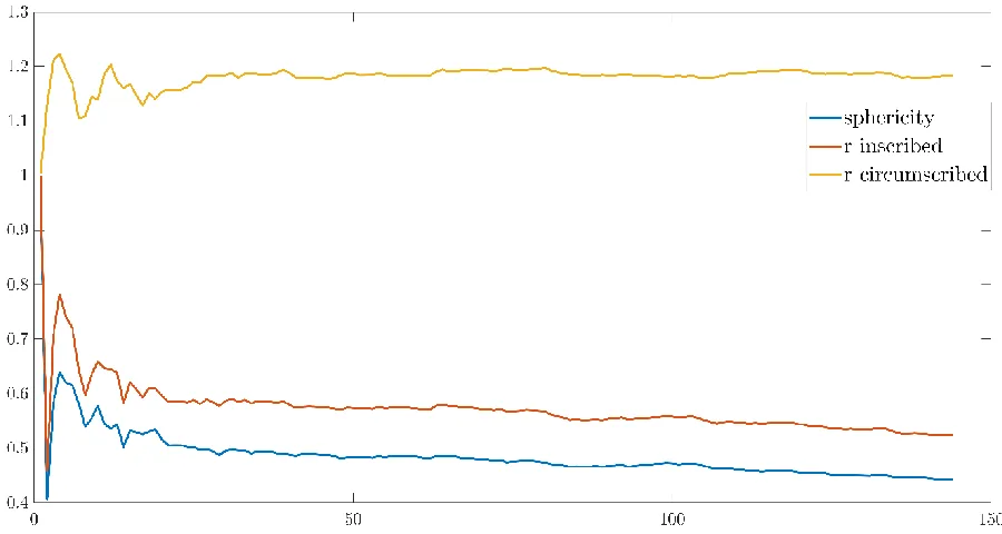
4.1. Number of Fourier coefficients
The pressure signal during 1 period (one breath from the maximal pressure obtained during the previous inspiration up to the next inspiration) was expanded into cosines and sines up to the Nth component. The number of components needed for the stabilization of the parameters was determined for one phase diagram of every segment for three subjects. The results of the analysis are shown for one phase diagram of one subject. Figure 11 displays the used phase diagram using the raw data. Stabilization of the parameters seem to occur around 25 Fourier components, see figures 12 to 14. However, stabilization of the parameters of other phase diagrams that were investigated, occurred around 50 components. Therefore, the parameters of subject 2, 3, and 4 were computed for both 25 and 50 Fourier components. These are shown for the triangularity in figures 15 and 16, as this parameters showed more difference between 25 and 50 Fourier components compared to Aex1 and sphericity. The parameter values differed minimally for 50 compared to 25 Fourier components. This implicates that there is no additional information in the extra 25 Fourier components with regard to the determined parameters. Therefore, for further analysis 25 Fourier components will be used.
22
23
Figure 15: Triangularity of subject 2 with 25 Fourier components. Subject 2 is a 7 years old male with a 14% lung function decline at 3 minutes after exercise.
24
4.2. Scaling of Fourier vector
In figure 17, the influence of scaling the Fourier vector is shown for two phase diagrams. The phase diagrams were aligned to 0,0 which corresponds to the end of expiration, with expiration being positive. Observing figure 17, the appearance of the phase diagram seems similar for the scaled and unscaled Fourier vector. Scaling the Fourier vector results in a proportionally decrease of the total phase diagram. As scaling has no influence on the appearance of phase diagram and the parameters Aex1, sphericity, and triangularity only dependent on the appearance of the phase diagram, these parameters are not influenced by scaling.
25
4.3. Influence mean Fourier vector on dot product
It was hypothesized that the phase diagrams close to the final spirometry measurement correspond to normal tidal breathing flow volumes curves as the lung function is nearly maximal at this point due to administration of a bronchodilator. As it is unknown how many phase diagrams should be taken into consideration for the mean Fourier vector, the influence of including additional phase diagrams was investigated. First, all phase diagrams after the final lung function measurement were used to calculate the mean Fourier vector and the dot product between all individual phase diagrams and this mean Fourier vector were determined. Both the dot product values and the lung function values were plotted and a linear and quadratic equation with their norm of residuals were determined. Thereafter, all phase diagrams 15, 30, 45 and 60 seconds prior to the final lung function measurement were taken into consideration for the mean Fourier vector. Subsequently, if the norm of residuals of the equation was still declining, 15 additional seconds concerned for the mean were used up to the moment of increasing norm of residuals.
Figures 18 and 19 show the values of the dot product between the individual phase diagrams and the mean resulting in the minimal norm of residuals for subject 7 and 21. The results of these subjects is shown as subject 7 experienced a lung function change of 28.5% during the total measurement compared to a lung function change of 5.5% of subject 21. Noticeable is the difference in distribution of values between subject 7 and 21. The coherence differs per subject and is higher for subject 21 than 7 which also result in a lower norm of residuals of respectively 1.36 and 2.17 concerning the quadratic functions. There is no consistency observed in the relation between the FEV1 and the course of dot products during the total measurement for all subjects, as both the same and contradictory trends between FEV1 and the dot products have been observed.
26 In general, the norm of residuals of the quadratic equation approaching the dot product values was lower compared to the norm of residuals of the linear equation. Besides, using more Fourier vector close to the final spirometry measurement results in a lower norm of residuals and therefore a better approximation of the trend of the dot products during the total measurement of a subject. Nevertheless, every subject has an optimal number of phase diagrams resulting in a minimal norm of residuals. This optimal number was determined using interval of 15 seconds and therefore there is an opportunity to lower the norm of residuals by adding more phase diagrams one by one instead of 15 second intervals. However, this also depends on the coherence of the included phase diagrams for the calculation of the mean Fourier vector. In general, the norm of residuals decreases by including phase diagrams for the mean Fourier vector of which the dot product value is within the range of the already included phase diagrams. So, in order to reduce the norm of residuals the appearance of the phase diagrams must be comparable to previously included phase diagrams. Subsequently, the norm of residuals was, in general, higher if the dot product values during the total pressure measurements were more dispersed. This could be explained as the norm of residuals is an indication of the agreement between the values and the equation. So, if the values are more in line with the equation, the norm of residuals is lower and therefore more dispersed values negatively affect the norm of residuals. Table 2 gives an overview of the phase diagram used to calculate mean Fourier vector, the corresponding time interval, and the linear and quadratic functions with their norm of residuals. Regarding subject 12, calculation of the mean Fourier vector using all phase diagrams, compared to only those after the final spirometry, results in a lower norm of residuals. Subject 12 had only 37 representative phase diagrams with just 4 phase diagrams after administration of a β2-agonist. The norm of residuals of the other subjects was minimal by using up to 120 seconds before the final spirometry for the mean Fourier vector with up to 60 seconds for most subjects. As administration of a β2-agonist results in an increased airflow within 3 to 5 minutes after administration, it is presumed that the appearance of phase diagrams more than 2 minutes before the final should not be taken into consideration for the mean Fourier vector [60].
27
Table 2: Overview of function with the minimal norm of residuals per subject and the corresponding phase diagrams used for the mean.
Subject Phase diagrams
Time Linear Norm of
residual s
Quadratic Norm of residual s 2 162-171 From spiro y=9.90e-6x + 0.85 1.486 y=7.82e-7x2 – 0.00071x
+ 0.98
1.407 3 116-136 60 sec
before spiro
y=-9.88e-5x + 0.68 2.507 y=5.60e-7x2 – 0.00062x + 0.80
2.464
5 92-135 60 sec
before FOT
y=-3.51e-5x + 0.84 1.410 y=5.24e-7x2 – 0.00059x + 0.94
1.354 6 188-219 60 sec
before spiro
y=9.86e-5x + 0.80 1.878 y=-1.23e-7x2 – 0.00024x + 0.77
1.873 7 139-167 75 sec
before spiro
y=-0.00025x + 0.91 2.174 y=-7.95e-7x2 – 0.00018x + 0.90
2.170 10 126-148 90 sec
before spiro
y=0.00019x + 0.81 1.146 y=-1.37e-7x2 + 0.00033x + 0.78
1.142
12 1-37 Total
dataset
y=-1.10e-5x + 0.83 0.696 y=6.03e-7x2 – 0.00072x + 0.96
0.653 13 103-116 45 sec
before spiro
y=-0.00013x + 0.87 1.428 y=2.72e-7x2 – 0.00038x + 0.92
1.424 14 84-126 75 sec
before spiro
y=-8.12e-5x + 0.91 1.258 y=-2.17e-7x2 + 0.00015x + 0.87
1.251
15 50-59 45 sec
before spiro
y=0.00014x + 0.60 1.504 y=-6.31e-8x2 + 0.00020x + 0.59
1.503
16 73-79 60 sec
before spiro
y=0.00010x + 0.72 1.506 y=5.82e-7x2 – 0.00044x + 0.81
1.493 17 92-139 120 sec
before spiro
y=6.87e-5x + 0.73 2.168 y=-9.18e-7x2 + 0.00017x + 0.71
2.167 18 90-130 30 sec
before spiro
y=-9.31e-5x + 0.84 1.876 y=5.67e-7x2 – 0.00076x + 0.98
1.808 19 97-104 From spiro y=-1.58e-5x + 0.61 1.663 y=-4.43e-7x2 +
0.00043x + 0.53
1.618 21 179-232 60 sec
before spiro
y=-3.42e-8x +0.92 1.366 y=-9.87e-7x2 + 0.00011x + 0.90
1.362 22 207-237 From spiro y=9.66e-5x + 0.84 1.591 y=-2.66e-7x2 +
0.00030x +0.78
1.567 24 143-171 30 sec
before spiro
y=3.81e-5x + 0.78 2.653 y=3.85e-7x2 – 0.00040x + 0.86
2.622 25 90-110 45 sec
before spiro
y=-7.64e-5x + 0.84 1.511 y=-1.11e-7x2 + 4.03e-5x + 0.82
28
4.4. Parameter calculation
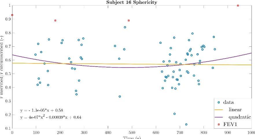
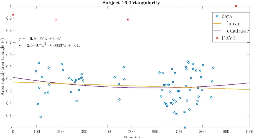
For the calculation of the sphericity, triangularity and the Aex1 the equations (7), (8), and (11) were used. These parameters were calculated for every single phase diagram and investigated whether the trend in the values of the parameters was comparable to the FEV1 and therefore related to lung function changes. The linear and quadratic functions with norm of residuals were determined per parameter per subject. Most quadratic function were parabolic with either a minimum or maximum. Unfortunately, no link between the parabolic appearances of the parameters was observed between the parameter values per subjects. In addition, the lung function change showed no association with the parabola having a minimum of maximum. All parameters of subject 14 showed linearly related values during the total pressure measurement. The linear and quadratic functions with norm of residuals can be observed in tables 3 to 5. As subject 12 only has 37 representative phase diagrams, the subject with the lowest norm of residuals hereafter is subject 16. Figures 20 to 22 display the parameter values of subject 16 during the total measurement.
Table 3: Linear and quadratic functions of Aex1 per subject. Subject Linear Norm of
residuals
Quadratic Norm of
residuals 2 y=0.23x + 2.1e2 7527.7 y=-0.0032x2 + 3.2x - 3.1e2 7270.3 5 y=2.4x - 2.5e2 57897 y=0.11x2 – 9.2x + 1.7e3 57298
6 y=0.62x – 20 19917 y=0.003x2-2.8x+7.2e2 19611
7 y=25x + 1.1e3 1.5409e6 y=-0.23x2 + 2.4e2x – 3.6e4 1.5332e6 10 y=-0.067x + 2.4e2 1390.1 y=8.2e-5x2 – 0.15x + 2.6e2 1389.1 12 y=-0.021x + 3.1e2 2552.2 y=-0.00096x2 + 1.1x + 99 2523 13 y=0.94x + 1.1e2 9396.2 y=0.002x2 – 0.9x + 4.2e2 9357.4 14 y=0.3x + 0.85 4191.6 y=4.2e-6x2 + 0.3x + 86 4191.6
15 y=4.5x + 85 80782 y=0.012x2 – 6.4x + 2e3 80681
29 During the determination of the Aex1 values of the 18 subjects, several subjects displayed a few values which can be interpreted as outliers. Investigation of the phase diagrams of these considered outliers indicated that these phase diagrams displayed artefacts or unrepresentative breaths instead of breaths. In 4.7 different methods for removal of these unrepresentative phase diagrams are proposed.
Table 4: Linear and quadratic functions of sphericity per subject.
Subject Linear Norm of residuals
Quadratic Norm of
residuals 2 y=-8.2e-6x + 0.57 1.7082 y=3.7e-7x2- 0.00035x + 0.63 1.6928 3 y=-2.2e-5x + 0.58 1.9927 y=1.5e-7x2 - 0.00019x + 0.62 1.9888 5 y=7.1e-5x +0.5 1.743 y=-2.6e-7x2 + 0.00035x + 0.45 1.7318 6 y=-5.4e-5x + 0.63 2.1022 y=-3.7e-7x2 +0.00037x + 0.54 2.0575 7 y=-0.00016x + 0.66 1.9233 y=-3.5-7x2 + 0.00017x +0.61 1.9093 10 y=0.00021x + 0.4 0.9978 y=-1.3e-8x2 + 0.00022x + 0.4 0.9978 12 y=-0.00011x + 0.65 0.7054 y=-2.7e-7x2 +0.0002x + 0.59 0.6973 13 y=-0.00013x + 0.61 1.5987 y=2.9e-7x2 – 0.0004x + 0.66 1.5937 14 y=-1.3e-5x + 0.66 1.4063 y=-8.2e-9x2 – 4.9e-6x + 0.66 1.4063 15 y=-3.5e-5x + 0.53 1.1897 y=-3.2e-7x2 + 0.00027x + 0.48 1.1844 16 y=-1.3e-5x + 0.58 1.2071 y=4e-7x2 – 0.00039x + 0.64 1.1999 17 y=-2.5e-5x + 0.53 1.8158 y=3e-7x2 – 0.00035x + 0.6 1.8038 18 y=-9.3e5x + 0.62 2.1286 y=3.9e-7x2 – 0.00056x + 0.72 2.1001 19 y=-3.4e-5x + 0.57 1.9229 y=-8.2e7x2 + 0.00079x + 0.43 1.7831 21 y=9.9e-5x + 0.62 1.3693 y=-2.4e-7x2 + 0.00036x + 0.57 1.3407 22 y=-6.5e-5x + 0.57 2.2536 y=-3.1e-7x2 + 0.00027x + 0.5 2.2314 24 y=3.8e-5x + 0.62 1.5045 y=1.1e-7x2 – 9e-5x + 0.65 1.4998 25 y=1.8e-5x + 0.53 1.6644 y=-6.2e-9x2 + 2.5e-5x + 0.53 1.6644
Figure 20: Aex1 of the 13.5 years old male subject 16 who experienced a minimal lung function at 3 and 6 minutes
30
Table 5: Linear and quadratic functions of triangularity per subject.
Subject Linear Norm of residuals
Quadratic Norm of
residuals 2 y=-2.15e-5x + 0.37 1.5712 y=3e-7x2 - 0.0003x + 0.42 1.5604 3 y=-5.4e-5x + 0.43 1.3937 y=-1.3e-7x2 + 9.2e-5x + 0.4 1.3906 5 y=4.5e-5x + 0.27 1.7449 y=6.2e-8 – 2e-5x + 0.29 1.7443 6 y=-0.00011x + 0.46 1.7887 y=-3e-7x2 + 0.00024x + 0.38 1.7544 7 y=-0.00014x + 0.45 1.7711 y=-6.1e-8x2 - 8.3e-5x + 0.44 1.7715 10 y=0.00022x + 0.15 1.3951 y=1e-7x2 + 0.00012x + 0.17 1.3937 12 y=-8.9e-5x + 0.4 0.7551 y=-2.1-8x2 -6.5e-5x + 0.39 0.7550 13 y=-0.0001x + 0.36 1.4882 y=-3.9e-7x2 + 0.00025x + 0.3 1.4789 14 y=-8.9e-5x + 0.43 1.5583 y=3.8e-9x2 – 9.3e-5x + 0.43 1.5583 16 y=-6.1e-5x + 0.37 1.0799 y=2.5e-7x2 – 0.0003x + 0.41 1.0767 17 y=2.9e-5x + 0.29 1.8411 y=8e-7x2 – 0.00082x + 0.46 1.7565 18 y=-6.5e-5x + 0.36 1.8997 y=2.2e-7x2 – 0.00033x + 0.41 1.8894 19 y=-9.7e-5x + 0.37 1.5511 y=-5.9e-7x2 + 0.00049x + 0.27 1.4631 21 y=0.00015x + 0.41 1.8236 y=-4.2e-7x2 + 0.0006x + 0.32 1.7615 22 y=-0.00015x + 0.42 2.2111 y=-8.8e-8x2 – 5.2e-5x + 0.4 2.2093 24 y=7.6e-5x + 0.43 2.0929 y=6.8e-7x2 – 0.0007x + 0.58 1.9675 25 y=-7.2e-5x + 0.39 1.7259 y=8.9e-8x2 – 0.00017x + 0.4 1.725
31
32
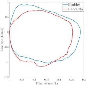
4.5. Healthy versus unhealthy
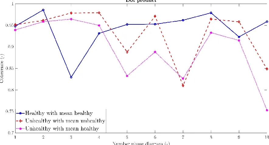
The phase diagrams of subject 21 up to 3 minutes after exercise were investigated to select 10 healthy phase diagrams, see Appendix E.1. Subject 21 had a minimal change of 6% of the lung function during the measurement which occurred at 1 minute after exercise. The phase diagrams of subject 7 between 3 and 6 minutes after exercise were investigated to select 10 unhealthy phase diagrams, see Appendix E.2. Subject 7 was examined as his lung function changes 28% during the measurement, with a minimal value at 3 minutes after exercise. The dot product between two Fourier vectors provides an indication of the coherence. To investigate if there is a difference between healthy and unhealthy phase diagrams, several dot products were calculated. The dot product between healthy phase diagrams and their mean phase diagram was calculated to check whether the appearance of the healthy phase diagrams was similar. The dot product between unhealthy phase diagrams and their mean phase diagram was calculated to check the variation between the unhealthy phase diagrams. Thereafter, the dot product between unhealthy phase diagrams and the mean of the 10 healthy phase diagrams was calculated to investigate if they differ in appearance, see figure 23 for the results.
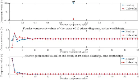
[image:39.595.75.517.340.578.2]The coherence between every healthy phase diagrams and their mean phase diagram is above 0.8, see figure 23. This is comparable to the coherence between unhealthy phase diagrams and their mean phase diagram, observable in figure 24. Regarding figure 25, the range of coherence between the unhealthy phase diagrams and the mean of 10 healthy phase diagrams is unfortunately also above 0.8. This indicates that the dot product between the mean of 10 healthy phase diagrams and a phase diagram is unable to discriminate between a healthy and unhealthy phase diagram. To investigate in more detail if there is a difference between healthy and unhealthy phase diagrams, the Fourier coefficients of both healthy and unhealthy phase diagrams were compared for every single and the mean phase diagrams.
33 In figure 24, the a0 coefficient of the Fourier vector is displayed for both healthy and unhealthy phase diagrams. The range of the healthy and unhealthy a0 values seems comparable and therefore it is assumptive that a0 unsuitable as discriminator between healthy and unhealthy.
Figure 24: First Fourier coefficient, a0, for the healthy phase diagrams in blue and unhealthy in red. For the 10 selected healthy phase diagrams, subject 21 who is a 9.5 years old experienced a 6% decline at 1 min after exercise. The 10 selected unhealthy phase diagrams were between 3 and 6 minutes after exercise of the 8.5 years old male subject 7 as he experienced a 28% decline at 3 minutes.
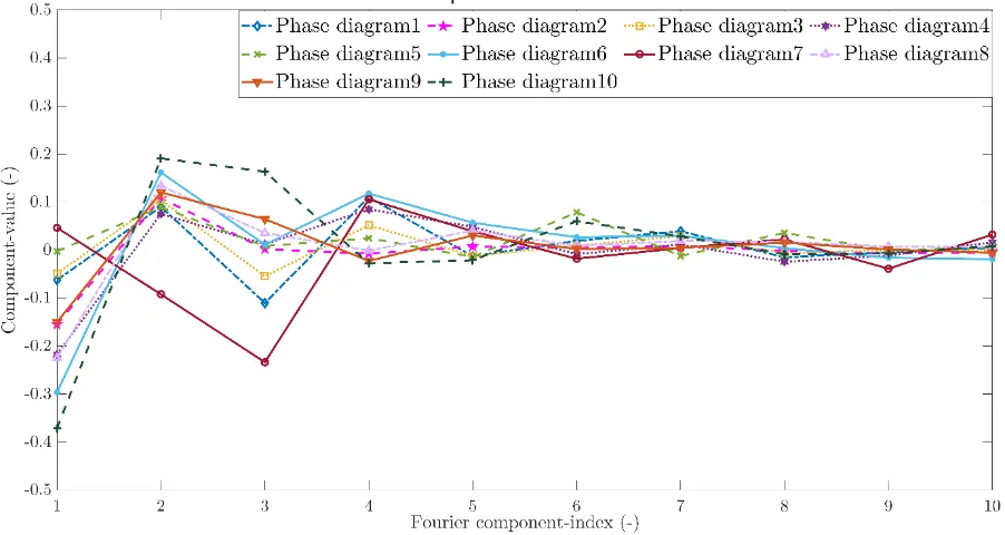
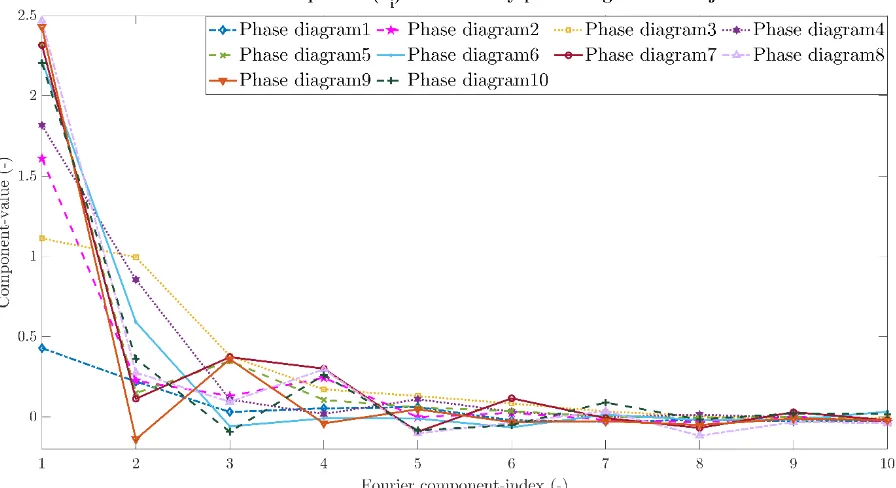
34 Figure 25 indicates the contribution of each cosine term for each healthy phase diagram. Noticeable is the decline in contribution of the cosine term as more terms were added. This is also observable for the unhealthy phase diagrams and the sines terms. Therefore, the contribution of the healthy and unhealthy phase diagrams of both sine and cosine components is shown up to 10 components in figures 26 to 29.
[image:41.595.73.524.166.406.2]Figure 26: The 10 a's indicating the contribution of the cosine term for the healthy phase diagrams which were selected at the minimal lung function around 1 minute after exercise of subject 21. The number of the Fourier coefficient is indicated on the x-axis and the contribution of the cosine term on the y-axis. Each colour represents one healthy phase diagram.
[image:41.595.72.523.482.722.2]35
Figure 28: The 10 b's indicating the contribution of the sine term for the healthy phase diagrams which were selected at the minimal lung function around 1 minute after exercise of subject 21. The number of the Fourier coefficient is indicated on the x-axis and the contribution of the sine term on the y-axis. Each colour represents one healthy phase diagram.
[image:42.595.72.525.400.642.2]![Figure 1: The osmotic theory describing the pathogenesis of exercise induced bronchoconstriction [20]](https://thumb-us.123doks.com/thumbv2/123dok_us/9658532.467892/11.595.382.522.106.367/figure-osmotic-theory-describing-pathogenesis-exercise-induced-bronchoconstriction.webp)
![Figure 3: Flow volume curves of a healthy subject on the left and an asthmatic subject with a moderate airflow limitation on the right [21]](https://thumb-us.123doks.com/thumbv2/123dok_us/9658532.467892/15.595.86.493.181.432/figure-healthy-subject-asthmatic-subject-moderate-airflow-limitation.webp)