Metabolism, migration and memory in cytotoxic T cells
David Finlay and Doreen Cantrell
Division of Cell Biology and Immunology, University of Dundee, Dundee, UK.
Abstract
The transcriptional and metabolic programs that control CD8+ T cells are regulated by a diverse network of serine/threonine kinases. The view has been that the kinases AKT and mammalian target of rapamycin (mTOR) control T cell metabolism. Here, we challenge this paradigm and discuss an alternative role for these kinases in CD8+ T cells, namely to control cell migration. Another emerging concept is that AMP-activated protein kinase (AMPK) family members control T cell metabolism and determine the effector versus memory fate of CD8+ T cells. We speculate that one link between metabolism and immunological memory is due to the acquired ability of kinases that evolved to control T cell metabolism to control the expression of key transcription factors that regulate CD8+ T cell effector function and migratory capacity.
Introduction
Cytotoxic T cells that express the CD8 co-receptor and recognize peptide–MHC class I complexes have a key role in clearing viral infections. During a primary immune response to viruses, naïve CD8+ T cells expressing pathogen-specific T cell receptors (TCRs) clonally expand and differentiate into effector CD8+ T cells that control the primary infection. This differentiation process produces effector cytotoxic T lymphocytes (CTLs) that can destroy virally infected cells via the targeted secretion of perforin and granzymes from lytic
granules. Once the primary infection is cleared there is a contraction phase when most of the effector CTLs die by apoptosis. However, an effective immune response also produces a stable population of antigen-specific long-lived memory CD8+ T cells that can respond rapidly to clear secondary infections1-3.
The transcriptional programs that determine the effector versus memory fate of T cells are controlled by antigen receptors, co-receptors, cytokines and chemokines. These molecules also co-ordinate T cell metabolism and ensure that during an immune response T cells increase their uptake of glucose, amino acid and iron and switch to glycolysis to increase cellular energy production and nutrient uptake for the biosynthetic demands of their effector functions. One other important consideration is that activation of CD8+ T cells induces essential changes in their migratory patterns to re-direct effector CTLs to sites of inflammation and concomitantly reduce their capacity to home to secondary lymphoid tissues. The challenge is thus to understand the molecular pathways that synchronize metabolism and migration with effector and memory T cell differentiation.
In this Opinion article, we explore how serine/threonine kinases such as AKT (also known as PKB) and members of the AMP-activated protein kinase (AMPK) family, such as LKB1 (also known as STK11) and AMPK, co-ordinate these processes. We challenge the dogma that AKT has an obligatory role in controlling T cell metabolism and survival, and discuss how LKB1 is perhaps more critical. We introduce the concept that the physiological role for
Author Manuscript
Nat Rev Immunol. Author manuscript; available in PMC 2012 December 13.
Published in final edited form as:
Nat Rev Immunol. 2011 February ; 11(2): 109–117. doi:10.1038/nri2888.
Europe PMC Funders Author Manuscripts
AKT is to co-ordinate the repertoire of adhesion and chemokine receptors expressed by CD8+ T cells and hence regulate their trafficking and migration. We discuss the emerging idea that changes in T cell metabolism dictate the decision of T cells to produce memory versus terminally differentiated effector T cells and consider whether there really is any evidence for such a link or whether the link between metabolism and immunological memory reflects that kinases that evolved to control cell metabolism have acquired the ability to control T cell migration. The ability of such kinases to control T cell migration then influences the fate of CD8+ T cells in terms of the decision to produce memory versus terminally differentiated effector CD8+ T cells.
Metabolism and CTLs
It was recognised over 30 years ago that it is important for CTL to control their cellular metabolism and to co-ordinate oxidative phosphorylation and glycolytic energy pathways4-6. Quiescent naïve and/or memory CD8+ T cells will only require energy to prevent cell atrophy and for survival and migration. By contrast, effector CTLs will have higher energy demands because they need to proliferate rapidly and produce effector cytokines. It is thus essential that CD8+ T cells can increase cellular energy production and nutrient uptake to satisfy increased biosynthetic demands as and when they occur7. One mechanism that ensures that CD8+ T cells regulate their metabolism to meet increased rates of biosynthesis is that extrinsic signals from antigen receptors and cytokines induce and then maintain cell surface expression of amino acid transporters, the transferrin receptor and glucose
transporters8-10. This ensures that nutrient uptake is increased to meet the metabolic and biosynthetic needs of the T cell as it responds to either developmental or pathogenic cues.
One particularly important metabolic change that occurs in activated T cells is that the cells switch from oxidative phosphorylation to aerobic glycolysis11; that is, they use an oxygen-independent mechanism to produce ATP from glucose12. Effector CTLs may have to migrate, survive and produce effector cytokines in the hypoxic environment at sites of inflammation. Moreover, lymphoid tissue is also relatively hypoxic with the oxygen tension ranging from 1– 5%13. A switch to aerobic glycolysis may thus help CTLs proliferate and mediate their effector function in relatively hypoxic conditions. However, the problem with glycolysis is that it is a relatively inefficient route to produce ATP as it only yields two molecules of ATP for every molecule of glucose. This is in contrast to oxidative
phosphorylation that yields 30 molecules of ATP from each molecule of glucose. Glycolysis thus requires that cells can sustain high levels of glucose uptake. In CTLs the key process to ensure sustained high-level glucose uptake is the upregulation of the expression and function of glucose transporter type 1 (GLUT1)8. It is also essential that T cells increase the activity/ expression of rate-limiting glycolytic enzymes, such as hexokinase 1, hexokinase 2 and phosphofructokinase 112, 14. The dependence of CTLs on aerobic glycolysis thus makes these cells dependent on exogenous glucose7, 15. Nevertheless, the advantage of glycolysis, and hence the most probable reason that the glycolytic switch occurs in CTLs, is that glycolysis promotes the use of glucose as a source of carbon for the synthesis of nucleic acid and phospholipids12. An increase in the production of such biosynthetic precursors would be favourable for CTLs, which have to maintain high levels of protein and lipid synthesis to function as effector CD8+ T cells.
AKT and T cell metabolism: the dogma
How do T cells increase their metabolism in response to immune stimulation? The current dogma is that the serine/threonine kinase AKT has a key role. This kinase is rapidly activated in response to TCR triggering or by cytokines such as interleukin-2 (IL-2) and IL-1516-18. AKT activation in T cells is regulated by stimuli that increase
phosphatidylinositol-3,4,5-trisphosphate (PtdIns(3,4,5)P3) levels and requires the serine/
Europe PMC Funders Author Manuscripts
threonine kinase 3-phosphoinositide-dependent protein kinase 1 (PDK1)18, 19 (Figure 1). The activation of AKT requires its phosphorylation on Thr308 by PDK1 and on Ser473 by mammalian target of rapamycin (mTOR) complex 2 (mTORC2)20, 21. PDK1 and AKT both have a pleckstrin homology (PH) domain that binds PtdIns(3,4,5)P3 with high affinity. This PtdIns(3,4,5)P3binding to AKT is required for PDK1 to phosphorylate AKT on Thr308. However, the binding PtdIns(3,4,5)P3 to PDK1 is not essential for the catalytic activity of PDK1, although it does promote localization of the enzyme to the plasma membrane and support efficient induction of high levels of AKT activity18, 22.
The model that AKT controls metabolic and survival programs in T cells stems from experiments showing that phosphoinositide 3-kinase (PI3K) inhibitors, which prevent AKT activation, prevent increases in both glucose and amino acid uptake by activated T
cells16, 23. In addition, constitutively active mutants of AKT can promote glucose uptake by controlling the expression of GLUT124. The mechanism for AKT control of the expression of glucose transporters is not fully understood but AKT substrates include the RAB GTPase-activating proteins AS160 (AKT substrate of 160 kDa) and TBC1D1, which can control the translocation of glucose transporters to the cell membrane25-27. Further evidence that PtdIns(3,4,5)P3–PDK1-mediated activation of AKT controls T cell metabolism was obtained from studies of T cell progenitors in the thymus. In these cells, AKT stimulation is essential for the increases in glucose uptake and expression of amino acid transporters and transferrin receptor required to support the survival and proliferation of these cells during thymopoiesis9, 28. T cell progenitors that do not express PDK1 or AKT thus fail to express these important nutrient receptors and do not develop, since they cannot meet the metabolic demands of thymopoiesis.
AKT and T cell metabolism: the reality
The consensus view of AKT is that it has a universal role in controlling T cell metabolism. It is thus assumed that the metabolic role for AKT in T cell progenitors is recapitulated in all peripheral T cells. Indeed, many of the extrinsic stimuli that control peripheral T cell metabolism activate PI3K and AKT 17, 29, 30. There are also numerous experiments that have used PI3K inhibitors in different T cell populations that support the idea that the PI3K, and hence AKT, are important for glucose uptake and/or protein synthesis2331, 32. However, a criticism of many of these studies is that one of the most commonly used PI3K inhibitors LY294002 has off-target effects and can inhibit mTOR and the PIM family of serine/ threonine kinases33, 34. A second criticism is that there is frequently an assumption that all PI3K-dependent responses are mediated by AKT and very little consideration is given to the role of other downstream effector molecules of PtdIns(3,4,5)P3, such as Tec family tyrosine kinases and guanine-nucleotide-exchange factors for Rho GTPases35.
In this context, evidence for AKT-independent pathways that control T cell metabolism, survival and proliferation have now been documented. While constitutively active AKT has been shown to stimulate glucose uptake and to promote cell growth and survival36, active AKT differentially controls T cell subsets, promoting the survival of CD4+ but not CD8+ T cells37. Another example of how T cell metabolism and survival can be independent of AKT comes from in vivo studies of the role of PDK1. For example, deletion of PDK1 does not prevent IL-7 from supporting CD4+ T cell survival38. One other in vivo demonstration of the AKT independence of T cell metabolism comes from studies of the role of PDK1 in the pathology caused by deletion of the tumour suppressor PTEN (phosphatase and tensin homologue deleted on chromosome 10)19. This molecule is a lipid phosphatase with specificity for the 3′-end of PtdIns(3,4,5)P3. The tissue-specific deletion of PTEN in thymic T cell progenitors using a Cre–lox approach increases cellular levels of PtdIns(3,4,5)P3 and is sufficient to strongly activate PDK1–AKT signalling. PTEN deletion also causes T cell lymphomagenesis, which results in the development of lymphoma cells that are large
Europe PMC Funders Author Manuscripts
blastoid cells indicative of unrestrained activation of metabolic pathways39. In normal T cell progenitors, PDK1–AKT signalling controls nutrient receptor expression and cell
metabolism and hence is essential for the proliferation and survival of these cells9. It was therefore surprising that deletion of PTEN bypassed the normal PDK1–AKT-controlled metabolic checkpoint in T cell progenitors, such that the growth and proliferation of PTEN-deficient T cells is not dependent on PDK1 and AKT signalling19. The reason this result is surprising is that PDK1 is required for malignant transformation of PTEN-deficient thymocytes19 and it was assumed that the requirement for PDK1 in the transformation of PTEN-deficient thymocytes was because PDK1-mediated activation of AKT was required to support the metabolism of these cells. The fact that the metabolism, survival and
proliferation of PTEN-deficient thymocytes can be independent of PDK1 and AKT
exemplifies how AKT does not have an equal and obligate role in controlling metabolism in all T cell populations.
AKT-independent mechanisms
What are the candidates to mediate AKT-independent control of T cell metabolism? It could be other members of the AGC family of serine/threonine kinases, such as 90 kDa ribosomal protein S6 kinase (p90-RSK), 70 kDa ribosomal protein S6 kinase 1 (p70-S6K) or serum/ glucocorticoid-regulated kinase (SGK), which share substrate specificity with AKT 40-42. Other candidates are the PIM family of serine kinases because they have been shown to have a role in T cell survival and also have a substrate specificity similar to AKT 43. Moreover, the PIM kinases are inhibited by the PI3K inhibitor LY29400234. The other emerging possible molecules that control T cell metabolism include the mitogen-activated protein (MAP) kinases extracellular-signal-regulated kinase 1 (ERK1) and ERK2, which have recently been described to be essential for the induction of glutamine uptake that accompanies T cell activation44.
It has also been reported that the serine/threonine kinase LKB1 is important for T cell metabolism. LKB1 is an evolutionarily conserved kinase that can regulate cellular responses to energy stress in many cell types and is required to synchronize cellular energy
checkpoints and cell division in Drosophila melangastor45. LKB1 phosphorylates and activates multiple AMPK family kinases including AMPK46, 47. In T cells, AMPK is phosphorylated and activated by LKB1 in response to increases in cellular AMP/ATP ratios but AMPK can also be activated in response to TCR triggering via a Ca2+ –calmodulin-dependent protein kinase pathway48. In many cell lineages, AMPK acts to restore cellular energy balance by inhibiting ATP-consuming processes and stimulating ATP-generating pathways49. It has thus been proposed that in T cells, increases in intracellular Ca2+ concentrations that activate calmodulin-dependent kinases stimulate AMPK to promote the conservation and accelerated production of ATP in anticipation of energy supplies becoming depleted by T cell activation.
What is the evidence that LKB1 and AMPK contribute to the regulation of the energy status of T cells? One compelling set of data is that LKB1 is essential for the survival and
development of thymic T cell progenitors that had undergo TCR β-selection and is also required for the survival and proliferation of peripheral CD8+ T cells50, 51. Importantly, LKB1 appears to be required for several key metabolic processes in T cells. For example, LKB1 controls the expression of CD98, a key subunit of L-system amino acid transporter 1 in T cells and is also required to sustain the phosphorylation of the ribosomal S6 subunit, a key regulator of protein synthesis in T cells51. Interestingly, LKB1 is an essential regulator of AMPK in energy-stressed T cells50, yet the loss of AMPK does not phenocopy the loss of LKB1. AMPK-deficient T cells have increased sensitivity to energy stress in vitro but can still develop normally in the thymus in vivo in contrast to LKB1-deficient T cells51, 52. This is most likely due to redundancy in vivo between different AMPK family members for the
Europe PMC Funders Author Manuscripts
control of T cell metabolism, and the loss of LKB1 expression circumvents this redundancy by simultaneously eliminating the activation of multiple AMPK family members. Therefore, contrary to the current dogma, AKT does not have an obligatory role controlling T cell metabolism and evidence is emerging that LKB1-regulated kinases have a role.
Control of CD8
+T cell migration
AKT and migration
Recent work has established that the activation of AKT downregulates the expression of the adhesion molecule CD62L (also known as L-selectin) and the chemokine receptors CC-chemokine receptor 7 (CCR7) and sphingosine 1-phosphate receptor 1 (S1P1) in CD8+ T cells: through this downregulation AKT determines the differential trafficking of naïve versus effector T cells18, 19, 53. Naïve and memory CD8+ T cells constantly circulate between the blood, lymphoid tissues and lymphatic vessels. By contrast, activated CD8+ T cells must suspend migration and reside in lymphoid tissue while they clonally expand and differentiate into effector CTLs. It is then critical that effector CTLs regain their motility, exit the lymphoid tissue and migrate to the site of infection to mount an immune response. The balanced and differential trafficking of naïve versus effector CD8+ T cells is thus critical for the function of CD8+ T cells during adaptive immune responses.
In this context, naïve and memory CD8+ T cells enter secondary lymphoid organs because they express CCR7 and can react to a gradient of CCR7 ligands that allows them to cross the endothelial cell barrier in specialized high endothelial venules (HEVs)17, 54. Naïve and memory CD8+ T cells also express high levels of CD62L, which mediates the first step of transendothelial migration — the capture and rolling of T cells on the endothelium of HEVs. Once in the lymph nodes the fibroblastic reticular cell (FRC) network and chemokines such as the CCR7 ligands CCL19, CCL21 and sphingosine 1-phosphate (S1P) coordinate T cell migration54, 55. Naive T cells then spend several hours in the lymph node before exiting into the efferent lymphatic and returning to the circulation. The egress of T cells from lymph nodes is determined by S1P155, 56. Activated T cells are retained in the lymph node because they down-regulate or inactivate the expression of S1P155-57. They then undergo a period of clonal expansion and differentiation to effector CTLs at which point the cells regain S1P1 function and exit to the periphery. Effector CTLs then migrate to sites of inflammation or infection and have a reduced ability to home to peripheral lymph nodes compared with naïve and memory CD8+ T cells. The changes in the trafficking behaviour of effector CTLs occur because they downregulate CCR7, CD62L and S1P1 expression and upregulate the
expression of pro-inflammatory adhesion molecules, such as VLA4 (very late antigen 4), ligands for P-selectin and E-selectin and pro-inflammatory chemokine receptors such as CXC-chemokine receptor 3 (CXCR3) and CCR554, 57, 58. The loss of CD62L, CCR7 and S1P1 expression by CTLs is an important mechanism that prevents these effector CTLs from re-entering secondary lymphoid organs and allows their redirection to peripheral tissues. Moreover, the importance of tightly regulating the expression of these receptors is evidenced by observations that changes in the turnover of S1P1, CCR7 or CD62L can markedly modify CTL-mediated immune responses58, 59.
Recently, some of the molecular mechanisms that control the expression of these key receptors have been described. One of the first clues came from observations that the cytokines IL-2 and IL-15 differed in their ability to downregulate CD62L expression on antigen-primed CD8+ T cells and diffferentially regulated AKT activation16, 17. Antigen-primed CD8+ T cells cultured in the presence of IL-2 sustain AKT activation at high levels and downregulate CD62L. By contrast, T cells cultured with IL-15 only sustained low-level AKT activation and failed to downregulate CD62L expression16, 17. These observations were initially correlative but subsequent work verified that CD62L, CCR7 and S1P1
Europe PMC Funders Author Manuscripts
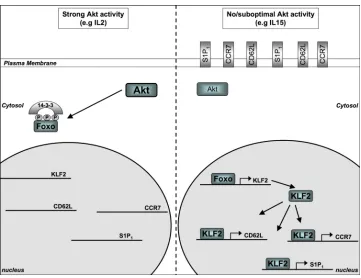
expression by CD8+ T cells reflected the level of AKT activation in the cell17-19 (Figure 2). Hence, effector CTLs have high levels of AKT activity, express low levels of CD62L, CCR7 and S1P1 mRNA and protein and cannot traffic from the blood to lymphoid tissue. By contrast, CTLs that cannot fully activate AKT retain expression of CD62L, CCR7 and S1P1 and retain the ability to traffic to lymph nodes18. Conversely, in naïve T cells, which normally express high levels of CD62L, CCR7 and S1P1, PDK1-mediated activation of AKT is sufficient to terminate the expression of these homing receptors and prevent the cells from homing to peripheral lymph nodes or spleen19. Therefore, lymph node homing by T cells is controlled by the level of AKT signalling, which controlsthe expression of key adhesion and chemokine receptors —— that direct T cells into the secondary lymphoid tissues.
Foxo, mTOR and T cell migration
How does active AKT suppress the expression of CD62L? The answer to this question lies in the ability of AKT to control the activity of the Foxo family transcription factors FOXO1 and FOXO3A and hence to control the expression of the transcription factor Kruppel-like factor 2 (KLF2) (Figure 2)53. KLF2 directly regulates Cd62l, gene transcription60-62. KLF2-deficient naïve CD8+ T cells thus do not CD62L homing receptors and fail to home to secondary lymphoid organs and instead migrate to peripheral tissues. FOXO1 and FOXO3A are transcriptionally active in the nucleus of quiescent naïve T cells and drive the expression of KLF2. Following immune activation, AKT-mediated phosphorylation of FOXO
transcription factors results in their translocation from the nucleus to the cytoplasm63 where they form a complex with 14-3-3 scaffold proteins64. AKT-mediated inhibition of FOXO proteins thus terminates the expression of KLF2 and hence the expression of KLF2 gene targets, including Cd62l, S1p1 and Ccr718.
Another way AKT controls the expression of KLF2 is by regulating mTOR in the mTORC1 complex65. AKT phosphorylates and inactivates the RHEB GTPase-activating protein TSC2 (tuberous sclerosis complex 1) resulting in the accumulation of RHEB–GTP complexes, which activate mTORC1 (Figure 3). AKT also regulates mTORC1 by phosphorylating PRAS40 (40kDa proline-rich AKT1 substrate), thereby blocking PRAS40-mediated inhibition of mTORC166 (Figure 3). mTORC1 can control the translation of a subset of mRNAs, protein synthesis and ribosomal biogenesis65, 67. It is not known if these metabolic functions of mTOR are important in T cells. However, mTOR activation does downregulate the expression of KLF2 and therefore CD62L, CCR7 and S1P117. The molecular details of the control of KLF2 expression by mTOR in T cells remains to be fully determined but it appears to be independent of regulated FOXO phosphorylation or localization (Finlay D. and Cantrell D.A. unpublished data).
The salient fact about the link between mTORC1 and KLF2 is that the mTORC1 inhibitor rapamycin, which is widely used clinically as an immunosuppressant, causes CTLs to re-express KLF2, CD62L and CCR7 and regain the ability to home to lymph nodes17. It is generally believed that rapamycin suppresses immune responses through its suppression of T cell proliferation. However, it is now clear that rapamycin treatment can also re-direct the trafficking of effector CTLs from peripheral tissue to lymph nodes and spleen17. The ability to re-direct CTLs to secondary lymphoid tissue would result in their retention in secondary lymphoid organs and hence prevent immune destruction of target cells in peripheral tissues. It could also promote the destruction of antigen-primed antigen-presenting cells (APCs) by CTLs in lymph nodes, which could contribute to the clinical efficacy of rapamycin as an immunosuppressant.
Europe PMC Funders Author Manuscripts
Determining metabolism versus migration
Different AKT activation thresholds
The discussions above indicate how AKT can control the activity pathways that converge to control T cell migration (Figure 4). However, one significant observation about AKT is that it does not function as a simple on/off switch in T cells; rather the magnitude of AKT activity is relevant. This insight comes from studies of T cells homozygous for a knock-in mutant (Lys465Glu) of PDK1 that cannot bind PtdIns(3,4,5)P318, 22. The integrity of the PDK1 PH domain is not required for PDK1 catalytic activity but the loss of PtdIns(3,4,5)P3 -binding by PDK1 strongly reduces AKT phosphorylation and activation. The
PDK1Lys465Glu mutant can thus only support sub-maximal levels of AKT in activated T cells (approximately 10–20% of that in wild-type T cells)18. Intriguingly, the low levels of AKT activity in T cells expressing the PDK1Lys465Glu mutant are sufficient for T cell growth and proliferation and thus are clearly sufficient to support T cell metabolism during thymopoiesis and when T cells respond to cognate antigen. However, low levels of AKT activity in T cells expressing mutant PDK1 Lys465Glu are not sufficient to terminate the expression of KLF2 or to switch the chemokine and adhesion receptor profile of naïve T cells to that of CTLs.
These data suggest how the initial strength of TCR triggering might dictate the outcome of an immune response by controlling the ability of the T cells to migrate rather than by controlling their ability to proliferate. For example, the downregulation of S1P1 following CD8+ T cell activation is one the mechanisms that retains activated CD8+ T cells in lymph nodes and would only occur if there was strong activation of AKT. Activation with weak TCR agonists that cause weak activation of AKT might thus support T cell proliferation but would be unable to downregulate S1P1 expression and therefore be unable to retain cells in the lymph nodes. The premature exit of activated CD8+ T cells from lymph nodes into the blood has been observed to occur in response to weak stimulation68 and would curtail CD8+ T cell exposure to APCs and pro-inflammatory cytokines and hence would result in an attenuated immune response.
AMPK, mTOR and T cell memory
It has recently been proposed that changes in T cell metabolism might be at the core of the decision of CD8+ T cells to produce memory versus terminally differentiated effector CD8+ T cells69-71. This idea originates from experiments showing that in vivo treatment of mice with the mTOR inhibitor rapamycin increased the production of memory CD8+ T cells by accelerating the conversion of effector CD8+ T cells into the memory pool69, 71. It has also been shown that the activation of AMPK by treatment of mice with the drug metformin enhances the production of memory CD8+ T cells71. These two sets of observations might be linked because metformin interferes with oxidative phosphorylation mediated ATP synthesis through inhibition of the respiratory chain complex 1 and hence causes an increase in cellular AMP:ATP ratio. This energy stress can drive an LKB1-dependent activation of AMPK, which then phosphorylates TSC2 and raptor to coordinately suppress mTORC1 activity72 (Figure 3).
The T cell memory experiments with metformin were initiated by the observation that TNFR-associated factor 6 (TRAF6)-deficient CD8+ T cells cannot develop into memory CD8+ T cells and have reduced activity of AMPK71. However, it has not been directly assessed whether AMPK-deficient T cells can produce memory T cells. Moreover, evidence is emerging that metformin can control cellular metabolism independently of AMPK73, 74. Nevertheless, as the primary effect of metformin is to inhibit respiratory chain complex 174, the metformin data are a good indication that changes in cell metabolism can change T cell
Europe PMC Funders Author Manuscripts
behaviour. However, there are many issues to resolve concerning the affect of metformin on T cell memory. First, there has been no clear demonstration that the in vivo effects of metformin reflect the action of this drug specifically on T cells rather than other cells on the immune system. Second, it needs to be proven that metformin acts through AMPK. One other factor to be considered is that it should be clear from the previous discussions that mTOR (and hence AMPK) should not be considered solely as enzymes that control T cell metabolism. The ability of mTOR to control T cell migration by controlling the expression of KLF2, CD62L, CCR7 and S1P1 is an example of mTOR functions that can be
independent of any metabolic function of this enzyme17. Rapamycin treatment restores expression of KLF2 and its targets in IL-2-maintained CTLs and redirects CTL trafficking to secondary lymphoid tissue, even though IL-2-mediated control of CD8+ T cell survival and proliferation is not rapamycin sensitive17.
How could inhibition of mTORC1 with rapamycin or through activation of AMPK promote the transition of effector CTLs to memory CD8+ T cells? One explanation could be the ability of rapamycin to reprogram the trafficking of effector CTLs such that these cells regain the ability to enter secondary lymphoid tissue 17, 75. This would bring them into the proximity of stromal cells that produce cytokines, such IL-7 and IL-15, that are required for the survival and homeostasis of memory T cells. It is also relevant here that in CD8+ T cells, mTOR activation is required to sustain the expression of the transcription factor T-bet while suppressing the expression of eomesodermin (Figure 4)75. The molecular basis for mTOR-mediated control of T-bet and eomesodermin expression is not known nor is it clear if this is linked in any way to the role of mTOR as a metabolic regulator. However, the balance of T-bet and eomesodermin expression can determine the effector or memory fate of CD8+ T cells76, and the ability of mTOR to control the expression of these transcription factors gives some insights as to how levels of mTOR activity might determine CTL fate. Hence, T-bet is a key transcription factor in CTLs and is important for driving expression of the cytolytic differentiation program58. It is however intriguing that T-bet also controls the expression of CXCR3 and P-selectin ligands, which are crucial for the trafficking of effector CD8+ T cells to sites of inflammation58, 77. This raises the fundamental question of whether changes in CD8+ T cell metabolism determine the effector or memory fate of CD8+ T cells by directly controlling their differentiation. An alternative view is that enzymes that evolved to control T cell metabolism have evolved the ability to control T cell migration and it is their effects on migration and T cell homing that influences the fate of CD8+ T cells in terms of the decision to produce memory versus terminally differentiated effector CTLs. This latter view is probably too simplistic but if T cells migrate to different sites they will have the potential to adopt different fates because they will exist in a different cytokine and chemokine milieu. The most probable scenario is that mTOR controls CD8+ T cell memory by a combination of factors, including the control of T cell trafficking and the control of the expression of the key transcription factors that control CTL effector function.
What about the proposal that metabolism regulates memory? This concept stems from the recognition that mTOR can control immunological memory and the underlying dogma that mTOR controls T cell metabolism. However, there is no direct evidence that mTOR regulates T cell metabolism in the cellular models that demonstrate a role for mTOR in CTL function. Nevertheless, it should be remembered that mTOR activity will be determined by nutrient availability 78. Hence any change in the strength or quality of T cell activation during an immune response could cause suboptimal expression of nutrient receptors on T cells thereby limiting their glucose and amino acid uptake and limiting the strength of mTOR activation.
Europe PMC Funders Author Manuscripts
Differential AKT and mTOR signaling in CTLs
There is a lot of evidence that effector versus memory T cells can develop from a common naïve T cell progenitor 79-81 and that memory CD8+ T cells can develop from antigen-primed CD8+ T cells that have differentiated into effector CTLs82. However, if differential activation of mTOR signaling is a driver of effector versus memory CD8+ T cell fate then how can the progeny of a single cell generate heterogeneous mTOR signaling? How can an effector CD8+ T cell divert to a memory CD8+ T cell fate? Moreover, in the context of this discussion, is there a link with metabolism?
One plausible idea is that competition for essential cytokines, such as IL-2, IL-12 and IL-15, could be the mechanism that creates heterogeneity in mTOR signaling in T cells. It is quite reasonable to assume that levels of these cytokines are finite in any tissue. It is also well established that sustained signaling by these cytokines is essential for CD8+ T cell differentiation. Moreover, in the context of IL-2 signal transduction, the ‘strength’ and duration of exposure to IL-2 is critical83, 84. Hence, in vivo studies show that CD8+ T cells that express high levels of the high-affinity IL-2 receptor are more likely to develop into terminally differentiated effector CD8+ T cells, whereas cells with lower levels of IL-2 receptors give rise to long lived ‘memory’ CD8+ T cells84. Similarily, in vitro experiments show that antigen-primed CD8+ T cells cultured with high-dose IL-2 differentiate into effector CTLs, whereas cells cultured in low-dose IL-2 differentiate into ‘memory’ CD8+ T cells83. Importantly, IL-15 could only support the production of ‘memory’ CD8+ T cells, reflecting the fact that IL-15 can only weakly activate AKT in antigen-primed CD8+ T cells 16, 17, 83.
So a simple idea of how a single antigen-primed naïve CD8+ T cell could produce progeny that were heterogenous for AKT activation is to consider that populations of T cells have to compete for cytokines, such as IL-2 (Figure 5). This could be a stochastic process initially or it could be directed by heterogeneity in the expression of the IL-2 receptor by CTLs84. Cells with a low level of IL-2 signaling would have lower levels of AKT and mTOR activity. As discussed, this would change the trafficking properties of these cells and any heterogeneity would be reinforced when the cells moved to different microenvironments. It is also relevant that IL-2 is a potent regulator of the expression of the receptors for key cytokines that determine CD8+ T cell fate: IL-2 can thus positively regulate IL-12 receptor expression85 and negatively control IL-7 receptor levels86. Therefore, in addition to altering T cell migratory properties due to lower AKT and mTOR activity, low dose IL-2 would cause CD8+ T cells to downregulate IL-12 receptor expression and increase their expression of IL-7 receptors. This would favour their differentiation towards a memory CD8+ T cell fate. Returning to the theme of metabolism, high levels of IL-2 signaling could support the high metabolic responses required for effector CTL responses, whereas low levels of IL-2 would not.
This is obviously an over-simplistic view and it is unlikely that the complexities of effector versus memory CD8+ T cell decisions can be explained on the basis of how T cells deal with a single cytokine. However, effector versus memory CD8+ T cell fate decisions could be explained by a model that places AKT and mTOR at the core of a pathway that integrates signals from a wide range of cytokines, co-stimulatory molecules and antigen receptors to direct CD8+ T cell fate. The key to this model is that the strength and duration of AKT, and hence mTOR, activation is determined by the balance of signals from these surface receptors so there are multiple routes to achieve differential AKT activation at any time as T cells respond to immune-mediated activation signals. In addition to the example of cytokine competition discussed above, another route could be changes in the levels of TCR
triggering. Effector CD8+ T cells at the sites of infection might sustain high levels of AKT
Europe PMC Funders Author Manuscripts
activity as a result of continual antigen receptor occupancy. Then, as pathogens are cleared from the body, the levels of TCR triggering would decrease, allowing the levels of active AKT to drop and thereby directing the CD8+ T cells to home back to secondary lymphoid tissues where competition for homeostatic cytokines would determine the number of memory CD8+ T cells that could be supported within a particular niche.
Concluding remarks
We still have much to learn about how CD8+ T cells control their metabolism to meet the energy demands of their effector function. However, we have begun to uncover an extraordinary amount about how the enzymes that evolved to control T cell metabolism control T cell migration and differentiation and determine the balance of effector versus memory CD8+ T cell development. We have also been able to see how changes in the strength of activation of serine/threonine kinases can create heterogeneity in signal
transduction pathways needed to allow a single T cell to generate progeny that have multiple fates. This work gives important insights about pharmacological strategies that might be able to manipulate immune responses to ensure effective vaccination and/or stem the T cell pathology caused by effector CTLs.
Glossary terms
Perforin A component of cytolytic granules that participates in the permeabilization of plasma membranes, allowing granzymes and other cytotoxic components to enter target cells.
Granzymes Secreted serine proteases that enter target cells through perforin pores, ranzymes activate intracellular caspases and lead to target-cell apoptosis.
Glycolysis A metabolic process that occurs in the cytoplasm and breaks down one molecule of glucose into two molecules of pyruvate, resulting in the production of ATP.
Transferrin receptor
Also known as CD71, this receptor regulates the cellular import of iron by binding the ironcarrier protein transferrin. In addition, it mediates clearance of circulating IgA1 by renal mesangial cells.
Oxidative phosphorylation
A metabolic process that encompasses two sets of reactions that occur in the mitochondria. The first reaction involves the
conversion of intermediate molecules (pyruvate and fatty acids) to acetyl coenzyme A (acetyl-CoA) and the degradation of acetyl-CoA to carbon dioxide in the tricarboxylic-acid cycle, yielding free electrons that are carried by NADH and FADH2. The second reaction involves the transfer of electrons from NADH and FADH2 to the electron-transport chain, resulting in the movement of protons out of the mitochondrial matrix. The resulting electrochemical potential is used by the F1F0 ATP synthase to synthesize ATP.
Aerobic glycolysis Gycolysis is an anerobic metabolic pathway converting glucose to pyruvate that can be either metabolized by oxidative
phosphorylation in the presence of O2 or converted to lactate. The term aerobic glycolysis describes the conversion of glucose to lactate although oxygen is not limiting. For example, most cancer cells predominantly produce energy by a high rate of aerobic glycolysis followed by lactic acid fermentation in the cytosol, rather
Europe PMC Funders Author Manuscripts
than by a comparatively low rate of glycolysis followed by oxidation of pyruvate in mitochondria like most normal cells (termed the Warburg effect).
Rate-limiting glycolytic enzymes
There are three enzymatic steps in the glycolytic pathway that are essentially irreversible catalysed by hexokinase,
phosphofructokinase and pyruvate kinase. Allosteric, transcriptional and post-translational regulation of these enzymes is crucial for the regulation of glycolysis. Phosphofructokinase catalyses the main rate-limiting step of glycolysis and is the most important control point.
Mammalian target of rapamycin
(mTOR). A conserved serine/threonine protein kinase that regulates cell growth and metabolism, as well as cytokine and growth-factor expression, in response to environmental cues. mTOR receives stimulatory signals from RAS and phosphoinositide 3-kinase (PI3K) downstream of growth factors and nutrients, such as amino acids, glucose and oxygen.
Cre–lox approach A site-specific recombination system that is used to delete or rearrange genes by Cre recombinase activity. Two short DNA sequences (LoxP sites) are engineered to flank the target DNA. Depending on the orientation of the LoxP sites, expression of Cre recombinase leads to excision or inversion of the intervening sequence.
β-selection A process leading, through a cell autonomous signalling cascade, to the proliferation and survival of thymocytes that have successfully recombined the β-chain of the T cell receptor (TCR) locus to express a functional pre-TCR on their cell surface.
L-system amino acid transporter 1
A heterodimeric membrane transport protein that preferentially transports neutral branched (valine, leucine, isoleucine) and aromatic (tryptophan, tyrosine) amino acids.
14-3-3 scaffold proteins
A family of conserved proteins that is present in all eukaryotic organisms. These proteins are involved in diverse cellular processes, such as apoptosis and stress, as well as in intracellular signalling and cell-cycle regulation. They function as adaptors in protein interactions and can regulate protein localization and enzymatic activity. Approximately 100 binding partners have been reported for the 14-3-3 proteins.
Rapamycin An immunosuppressive drug that, in contrast to calcineurin inhibitors (such as cyclosporin A and FK506), does not prevent T cell activation but blocks interleukin-2-mediated clonal expansion by blocking mTOR (mammalian target of rapamycin). Rapamycin-mediated mTOR inhibition also affects T cell trafficking but does not interfere with the function and expansion of naturally occurring regulatory T cells.
References
1. Joshi NS, Kaech SM. Effector CD8 T cell development: a balancing act between memory cell potential and terminal differentiation. J Immunol. 2008; 180:1309–1315. [PubMed: 18209024]
Europe PMC Funders Author Manuscripts
2. Parish IA, Kaech SM. Diversity in CD8(+) T cell differentiation. Curr Opin Immunol. 2009; 21:291–297. [PubMed: 19497720]
3. Jenkins MR, Griffiths GM. The synapse and cytolytic machinery of cytotoxic T cells. Curr Opin Immunol. 2010; 22:308–313. [PubMed: 20226643]
4. Greiner EF, Guppy M, Brand K. Glucose is essential for proliferation and the glycolytic enzyme induction that provokes a transition to glycolytic energy production. J Biol Chem. 1994; 269:31484–31490. [PubMed: 7989314]
5. MacLennan IC, Golstein P. Requirement for hexose, unrelated to energy provision, in T-cell-mediated cytolysis at the lethal hit stage. J Exp Med. 1978; 147:1551–1567. [PubMed: 308086] 6. MacDonald HR. Energy metabolism and T-cell-mediated cytolysis. II. Selective inhibition of
cytolysis by 2-deoxy-D-glucose. J Exp Med. 1977; 146:710–719. [PubMed: 302305]
7. Cham CM, Driessens G, O’Keefe JP, Gajewski TF. Glucose deprivation inhibits multiple key gene expression events and effector functions in CD8+ T cells. Eur J Immunol. 2008; 38:2438–2450. [PubMed: 18792400]
8. Jacobs SR, et al. Glucose uptake is limiting in T cell activation and requires CD28-mediated Akt-dependent and inAkt-dependent pathways. J Immunol. 2008; 180:4476–4486. [PubMed: 18354169] 9. Kelly AP, et al. Notch-induced T cell development requires phosphoinositide-dependent kinase 1.
Embo J. 2007; 26:3441–3450. [PubMed: 17599070]
10. Fox CJ, Hammerman PS, Thompson CB. Fuel feeds function: energy metabolism and the T-cell response. Nat Rev Immunol. 2005; 5:844–852. [PubMed: 16239903]
11. Brand K, Leibold W, Luppa P, Schoerner C, Schulz A. Metabolic alterations associated with proliferation of mitogen-activated lymphocytes and of lymphoblastoid cell lines: evaluation of glucose and glutamine metabolism. Immunobiology. 1986; 173:23–34. [PubMed: 3492437] 12. Vander Heiden MG, Cantley LC, Thompson CB. Understanding the Warburg effect: the metabolic
requirements of cell proliferation. Science. 2009; 324:1029–1033. [PubMed: 19460998] 13. Caldwell CC, et al. Differential effects of physiologically relevant hypoxic conditions on T
lymphocyte development and effector functions. J Immunol. 2001; 167:6140–6149. [PubMed: 11714773]
14. Marko AJ, Miller RA, Kelman A, Frauwirth KA. Induction of Glucose Metabolism in Stimulated T Lymphocytes Is Regulated by Mitogen-Activated Protein Kinase Signaling. PLoS One. 2010; 5:e15425. [PubMed: 21085672]
15. Peng T, Golub TR, Sabatini DM. The immunosuppressant rapamycin mimics a starvation-like signal distinct from amino acid and glucose deprivation. Mol Cell Biol. 2002; 22:5575–5584. [PubMed: 12101249]
16. Cornish GH, Sinclair LV, Cantrell DA. Differential regulation of T-cell growth by IL-2 and IL-15. Blood. 2006; 108:600–608. [PubMed: 16569767]
17. Sinclair LV, et al. Phosphatidylinositol-3-OH kinase and nutrient-sensing mTOR pathways control T lymphocyte trafficking. Nat Immunol. 2008; 9:513–521. [PubMed: 18391955]
18. Waugh C, Sinclair L, Finlay D, Bayascas J, Cantrell D. PI(3,4,5)P3 binding to Phosphoinositide dependent kinase 1 regulates a Protein Kinase B/Akt signalling threshold that dictates T cell migration not proliferation. Mol Cell Biol. 2009; 29:5952–5962. [PubMed: 19703999] 19. Finlay DK, et al. Phosphoinositide-dependent kinase 1 controls migration and malignant
transformation but not cell growth and proliferation in PTEN-null lymphocytes. J Exp Med. 2009; 206:2441–2454. [PubMed: 19808258]
20. Pearce LR, Komander D, Alessi DR. The nuts and bolts of AGC protein kinases. Nature reviews. 2010; 11:9–22.
21. Sarbassov DD, Guertin DA, Ali SM, Sabatini DM. Phosphorylation and regulation of Akt/PKB by the rictor-mTOR complex. Science. 2005; 307:1098–1101. [PubMed: 15718470]
22. Bayascas JR, et al. Mutation of the PDK1 PH domain inhibits protein kinase B/Akt, leading to small size and insulin resistance. Mol Cell Biol. 2008; 28:3258–3272. [PubMed: 18347057] 23. Frauwirth KA, et al. The CD28 signaling pathway regulates glucose metabolism. Immunity. 2002;
16:769–777. [PubMed: 12121659]
Europe PMC Funders Author Manuscripts
24. Rathmell JC, Elstrom RL, Cinalli RM, Thompson CB. Activated Akt promotes increased resting T cell size, CD28-independent T cell growth, and development of autoimmunity and lymphoma. Eur J Immunol. 2003; 33:2223–2232. [PubMed: 12884297]
25. Zhou QL, et al. Akt substrate TBC1D1 regulates GLUT1 expression through the mTOR pathway in 3T3-L1 adipocytes. Biochem J. 2008; 411:647–655. [PubMed: 18215134]
26. Taylor EB, et al. Discovery of TBC1D1 as an insulin-, AICAR-, and contraction-stimulated signaling nexus in mouse skeletal muscle. J Biol Chem. 2008; 283:9787–9796. [PubMed: 18276596]
27. Sano H, et al. Insulin-stimulated phosphorylation of a Rab GTPase-activating protein regulates GLUT4 translocation. J Biol Chem. 2003; 278:14599–14602. [PubMed: 12637568]
28. Juntilla MM, Koretzky GA. Critical roles of the PI3K/Akt signaling pathway in T cell development. Immunology letters. 2008; 116:104–110. [PubMed: 18243340]
29. Garcon F, et al. CD28 provides T-cell costimulation and enhances PI3K activity at the immune synapse independently of its capacity to interact with the p85/p110 heterodimer. Blood. 2008; 111:1464–1471. [PubMed: 18006698]
30. Costello PS, Gallagher M, Cantrell DA. Sustained and dynamic inositol lipid metabolism inside and outside the immunological synapse. Nat Immunol. 2002; 3:1082–1089. [PubMed: 12389042] 31. Barata JT, et al. Activation of PI3K is indispensable for interleukin 7-mediated viability,
proliferation, glucose use, and growth of T cell acute lymphoblastic leukemia cells. J Exp Med. 2004; 200:659–669. [PubMed: 15353558]
32. Fung MM, Rohwer F, McGuire KL. IL-2 activation of a PI3K-dependent STAT3 serine phosphorylation pathway in primary human T cells. Cellular signalling. 2003; 15:625–636. [PubMed: 12681450]
33. Bain J, et al. The selectivity of protein kinase inhibitors: a further update. Biochem J. 2007; 408:297–315. [PubMed: 17850214]
34. Jacobs MD, et al. Pim-1 ligand-bound structures reveal the mechanism of serine/threonine kinase inhibition by LY294002. J Biol Chem. 2005; 280:13728–13734. [PubMed: 15657054]
35. Cantrell DA. Phosphoinositide 3-kinase signalling pathways. Journal of cell science. 2001; 114:1439–1445. [PubMed: 11282020]
36. Rathmell JC, et al. Akt-directed glucose metabolism can prevent Bax conformation change and promote growth factor-independent survival. Mol Cell Biol. 2003; 23:7315–7328. [PubMed: 14517300]
37. Saibil SD, et al. CD4+ and CD8+ T cell survival is regulated differentially by protein kinase Ctheta, c-Rel, and protein kinase B. J Immunol. 2007; 178:2932–2939. [PubMed: 17312138] 38. Park SG, et al. The kinase PDK1 integrates T cell antigen receptor and CD28 coreceptor signaling
to induce NF-kappaB and activate T cells. Nat Immunol. 2009; 10:158–166. [PubMed: 19122654] 39. Hagenbeek TJ, Spits H. T-cell lymphomas in T-cell-specific Pten-deficient mice originate in the
thymus. Leukemia. 2008; 22:608–619. [PubMed: 18046443]
40. Brunet A, et al. Protein kinase SGK mediates survival signals by phosphorylating the forkhead transcription factor FKHRL1 (FOXO3a). Mol Cell Biol. 2001; 21:952–965. [PubMed: 11154281] 41. Sapkota GP, et al. BI-D1870 is a specific inhibitor of the p90 RSK (ribosomal S6 kinase) isoforms
in vitro and in vivo. Biochem J. 2007; 401:29–38. [PubMed: 17040210]
42. Zhang HH, Lipovsky AI, Dibble CC, Sahin M, Manning BD. S6K1 regulates GSK3 under conditions of mTOR-dependent feedback inhibition of Akt. Mol Cell. 2006; 24:185–197. [PubMed: 17052453]
43. Fox CJ, Hammerman PS, Thompson CB. The Pim kinases control rapamycin-resistant T cell survival and activation. J Exp Med. 2005; 201:259–266. [PubMed: 15642745]
44. Carr EL, et al. Glutamine uptake and metabolism are coordinately regulated by ERK/MAPK during T lymphocyte activation. J Immunol. 2010; 185:1037–1044. [PubMed: 20554958] 45. Lee JH, et al. Energy-dependent regulation of cell structure by AMP-activated protein kinase.
Nature. 2007; 447:1017–1020. [PubMed: 17486097]
46. Alessi DR, Sakamoto K, Bayascas JR. LKB1-dependent signaling pathways. Annual review of biochemistry. 2006; 75:137–163.
Europe PMC Funders Author Manuscripts
47. Hardie DG. AMP-activated/SNF1 protein kinases: conserved guardians of cellular energy. Nature reviews. 2007; 8:774–785.
48. Tamas P, et al. Regulation of the energy sensor AMP-activated protein kinase by antigen receptor and Ca2+ in T lymphocytes. J Exp Med. 2006; 203:1665–1670. [PubMed: 16818670]
49. Towler MC, Hardie DG. AMP-activated protein kinase in metabolic control and insulin signaling. Circulation research. 2007; 100:328–341. [PubMed: 17307971]
50. Cao Y, et al. The serine/threonine kinase LKB1 controls thymocyte survival through regulation of AMPK activation and Bcl-XL expression. Cell research. 2010; 20:99–108. [PubMed: 20029389] 51. Tamas P, et al. LKB1 is essential for the proliferation of T-cell progenitors and mature peripheral T
cells. Eur J Immunol. 2010; 40:242–253. [PubMed: 19830737]
52. Mayer A, Denanglaire S, Viollet B, Leo O, Andris F. AMP-activated protein kinase regulates lymphocyte responses to metabolic stress but is largely dispensable for immune cell development and function. Eur J Immunol. 2008; 38:948–956. [PubMed: 18350549]
53. Finlay D, Cantrell D. Phosphoinositide 3-kinase and the mammalian target of rapamycin pathways control T cell migration. Annals of the New York Academy of Sciences. 1183:149–157. [PubMed: 20146713]
54. Mora JR, von Andrian UH. T-cell homing specificity and plasticity: new concepts and future challenges. Trends Immunol. 2006; 27:235–243. [PubMed: 16580261]
55. Pham TH, Okada T, Matloubian M, Lo CG, Cyster JG. S1P1 receptor signaling overrides retention mediated by G alpha i-coupled receptors to promote T cell egress. Immunity. 2008; 28:122–133. [PubMed: 18164221]
56. Cyster JG. Chemokines, sphingosine-1-phosphate, and cell migration in secondary lymphoid organs. Annu Rev Immunol. 2005; 23:127–159. [PubMed: 15771568]
57. Bankovich AJ, Shiow LR, Cyster JG. CD69 suppresses sphingosine 1-phosophate receptor-1 (S1P1) function through interaction with membrane helix 4. J Biol Chem. 2010; 285:22328– 22337. [PubMed: 20463015]
58. Taqueti VR, et al. T-bet controls pathogenicity of CTLs in the heart by separable effects on migration and effector activity. J Immunol. 2006; 177:5890–5901. [PubMed: 17056513] 59. Unsoeld H, Voehringer D, Krautwald S, Pircher H. Constitutive expression of CCR7 directs
effector CD8 T cells into the splenic white pulp and impairs functional activity. J Immunol. 2004; 173:3013–3019. [PubMed: 15322160]
60. Sebzda E, Zou Z, Lee JS, Wang T, Kahn ML. Transcription factor KLF2 regulates the migration of naive T cells by restricting chemokine receptor expression patterns. Nat Immunol. 2008; 9:292– 300. [PubMed: 18246069]
61. Bai A, Hu H, Yeung M, Chen J. Kruppel-like factor 2 controls T cell trafficking by activating L-selectin (CD62L) and sphingosine-1-phosphate receptor 1 transcription. J Immunol. 2007; 178:7632–7639. [PubMed: 17548599]
62. Carlson CM, et al. Kruppel-like factor 2 regulates thymocyte and T-cell migration. Nature. 2006; 442:299–302. [PubMed: 16855590]
63. Fabre S, et al. Stable activation of phosphatidylinositol 3-kinase in the T cell immunological synapse stimulates Akt signaling to FoxO1 nuclear exclusion and cell growth control. J Immunol. 2005; 174:4161–4171. [PubMed: 15778376]
64. Brunet A, et al. Akt promotes cell survival by phosphorylating and inhibiting a Forkhead transcription factor. Cell. 1999; 96:857–868. [PubMed: 10102273]
65. Sarbassov DD, Ali SM, Sabatini DM. Growing roles for the mTOR pathway. Current opinion in cell biology. 2005; 17:596–603. [PubMed: 16226444]
66. Vander Haar E, Lee SI, Bandhakavi S, Griffin TJ, Kim DH. Insulin signalling to mTOR mediated by the Akt/PKB substrate PRAS40. Nature cell biology. 2007; 9:316–323.
67. Salmond RJ, Emery J, Okkenhaug K, Zamoyska R. MAPK, phosphatidylinositol 3-kinase, and mammalian target of rapamycin pathways converge at the level of ribosomal protein S6
phosphorylation to control metabolic signaling in CD8 T cells. J Immunol. 2009; 183:7388–7397. [PubMed: 19917692]
68. Zehn D, Lee SY, Bevan MJ. Complete but curtailed T-cell response to very low-affinity antigen. Nature. 2009; 458:211–214. [PubMed: 19182777]
Europe PMC Funders Author Manuscripts
69. Araki K, et al. mTOR regulates memory CD8 T-cell differentiation. Nature. 2009; 460:108–112. [PubMed: 19543266]
70. Prlic M, Bevan MJ. Immunology: A metabolic switch to memory. Nature. 2009; 460:41–42. [PubMed: 19571872]
71. Pearce EL, et al. Enhancing CD8 T-cell memory by modulating fatty acid metabolism. Nature. 2009; 460:103–107. [PubMed: 19494812]
72. Shaw RJ. LKB1 and AMP-activated protein kinase control of mTOR signalling and growth. Acta physiologica (Oxford, England). 2009; 196:65–80.
73. Foretz M, et al. Metformin inhibits hepatic gluconeogenesis in mice independently of the LKB1/ AMPK pathway via a decrease in hepatic energy state. J Clin Invest. 2010; 120:2355–2369. [PubMed: 20577053]
74. Ota S, et al. Metformin suppresses glucose-6-phosphatase expression by a complex I inhibition and AMPK activation-independent mechanism. Biochemical and biophysical research
communications. 2009; 388:311–316. [PubMed: 19664596]
75. Rao RR, Li Q, Odunsi K, Shrikant PA. The mTOR kinase determines effector versus memory CD8+ T cell fate by regulating the expression of transcription factors T-bet and Eomesodermin. Immunity. 2010; 32:67–78. [PubMed: 20060330]
76. Intlekofer AM, et al. Effector and memory CD8+ T cell fate coupled by T-bet and eomesodermin. Nat Immunol. 2005; 6:1236–1244. [PubMed: 16273099]
77. Lord GM, et al. T-bet is required for optimal proinflammatory CD4+ T-cell trafficking. Blood. 2005; 106:3432–3439. [PubMed: 16014561]
78. Hay N, Sonenberg N. Upstream and downstream of mTOR. Genes & development. 2004; 18:1926–1945. [PubMed: 15314020]
79. Chang JT, et al. Asymmetric T lymphocyte division in the initiation of adaptive immune responses. Science. 2007; 315:1687–1691. [PubMed: 17332376]
80. Ahmed R, Bevan MJ, Reiner SL, Fearon DT. The precursors of memory: models and controversies. Nat Rev Immunol. 2009; 9:662–668. [PubMed: 19680250]
81. Gerlach C, et al. One naive T cell, multiple fates in CD8+ T cell differentiation. J Exp Med. 2010; 207:1235–1246. [PubMed: 20479114]
82. Bannard O, Kraman M, Fearon DT. Secondary replicative function of CD8+ T cells that had developed an effector phenotype. Science. 2009; 323:505–509. [PubMed: 19164749]
83. Manjunath N, et al. Effector differentiation is not prerequisite for generation of memory cytotoxic T lymphocytes. J Clin Invest. 2001; 108:871–878. [PubMed: 11560956]
84. Kalia V, et al. Prolonged interleukin-2Ralpha expression on virus-specific CD8+ T cells favors terminal-effector differentiation in vivo. Immunity. 2010; 32:91–103. [PubMed: 20096608] 85. Athie-Morales V, Smits HH, Cantrell DA, Hilkens CM. Sustained IL-12 signaling is required for
Th1 development. J Immunol. 2004; 172:61–69. [PubMed: 14688310]
86. Xue HH, et al. IL-2 negatively regulates IL-7 receptor alpha chain expression in activated T lymphocytes. Proc Natl Acad Sci U S A. 2002; 99:13759–13764. [PubMed: 12354940]
Europe PMC Funders Author Manuscripts
Online summary
-AKT does not have an obligatory role in controlling T cell metabolism
-Members of the AMPK family of protein kinases have a role in T cell metabolism
-AKT controls T cell migration by regulating the expression of multiple adhesion and chemokine receptors that are crucial for homing to lymphoid tissues.
-Distinct magnitudes of AKT activity are required for different AKT functions; T cell proliferation versus T cell migration.
-The energy sensing kinases mTOR and AMPK may control T cell memory through a mechanism involving the control of T cell migration rather than T cell metabolism.
-Differential levels of mTOR/AKT activity generated through differences in T cell receptor and/or cytokine signalling strength could be the basis for effector versus memory cell formation.
Europe PMC Funders Author Manuscripts
Figure 1. Mechanism of activation of AKT
When there are low levels of phosphatidylinositol-3,4,5-trisphosphate (PtdIns(3,4,5)P3) in the plasma membrane, AKT is in an inactive conformation and cannot be phosphorylated by it upstream activating kinase 3-phosphoinositide-dependent protein kinase 1 (PDK1) (not shown). When PtdIns(3,4,5)P3 levels increase in the plasma membrane, for example following the activation of phosphoinositide 3-kinase (PI3K), AKT binds PtdIns(3,4,5)P3 through its phecktrin homology (PH) domain. Binding of PtdIns(3,4,5)P3 the PH domain induces a conformational change within the kinase domain of AKT allowing PDK1 to phosphorylate the critical residue required for AKT kinase activity, threonine 308 (Thr308). Mammalian target of rapamycin complex 2 (mTORC2) also phosphorylates AKT at the C terminal serine 473 (Ser473) site to fully activate its kinase activity. PDK1 has a PH domain that can bind PtdIns(3,4,5)P3 but the binding of PtdIns(3,4,5)P3 to PDK1 is not essential for PDK1 catalytic activity.
Europe PMC Funders Author Manuscripts
Figure 2. AKT control of adhesion and chemokine receptor expression
When AKT is strongly activated (for example in cytotoxic T lymphocytes (CTLs) that are cultured with interleukin-2 (IL-2)) FOXO transcription factors, which regulate the transcription of target genes such as the transcription factor Kruppel-like factor 2 (KLF2), are sequestered in the cytoplasm where they bind 14-3-3 proteins thus preventing FOXO transcriptional activity. When AKT is inactive or activated suboptimally (for example in quiescent naïve T cells or CTLs cultured with IL-15) FOXO transcription factors can gain access to the nucleus and drive the expression of KLF2. KLF2 in turn induces the expression of multiple adhesion and chemokine receptors, such as CD62L, and sphingosine
1-phosphate receptor 1 (S1P1), which control migration of T cells.
Europe PMC Funders Author Manuscripts
Figure 3. Regulation of mTORC1 activity
mTOR activity is regulated by the balance of multiple signalling pathways. phosphoinositide 3-kinase (PI3K)–AKT signalling activates mammalian target of rapamycin complex 1 (mTORC1) through the phosphorylation of the GTPase-activating protein TSC2 (tuberous sclerosis complex 1) at position Ser1462. This phosphorylation event results in the dissociation of the TSC1–TSC2 complex and inhibits the intrinsic GTPase activity of the small G protein RHEB, resulting in the accumulation of GTP-bound RHEB, which is a positive regulator of mTORC1. AKT also phosphorylates 40kDa proline-rich AKT1 substrate (PRAS40) at position Thr246, resulting in its dissociation from mammalian target of rapamycin (mTOR) thus blocking PRAS40-mediated mTORC1 inhibition. AMP-activated protein kinase (AMPK) activity inhibits mTORC1 by phosphorylating both TSC2 and the mTORC1 component raptor. Phosphorylation of TSC2 (at Thr1227 and Ser1345) by AMPK positively regulates its GAP activity, leading to RHEB-mediated GTP hydrolysis, thereby inhibiting mTORC1 activity. Phosphorylation of raptor (at Ser722 and Ser792) facilitates its binding to 14-3-3 proteins (not shown) and the subsequent inhibition of mTORC1 function.
Europe PMC Funders Author Manuscripts
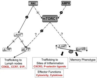
Figure 4. mTORC1 regulation of effector versus memory CD8+ T cell fate
Mammalian target of rapamycin complex 1 (mTORC1) controls CD8+ T cell migration by regulating the expression of Kruppel-like factor 2 (KLF2), which controls the expression of chemokine and adhesion molecules that are crucial for lymph node homing. mTORC1 also controls the expression of T-bet, which controls the expression of the CXC-chemokine receptor 3 (CXCR3) and P-selectin ligands, both of which are crucial for the trafficking of effector CD8+ T cells to sites of inflammation. In addition, mTORC1 regulates the balance of T-bet versus eomesodermin expression, transcription factors that are known regulators of effector versus memory CD8+ T cell function. T-bet is required for cytotoxicity and the production of effector cytokines whereas eomesodermin expression is required for the acquisition of the memory phenotype. AMPK, AMP-activated protein kinase; CCR7, CC-chemokine receptor 7; IL-7R, interleukin-7 receptor; S1P1, sphingosine-1-phosphate receptor 1.
Europe PMC Funders Author Manuscripts
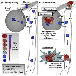
Figure 5. Model for how differential AKT and mTOR signalling controls effector versus memory CD8+ T cell formation
A | Naïve CD8+ T cells (TN) circulate between the blood, lymph nodes and efferent lymphatics. Crucial to this trafficking cycle is the expression of CD62L (which mediates adhesion to high endothelial venules (HEVs)), CC-chemokine receptor 7 (CCR7; directs transendothelial migration into lymph nodes) and sphingosine 1-phosphate receptor 1 (S1P1; directs T cell migration into the efferent lymphatics). B | During an infection, CD8+ T cells, which have been activated by their cognate antigen presented to them on antigen presenting cells, within the lymph node cease trafficking, proliferate and differentiate to produce CTL. After a period, CTLs exit the lymph node and any cells that no longer express CD62L or CC-chemokine receptor 7 (CCR7) will be unable to re-enter secondary lymphoid tissue. Moreover, such strongly activated CTL will upregulate the expression of pro-inflammatory chemokine receptors such as CXC-chemokine recpetor 3 (CXCR3) and P-selectin and E-selectin ligands allowing them to traffic across inflamed endothelium to sites of infection and inflammation. As the infection resolves, cytokines become limiting at the site of inflammation and in the absence of these survival signals effector CTL will undergo apoptosis (not shown). The loss of cytokine signalling will also cause loss of AKT/mTOR signalling and could allow re-expression of CD62L and CCR7 on some CTL and hence allow these cells to return to secondary lymphoid tissues. This would bring them into proximity of the stromal cells that make the homeostatic cytokines IL-7 and IL-15. Such cells will have the potential to generate memory T cells. An alternate possibility is that during the initial immune activation any cells that only weakly activate AKT/mTOR signalling within the lymph nodes could retain the expression of CD62L and CCR7 and hence immediately resume a pattern of homing to secondary lymphoid organs which would favour their development into memory T cells (TM).