Original Article
Effect of EPO gene modified human amniotic
mesenchymal stem cell transplantation on
renal function of rats with acute kidney injury
Qi Liu, Yi-Fan Zhang, Qiong Ye
Institute of Nephrology, Zhejiang Province Wenzhou Central Hospital, Wenzhou 325000, Zhejiang, China
Received November 16, 2015; Accepted April 5, 2016; Epub August 15, 2016; Published August 30, 2016
Abstract: To investigate the effect of EPO gene modified human amniotic mesenchymal stem cell transplantation on renal function and renal cell apoptosis in ischemic-reperfusion-induced acute kidney injury (AKI) rats. Human amniotic mesenchymal stem cells were cultured in vitro; retroviral virus PLXSN was used as the vector to mediate erythropoietin (EPO) gene transfection; it was divided into three groups: control group, negative transfection group, EPO transfection group. EPO protein in human amniotic mesenchymal stem cells was detected by Western blot after transfection; 68 SD rats were selected (male: female 1:1), weighing 220-260 g. Non-invasive artery clamp was used to clamp bilateral renal pedicle for 40 min; ischemia-reperfusion induced AKI model was established (after model
-ing there were a total of eight demyelination and death). The rats were divided into EPO-AM-MSCs (EPO-amniotic membrane-mesenchymal stem cells) group, AM-MSCs group and the control group, 20/group. On day 3, 28 after the transplantation left kidney specimens of 5 rats in three groups were stained by HE staining to observe renal tissue damage; TUNEL assay was used to detect renal cell apoptosis; on day 28 after transplantation fluorescence microscope was used to observe PKH-26 labeled AM-MSCs survival and distribution. In 1, 3, 14, 28 days after modeling serum creatinine (SCr) and blood urea nitrogen (BUN) were detected. At 3, 28 days after transplantation, HE staining examination showed that in EPO-AM-MSCs group tubular damage scores were 2.1 ± 0.12, 1.2 ± 0.10; in AM-MSCs group, tubular damage scores were 3.1 ± 0.12, 2.1 ± 0.11; in the control group tubular damage scores were 3.9 ± 0.16, 2.8 ± 0.13; the differences between two groups were statistically significant (P < 0.05). At 28 days after transplantation, PKH-26 positive cells: EPO-AM-MSCs group > AM-MSCs transplantation group > the control group; The difference between the groups was statistically significant (P < 0.05). At 3, 28 days after transplanta
-tion, TUNEL-positive apoptotic cells: the control group > AM-MSCs group > EPO-AM-MSCs group; The difference was statistically significant (P < 0.05) between groups. At 3, 14, 28 days after transplantation, serum levels of creatinine and blood urea nitrogen: EPO-AM-MSCs group < AM-MSCs group < control group (P < 0.05). EPO gene-modified human amniotic mesenchymal stem cell transplantation had a significant repair effect on rat acute kidney injury.
Keywords: EPO, human amniotic mesenchymal stem cells, transplantation, acute kidney injury, renal function,
apoptosis
Introduction
Acute kidney injury (AKI) is a more serious and widespread kidney disease, seriously threaten -ing human health, with high morbidity and mor -tality. Currently the main treatment in clinical is hemodialysis and kidney transplantation. But the cost of medical care of hemodialysis is high, and it only can partially replace kidney function; kidney transplantation is also facing a shortage of organs [1-4]. Therefore, to explore more effective treatment to prevent disease progression of renal injury is an important issue
of organ damage arising from the source [9, 10]. Therefore, we established rat acute kidney injury model and used EPO gene modified human amniotic mesenchymal stem cell trans -plantation to study the repair effect of stem cell on acute kidney injury.
Materials and methods
Design: a randomized controlled animal study
Time and place: Experiments are completed between February 2013 and 2014 October in Tianjin Medical University General Hospital ani -mal laboratory.
Experimental animals and major reagents, instruments
Human amniotic membrane was provided by Obstetrics and Gynecology department, Tianjin Medical University General Hospital, peeled from fresh placenta in full-term caesarean. Maternal prenatal hepatitis B virus, hepatitis C virus, syphilis and human immunodeficiency virus were negative. Before the experiment maternal were informed consent, and signed informed consent. 70 SD rats, male and female 1:1, weighting 180-220 g, were purchased from the Animal Laboratory (animal quality cer -tification number: SCXK20090028) in Chinese Academy of Medical Sciences; animal disposal methods were in line with animal ethic require -ments. DMEM/F12 medium, 5% fetal bovine serum (FBS) (Gibco BRL company, USA); PKH-26, glutamine (Sigma); 25% trypsin (GibcoBRL Co.); (Sigma, USA); Trizol, Lipofectamine TM 2000 (Invitrogen Corporation): Western blot chemiluminescence reagent (Santa cruz Co- rporation); rabbit anti-human EPO antibodies, HRP goat anti-rabbit IgG, PVDF membrane (Pierce Corporation); 5 μL micro-syringe (Ha-milton, United States); The main experimental apparatus included a carbon dioxide incubator (UK RS Biotech Inc.); Clean Benches (Suzhou Aetna air Technology Corporation) and fluores -cence inverted microscope (Nikon Corporation of Japan).
Preparation and labeling of human amniotic charge mesenchymal stem cells
Placenta acquisition had get informed consent; in accordance with Ref. [11]. Human amniotic membrane was separated and obtained. Am- niotic membrane was repeatedly washed with
D-Hank’s fluid to remove residual blood and mucus; amniotic membrane was cut into piec-es, digested with 0.25% trypsin at 37°C under 200 r/min for 10 min to remove human amni-otic epithelial cells; the digestion was discard-ed; after precipitation fresh digestion was added; experiment was repeated twice. Sa- mples were digested with the mixture of 0.01% of collagenase and DNA enzyme at 37°C under 200 r/min for 1 h. When tissue was digested into fluff, the digestion was filtered by a stain -less steel mesh, and the cell suspension was collected; cells were re-suspended in 10% FBS-DMEM medium, counted and placed in 25 cm2 flasks, incubated in a 37°C, 5% CO2 humidified incubator; After 40 h the medium was replaced and the non-adherent cells were discarded; when adherent cells covered 90% pf the bot-tom, cells were digested with 0.25% trypsin, and centrifuged to obtain Human amniotic membrane mesenchymal cells. Cell morpholo -gy was observed under an inverted microscope. Draw PKH-26 5 μL to 1.5 ml of EP tube; add 1ml complete medium, pipette uniform to pre-pare PKH-26 labeled solution. 90% confluent adherent cells were washed with PBS for 3 times, and incubated with 40 μL/cm2 labeled solution in a 37°C, 5% CO2, humidified incuba -tor for 2O min; then labeled solution was dis-carded and 37°C complete medium 5 ml was added; 10 min later, complete medium was dis-carded and cells were washed twice with fresh medium. After 24 h, PKH-26 labeled effects and morphological changes were observed under an inverted fluorescence microscope. Five horizons were randomly selected; PKH-26 positive cells and the total cells were counted and labeled rate was calculated. Labeled rate = the total number of positive cells within the visual field/total number of cells within the visu -al field × 100%. Before transplantation cells were washed with PBS for three times; the final concentration was 1 × 10¹⁰/L.
PcDNA3-EPO recombinant plasmid transfec-tion and testing
with fresh complete medium. On the next day medium was changed; cells were cultured with culture medium containing 400 mg/L G418 for 6 d, and then cultured with the medium con-taining 200 mg/L G418; 2 weeks later, positive clone formed; individual clones were selected for expanding culture and subculture. Expres- sion of EPO in EP0-AM-MSCswas detected by Western blot test: Well-grew AM-MSCs (1 × 10⁷) which were stably transfected with pcDNA3-EPO were mixed with RIPA buffer (containing 10 g/L PMSF) and centrifuged at 4°C after ice bath; the supernatant was collected. 100 g/L SDS-polyacrylamide gel electrophoresis was performed after protein concentration was determined. After electrophoresis, proteins were transferred to PVDF membranes, closed with blocking solution, incubated with poly -clonal rabbit anti-human EPO antibody (1:200); after washing the membrane, proteins were incubated with goat anti-rabbit HRP IgG (1:5000); after washing the membrane again, Western blot chemiluminescence reagent was added; 1 min later in a dark room, chemilumi-nescent white developing was performed with x-ray film; after washing an image analyzer was used for scanning.
Construction of ischemia-reperfusion-induced AKI model and cell transplantation
According to Reference [12], 70 SD rats were selected, male and female 1:1, weighing 250~300 g (after modeling totally 10 suffered to demyelination and death); the remaining 60 rats were randomly divided into three groups, 20/group. After intraperitoneal injection of 200 g/L urethane by 1.2 g/kg for anesthesia, ab-dominal incision was performed to expose kid-ney, with non-invasive artery clamp to clamp bilateral renal pedicle; 40 min later vessel clamp was released; after reperfusion cell transplantation was conducted. EPO-AM-MSCs group: 2 × 10⁶ EPO-AM-MSCs were re-suspend-ed in 100 ul of PBS; AM-MSCs group: 2 × 10⁶ AM-MSCs were re-suspended in 100 ul of PBS; PBS group: with 100 ul of PBS, transplanta-tions were performed respectively from upper, middle and lower renal parenchyma of the left kidney [13].
HE staining and histological observation and scoring
On day 3 and 28 after transplantation, left kid -ney specimens were drawn from 5 rats in each
group for HE staining. Ischemia-reperfusion-induced AKI mainly performed as renal tubular damage, including tubular necrosis, brush bor-der disappearance, tubular extension and tube formation. 0-normal kidney, 1-small injury (ran-ge < 5%), 2-moderate dama(ran-ge (ran(ran-ge 5%~25%), 3-moderate-severe injury (range 25%~75%), 4-severe injury (> 75%) [14].
Immunofluorescence to observe PKH-26 posi-tive cells
On day 28 after transplantation, tissue sec -tions were observed by fluorescence microsco -py. 10 fields were randomly selected for each slice in high magnification (× 200); PKH-26 positive cells in each horizon were calculated; the mean was taken as the number of PKH-26 positive cells in each group. Design, enforce-ment, and evaluation: experiment was designed by the first author; the intervention was con -ducted by the first author; assessment was per -formed by the second author. After formal train -ing, blinded assessment was conducted.
TUNEL apoptosis and renal function tests
On day 3, 28 after transplantation, 5 rats were randomly selected in three groups; thoracoto -my, left ventricular aortic cannulation and 4% paraformaldehyde perfusion were performed after anesthesia. The left kidney specimens were collected and paraffin slices were pre -pared; TUNEL assay was performed to count cells according to the German Roche kit opera -tions. After hydration, 37°C proteinase K diges -tion for 10 min, and marking fluid labeling at 37°C, biotinylated digoxin reaction was per -formed for 30 min before SABC, DAB coloring. Films were mounted; around the damaged area five high power fields were randomly selected and cells with the nucleus containing brown particles were counted at 1, 3, 14, 28 days after the transplantation; 5 rats were selected in three groups, and eyelid venous blood was drawn to detect serum creatinine (SCr) and blood urea nitrogen (BUN).
Statistical analysis
(Newman-kueuls method); P < 0.05 indicated that there were significant differences.
Results
Morphology of AM-MSCs under microscope At first, AM-MSCs showed adherent growth, morphological diversity, fusiform, polygons, stars, spindle or polygon (Figure 1A). After pas-saging to the second generation, AM-MSCs adherent speed increased; in eight hours cells were almost completely adherent; cells were uniform fibroblast-like cells, mostly showing single-radiation shape, long spindle or rotating nest-like growth, as shown in Figure 1B. Uniformity of AM-MSCs was good; purity was
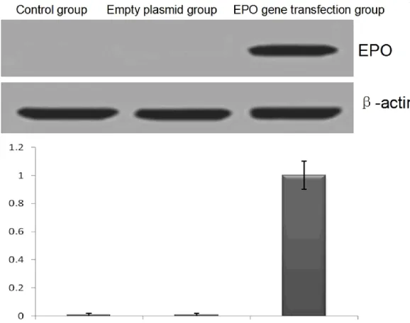
gene was stably integrated into the EPO trans -fected AM-MSCs, and the protein can be stably expressed, as shown in Figure 2.
HE staining and histological score
On day 3 and 28 after transplantation, renal tis -sue injury was shown in Figure 3. On day 3 and 28 after transplantation, in EPO-AM-MSCs group tubular damage scores were 2.1 ± 0.12, 1.2 ± 0.10; in AM-MSCs group scores were 3.1 ± 0.12, 2.1 ± 0.11; in the control group scores were 3.9 ± 0.16, 2.8 ± 0.13; between two groups differences were statistically significant (P < 0.05). Ischemia-reperfusion-induced acute kidney injury mainly was tubular necrosis, tubu
[image:4.612.88.525.72.194.2]-Figure 1. Morphology of AM-MSCs under microscope. A. Initially extracted AM-MSCs (inverted microscope, × 40), fusiform; B. AM-MSCs of the second generation (inverted microscope, × 100); C. At 24 h, PKH-26 labeled AM-MSCs (fluorescence microscope: × 100).
Figure 2. EPO protein expression in each group after 48 h.
over 98%. At 24 h, PKH-26 labeled AM-MSCs showed red fluorescence under a fluorescence microscope; FCM detection showed that cell labeling rate was 100%, as shown in Figure 1C.
EPO protein expression in each group
[image:4.612.91.380.255.483.2]lar formation, brush border disappearance and tubular expansion.
Comparison of PKH-26 positive cells
On day 28 after transplantation, PKH-26 posi -tive cells in EPO-AM-MSCs group were the most (25.3 ± 4.42), followed by AM-MSCs transplan -tation group (16.74 ± 3.45); in the control group, no PKH-26 positive cells were observed (00.0 ± 0.00); the difference between the groups were statistically significant (P < 0.05). The results showed that on day 28 after cell transplantation, viable EPO gene-modified AM-MSCs in renal tissue damage zone cells
were more than those in AM-MSCs cell trans-plantation, shown in Figure 4.
TUNEL to detect cell apoptosis
On day 3 after transplantation, the number of TUNEL positive apoptotic cells in EPO-AM-MSCs group was (23.65 ± 3.67), shown in
[image:5.612.96.526.74.303.2]Figure 5A; in AM-MSCs group, the number of TUNEL-positive apoptotic cells was (33.65 ± 4.32), shown in Figure 5B; in control group, the number of TUNEL positive apoptotic cells was (43.65 ± 5.67), shown in Figure 5C. On day 28 after transplantation, In EPO-AM-MSCs group, the number of TUNEL-positive apoptotic cells
Figure 3. Ischemia-reperfusion-induced acute kidney injury was mainly as tubular necrosis, tubular formation, brush
border disappearance and tubular expansion. Among EPO-AM-MSCs group, AM-MSCs group and control group there
[image:5.612.91.521.361.473.2]were statistically significant differences in renal tubular damage score on day 3, 28 after transplantation (P < 0.05).
was (13.42 ± 1.34), shown in Figure 5D; in AM-MSCs group, the number of TUNEL-positive apoptotic cells was (22.89 ± 2.75), shown in
Figure 5E; in control group, the number of TUNEL positive apoptotic cells was (36.74 ± 4.85), shown in Figure 5F; at the two time points, among the three groups there were sta-tistically significant differences (P < 0.05). Kidney function tests
At 1, 3, 14, 28 days after cell transplantation, SCr in EPO-AM-MSCs group was (35.8 ± 2.7), (26.2 ± 3.2), (15.9 ± 2.6), (10.1 ± 2.0) umol/L; SCr in AM-MSCs group was (42.3 ± 3.2), (33.4 ± 2.7), (25.2 ± 1.9), (16.8 ± 1.5) umol/L, respectively; SCr in the control group was (69.8 ± 4.9), (55.3 ± 2.0), (38.9 ± 3.6), (34.7 ± 3.1) umol/L; BUN in EPO-AM-MSCs group was respectively (16.4 ± 1.7), (14.3 ± 1.1), (11.2 ± 1.2), (8.3 ± 1.0) mmol/L; BUN in AM-MSCs group was (19.7 ± 1.8), (17.6 ± 1.3), (14.7 ± 1.6), (11.3 ± 1.2) mmol/L, respectively; BUN in the control group was (25.4 ± 2.2), (21.8 ± 1.5), (16.1 ± 2.2), (13.2 ± 1.1) mmol/L, shown in
Table 1; the differences were statistically sig -nificant (P < 0.05).
Discussion
Acute kidney injury is the kidney damage caused by renal structural or functional chang -es; it is a relatively common clinical syndrome, with high incidence and mortality [15-17]. Acute kidney injury can be induced by various rea -sons. The current treatment of kidney, such as dialysis and transplant, is facing various diffi -culties [18, 19]. In recent years, advances in stem cell technology enable human amniotic mesenchymal stem cells to effectively treat acute kidney injury [20, 21].
[image:6.612.91.523.71.278.2]A number of studies indicate that stem cell transplantation is an effective treatment meth-od in the repair of acute kidney injury. Now the more promising stem cells applied in renal replacement therapy mainly include umbilical cord mesenchymal stem cells, pluripotent stem cells, embryonic stem cells, human amniotic mesenchymal stem cells [13, 15]. Among them, Figure 5. On d3 and d28 after transplantation, TUNEL-positive apoptotic cells were the most in the control group,
[image:6.612.91.524.343.407.2]followed by AM-MSCs group; apoptotic cells were the fewest in EPO-AM-MSCs group (× 200).
Table 1. Comparison of the three groups in renal function (± s, n=5)
Groups SCr (umol/L) BUN (mmol/L)
1 d 3 d 14 d 28 d 1 d 3 d 14 d 28 d
Control group 69.8 ± 4.9 55.3 ± 2.0 38.9 ± 3.6 34.7 ± 3.1 25.4 ± 2.2 21.8 ± 1.5 16.1 ± 2.2 13.2 ± 1.1
AM-MSCs 42.3 ± 3.2* 33.4 ± 2.7* 25.2 ± 1.9* 16.8 ± 1.5* 19.7 ± 1.8* 17.6 ± 1.3* 14.7 ± 1.6 11.3 ± 1.2
the human amniotic mesenchymal stem cells are a class of stem cells with wide range of sources; they are easy to store and collect, with the advantages of multi-differentiation, avoid-ing the ethical controversy and the ability to self-renewal. With the progress of stem cell research, human amniotic mesenchymal stem cell transplantation for acute kidney injury dis -eases have become one of the hotspots of medical research [16, 17]. A number of studies have shown that human amniotic mesenchy -mal stem cells may secret cytokines and tro -phic factors by paracrine or autocrine manner to make some damaged cells repair them-selves; they also can fuse to the lesion tissue, replace or supply damage cells to restore dam -aged cells. Zhou et al. [13] found that in two weeks after the stem cell treatment, renal path-ological changes were significantly reduced in mice. Immunohistochemistry prompted that TGF-β, FN and VEGF were downregulated, and deposition of complement C3 in renal tissue was reduced. And a number of studies on dif-ferent animal models of progressive renal injury showed that mesenchymal stem cells had ben -eficial effects on renal tissue damage [15-17]. Recent studies have found that erythropoietin not only is a hematopoietic cytokine, but also has important protective effects on ischemia-reperfusion injury in a variety of tissues. The protection mechanism of Erythropoietin for acute kidney injury is complex, including anti-apoptotic, anti-oxidative, anti-inflammatory re-sponse, and promoting the regeneration of renal tubular cells; it is also involved in the interaction of a variety of cytokines, such as heme oxygenase-1, vascular endothelial growth factor, and heat shock proteins. Erythropoietin receptors are mainly expressed in the renal tubule and medullary collecting duct epithelial cells, mesangial cells; after binding to the erythropoietin, they play a protective role in ischemia-reperfusion injury; especially large dose of erythropoietin treatment before isch -emia can significantly reduce kidney damage; meanwhile studies have shown that erythropoi -etin is related with up-regulation of stem cell factor and c-Kit [22-24].
In this study, the therapeutic effect of EPO gene-modified human amniotic mesenchymal stem cell transplantation on ischemia-reperfu-sion-induced acute kidney injury (AKI) was
observed. The results showed that after EPO vector transfection, TUNEL assay showed that the average of apoptotic EPO gene-modified human amniotic mesenchymal stem cells decreased and average of cell proliferation increased; RT-PCR and Western blot found that after EPO transfection, in cord mesenchymal stem cells EPO gene protein and mRNA expres-sion levels were significantly enhanced. On d3 after transplantation, HE staining examination showed that tubular damage score in EPO-AM-MSCs group was lower than that in AM-EPO-AM-MSCs group and control group, and the difference was statistically significant (P < 0.05); mean -while at 3, 14, 28 days after transplantation, levels of serum creatinine and blood urea nitro-gen in EPO-AM-MSCs group were significantly lower than those in the AM-MSCs group and control group. In short, the umbilical cord mes-enchymal stem cell transplantation can pro -mote repair of acute kidney injury, mainly through promoting proliferation, anti-apoptosis and anti-inflammatory mechanism. Therefore, we believe that umbilical cord stem cell trans-plantation may offer a new approach for the treatment of acute kidney injury.
Disclosure of conflict of interest
None.
Address correspondence to: Yi-Fan Zhang, Institute
of Nephrology, Zhejiang Province Wenzhou Central Hospital, Wenzhou 325000, Zhejiang, China. Tel:
+86-0577-88070000; Fax: +86-0577-88070114; E-mail: [email protected]
References
[1] Cao Y, Qiu J, Wang B and Xi H. The analysis on
risk factors and clinical treatment of
cranioce-rebral injury concurrent withacute kidney inju
-ry. Cell Biochem Biophys 2015; 71: 199-204.
[2] Yang F, Zhang L, Wu H, Zou H and Du Y. Clinical analysis of cause, treatment and prognosis in acute kidney injury patients. PLoS One 2014;
9: e85214.
[3] Chionh CY and Cruz DN. Is acute peritoneal
di-alysis feasible for treatment of hospital-ac
-quired acute kidney injury? Semin Dial 2014;
27: 239-242.
[4] Gopinath S, Janga KC, Greenberg S and Shar
-ma SK. Tolvaptan in the treatment of acute hyponatremia associated with acute kidney in
[5] Adu D. Haemodialysis treatment for end stage chronic kidney disease and acute kidney injury in Africa. Ghana Med J 2013; 47: 1-2.
[6] Jamadarkhana P, Chaudhary A, Chhipa L, Dubey A, Mohanan A, Gupta R and Deshpande S. Treatment with a novel hypoxia-inducible factor hydroxylase inhibitor (TRC160334) ame
-liorates ischemic acute kidney injury. Am J
Nephrol 2012; 36: 208-218.
[7] Bianchi F, Sala E, Donadei C, Capelli I and La
Manna G. Potential advantages of acute kid
-ney injury management by mesenchymal stem cells. World J Stem Cells 2014; 6: 644-650.
[8] Kidder D. Mesenchymal stem cells attenuate ischemic acute kidney injury by inducing regu
-latory T cells through splenocyte interactions. Kidney Int 2014; 85: 981-982.
[9] Lee PY, Chien Y, Chiou GY, Lin CH, Chiou CH
and Tarng DC. Induced pluripotent stem cells
without c-Myc attenuate acute kidney injury via
downregulating the signaling of oxidative
stress and inflammation in ischemia-reperfu -sion rats. Cell Transplant 2012; 21:2569-2585.
[10] Westenfelder C and Togel FE. Protective ac
-tions of administered mesenchymal stem cells in acute kidney injury: relevance to clinical tri
-als. Kidney Int Suppl (2011) 2011; 1: 103-106.
[11] Shuang-zhi H, Ping S, Xi-ning P. Culture and identification of human amniotic mesenchy -mal stem cells. Chin Med Sci J 2010; 25: 211-214.
[12] Tögel F, Hu Z, Weiss K, Isaac J, Lange C and Westenfelder C. Administered mesenchymal
stem cells protect against ischemic acute re-nal failure through differentiation-independent
mechanisms. Am J Physiol Renal Physiol 2005;
289: F31-42.
[13] Zhou K, Zhang H, Jin O, Feng X, Yao G, Hou Y
and Sun L. Transplantation of human bone
marrow mesenchymal stem cell ameliorates
the autoimmune pathogenesis in MRL/lpr mice. Cell Mol Immunol 2008; 5: 417-424. [14] Mias C, Trouche E, Seguelas MH, Calcagno F,
Dignat-George F, Sabatier F, Piercecchi-Marti
MD, Daniel L, Bianchi P, Calise D, Bourin P, Pa-rini A and Cussac D. Ex vivo pretreatment with melatonin improves survival, proangiogenic/
mitogenic activity, and efficiency of mesenchy
-mal stem cells injected into ischemic kidney.
Stem Cells 2008; 26: 1749-1757.
[15] Ninichuk V, Gross O, Segerer S, Hoffmann R, Radomska E, Buchstaller A, Huss R, Akis N, Schlöndorff D and Anders HJ. Multipotent mes
-enchymal stem cells reduce interstitial fibrosis but do not delay progression of chronic kidney disease in collagen4A3-deficient mice. Kidney
Int 2006; 70: 121-129.
[16] Li F, Miao ZN, Xu YY, Zheng SY, Qin MD, Gu YZ and Zhang XG. Transplantation of human am
-niotic mesenchymal stem cells in the treat -ment of focal cerebral ischemia. Mol Med Rep 2012; 6: 625-630.
[17] Sun H, Hou Z, Yang H, Meng M, Li P, Zou Q, Yang L, Chen Y, Chai H, Zhong H, Yang ZZ, Zhao J, Lai L, Jiang X and Xiao Z. Multiple systemic
transplantations of human amniotic
mesen-chymal stem cells exert therapeutic effects in
an ALS mouse model. Cell Tissue Res 2014; 357: 571-582.
[18] De Smedt DM, Elseviers MM, Lins RL and Annemans L. Economic evaluation of different
treatment modalities in acute kidney injury.
Nephrol Dial Transplant 2012; 27: 4095-4101. [19] Fortenberry JD, Paden ML and Goldstein SL.
Acute kidney injury in children: an update on
diagnosis and treatment. Pediatr Clin North Am 2013; 60: 669-688.
[20] Chen YT, Sun CK, Lin YC, Chang LT, Chen YL, Tsai TH, Chung SY, Chua S, Kao YH, Yen CH, Shao PL, Chang KC, Leu S and Yip HK. Adipose-derived mesenchymal stem cell protects kid
-neys against ischemia-reperfusion injury
through suppressing oxidative stress and
in-flammatory reaction. J Transl Med 2011; 9:
51-67.
[21] Liu N, Tian J, Cheng J and Zhang J. Effect of
erythropoietin on the migration of bone mar
-row-derived mesenchymal stem cells to theacute kidney injury microenvironment. Exp
Cell Res 2013; 319: 2019-2027.
[22] Gobe GC, Bennett NC, West M, Colditz P, Brown L, Vesey DA and Johnson DW. Increased pro
-gression to kidney fibrosis after erythropoietin is used as a treatment for acute kidney injury. Am J Physiol Renal Physiol 2014; 306:
F681-692.
[23] Wang PR. Mouse adult renal progenitor cells in combination with erythropoietin or suramin-a potential new strategy for the treatment of acute kidney injury. Stem Cell Res Ther 2013;
4: 89.
[24] Liu N, Han G, Cheng J, Huang J and Tian J. Erythropoietin promotes the repair effect of acute kidney injury by bone-marrow mesenchy -malstem cells transplantation. Exp Biol Med