Original citation:
Winsper, Catherine, Marwaha, Steven, Lereya, Suzet Tanya, Thompson, Andrew D., Eyden, Julie and Singh, Swaran P.(2016) A systematic review of the neurobiological underpinnings of borderline personality disorder (BPD) in childhood and adolescence. Reviews in the
Neurosciences, 27 (8). pp. 827-847. doi:10.1515/revneuro-2016-0026
Permanent WRAP URL:
http://wrap.warwick.ac.uk/80218
Copyright and reuse:
The Warwick Research Archive Portal (WRAP) makes this work by researchers of the University of Warwick available open access under the following conditions. Copyright © and all moral rights to the version of the paper presented here belong to the individual author(s) and/or other copyright owners. To the extent reasonable and practicable the material made available in WRAP has been checked for eligibility before being made available.
Copies of full items can be used for personal research or study, educational, or not-for-profit purposes without prior permission or charge. Provided that the authors, title and full
bibliographic details are credited, a hyperlink and/or URL is given for the original metadata page and the content is not changed in any way.
Publisher’s statement:
“The final publication is available at www.degruyter.com ” https://doi.org/10.1515/revneuro-2016-0026
A note on versions:
The version presented here may differ from the published version or, version of record, if you wish to cite this item you are advised to consult the publisher’s version. Please see the ‘permanent WRAP URL’ above for details on accessing the published version and note that access may require a subscription.
For Preview Only
A Systematic Review of the Neurobiological Underpinnings of Borderline Personality Disorder (BPD) in Childhood and
Adolescence
Journal: Reviews in the Neurosciences
Manuscript ID RNS.2016.0026.R1 Manuscript Type: REVIEW
Date Submitted by the Author: n/a
Complete List of Authors: Winsper, Cathy Marwaha, Steven Lereya, Tanya Thompson, Andrew Eyden, Julie Singh, Swaran
Keywords: Systematic Review, Borderline Personality Disorder, Childhood, Adolescence
For Preview Only
A Systematic Review of the Neurobiological Underpinnings of Borderline Personality
Disorder (BPD) in Childhood and Adolescence
RUNNING HEAD: NEUROBIOLOGY OF YOUTH BPD
Catherine Winsper, Ph.D., Division of Mental Health and Wellbeing, Warwick Medical
School, University of Warwick;
Steven Marwaha, Ph.D., MRCPsych, MSc, MBBS,Division of Mental Health and Wellbeing,
Warwick Medical School, University of Warwick;
Suzet Tanya Lereya, Ph.D., Evidence Based Practice Unit (EBPU), University College London
and the Anna Freud Centre, London, UK;
Andrew Thompson, MD, MBBS, MedSci, FRCPsych,Division of Mental Health and Wellbeing
Warwick Medical School, University of Warwick;
Julie Eyden, MSc. Department of Psychology, University of Warwick;
Swaran P Singh, DM. Division of Mental Health and Wellbeing, Warwick Medical School,
University of Warwick.
Correspondence: Dr Cath Winsper, Warwick Medical School, University of
Warwick, Coventry, UK, CV4 7AL email: catherine.winsper@warwick.ac.uk; tel: 024 765
74886.
Funding: Prof. Singh receives funding from the National Institute for Health Research (NIHR)
Collaborations for Leadership in Applied Health Research and Care West Midlands
(CLAHRC-WM) initiative. The views expressed are those of the authors and not necessarily those of
the CLAHRC-WM collaborative organisations, the NIHR, or the Department of Health.
Declaration of Interest: C.W; S.M; S.T.L; A.T; J.E; & S.P.S report no conflict of interest.
Word count (excluding references and abstract):
Abstract: 244; Body text: 6, 951 3
For Preview Only
Abstract
Contemporary theories for the aetiology of Borderline Personality Disorder (BPD) take a
lifespan approach asserting that inborn biological predisposition is potentiated across
development by environmental risk factors. In this review we present and critically evaluate
evidence on the neurobiology of BPD in childhood and adolescence, compare this evidence
to the adult literature, and contextualise within a neurodevelopmental framework. A
systematic review was conducted to identify studies examining the neurobiological (i.e.,
genetic, structural neuroimaging, neurophysiological and neuropsychological) correlates of
BPD symptoms in children and adolescents aged 19 years or under. We identified, quality
assessed, and narratively summarised 34 studies published between 1980 and June 2016.
Similar to findings in adult populations, twin studies indicated moderate to high levels of
heritability of BPD, and there was some evidence for gene-environment interactions. Also
consistent with adult reports, some adolescents with BPD demonstrated structural (grey
and white matter) alterations in frontolimbic regions, and neuropsychological abnormalities
(i.e., reduced executive function and disturbances in social cognition). These findings
suggest that neurobiological abnormalities observed in adult BPD may not solely be the
consequence of chronic morbidity or prolonged medication use. They also provide tentative
support for neurodevelopmental theories of BPD by demonstrating that neurobiological
markers may be observed from childhood onwards, and interact with environmental factors
to increase risk of BPD in young populations. Prospective studies with a range of repeated
measures are now required to elucidate the temporal unfurling of neurobiological features,
and further delineate the complex pathways to BPD.
Keywords: Neurobiology; Borderline Personality Disorder; Childhood; Adolescence;
Systematic Review 3
For Preview Only
Introduction
Borderline Personality Disorder (BPD) is a serious and enduring mental disorder affecting
from 1 to 6% of the general population (Grant et al., 2008; Lenzenweger, Lane, Loranger, &
Kessler, 2007; Trull, Jahng, Tomko, Wood, & Sher, 2010). Contemporary theories for the
aetiology of BPD take a lifespan approach, proposing that an inborn tendency towards
emotionality is potentiated across early development by environmental risk factors
(Crowell, Beauchaine, & Linehan, 2009; Hughes, Crowell, Uyeji, & Coan, 2012). Within this
context, BPD is unlikely to appear de novo in early adulthood (Paris, 2013), but may be considered as the continuation of a collection of BPD precursor symptoms that first emerge
during childhood or early adolescence (Crowell et al., 2009; Winsper, Marwaha, et al.,
2015).
A growing body of studies have demonstrated the clinical utility, validity and
reliability of the adolescent (Kaess, Brunner, & Chanen, 2014; Sharp & Fonagy, 2015;
Winsper et al., 2016) and to a lesser extent child (Hawes, 2014; Winsper et al., 2016) BPD
phenotype. Recent systematic evidence indicates that the diagnostic stability of BPD in
adolescence is largely comparable (though slightly attenuated) to that in adulthood, and
that a considerable proportion of adolescents continue to manifest BPD symptoms up to 20
years later (Winsper, Marwaha, et al., 2015). Reflecting these findings, national treatment
guidelines and classification systems have recently confirmed the legitimacy of the BPD
diagnosis in adolescence (American Psychiatric Association, 2013; NICE, 2009; Tyrer,
Crawford, & Mulder, 2011). Nevertheless, many clinicians remain reluctant to diagnose the
disorder prior to age 18 (Griffiths, 2011; Laurenssen, Hutsebaut, Feenstra, Van Busschbach,
& Luyten, 2013), meaning that adolescents manifesting BPD symptoms may be
misdiagnosed (Paris, 2013) and opportunities for early intervention missed (Newton-Howes, 3
For Preview Only
Clark, & Chanen, 2015). Increasing awareness and understanding of the biological correlates
of youth BPD may help to reduce clinical reluctance, and further our understanding of the
aetiological mechanisms and pathological processes germane to the development of BPD
(Goodman, Mascitelli, & Triebwasser, 2013).
Our understanding of the potential neurobiological underpinnings of BPD in
adulthood has grown rapidly over the past few decades (Krause-Utz, Winter, Niedtfeld, &
Schmahl, 2014; van Zutphen, Siep, Jacob, Goebel, & Arntz, 2015). However, the extant
literature remains relatively underdeveloped in comparison to that of other mental
disorders (e.g., dementias, depression, and schizophrenia). Over recent years a number of
reviews have been published collating evidence on the genetic (Amad, Ramoz, Thomas,
Jardri, & Gorwood, 2014), and structural and functional brain abnormalities (Krause-Utz et
al., 2014; Ruocco, 2005; Ruocco, Amirthavasagam, & Zakzanis, 2012; van Zutphen et al.,
2015) associated with adult BPD. While reviews have pointed towards several
inconsistencies across studies (Ruocco, Amirthavasagam, Choi-Kain, & McMain, 2013), there
are some replicated findings (Krause-Utz et al., 2014; van Zutphen et al., 2015). Familial and
twin studies indicate a genetic component to adult BPD (Amad et al., 2014). Candidate
genes have been investigated largely within the serotonin system, though no clear gene has
been identified highlighting the need to consider epigenetic variability (Newton-Howes et
al., 2015) and “plasticity” genes (Amad et al., 2014). Neuroimaging studies suggest
structural and functional abnormalities in the frontolimbic network, including
hyper-reactivity of regions involved in emotional processing (e.g., insula, amygdala, hippocampus)
and diminished recruitment of regulatory control processes, e.g., anterior cingulate cortex,
medial frontal cortex, orbitofrontal cortex, and dorsolateral prefrontal cortex (Krause-Utz et
al., 2014; Christian Schmahl & Bremner, 2006). On a neurochemical level, altered function in 3
For Preview Only
neurotransmitter systems including the serotonin, glutamate and GABA systems has been
observed in BPD patients (Krause-Utz et al., 2014). In a synthesis of 10 studies, Ruocco
(2005) reported that adult BPD patients performed more poorly than controls across
several neuropsychological domains (i.e., cognitive flexibility, planning, attention, learning
and memory). Studies also suggest disturbances in social cognition including the recognition
(i.e., negative bias) of facial emotions (Domes, Schulze, & Herpertz, 2009), thoughts, and
intentions (Preißler, Dziobek, Ritter, Heekeren, & Roepke, 2010). Studies examining
mentalisation (i.e., recognition of the mental states of social interaction partners), however,
indicate that adults with BPD may have superior abilities (Arntz & Veen, 2001; Fertuck et al.,
2009; Franzen et al., 2011).
Examining the neurobiological correlates of BPD features in younger populations in
the early stages of the disorder may help reduce the likelihood of confounders, including
duration of illness, prolonged use of medication, and cumulative trauma experiences
(Chanen, Velakoulis, et al., 2008; Richter et al., 2014). Furthermore, considering younger
populations will allow for the prospective assessment of neurobiological and environmental
precursors (and their interactions) to shed light on the developmental pathways to BPD at
both the biological and behavioural level (Hughes et al., 2012).
In 2013, a narrative review examined aspects of the neurobiological basis of
adolescent-onset BPD (Goodman et al., 2013). Although the authors’ conclusions were very
tentative due to the limited number of available studies at the time, they emphasised the
importance of studying the biological basis of adolescent BPD to inform screening,
treatment and preventive strategies. In the current study we expand on this review by
taking advantage of the recent wave of research on BPD in childhood and adolescence, and 3
For Preview Only
present a systematic evaluation of all studies examining the neurobiological correlates of
BPD in individuals 19 years of age and under. Specifically, we aimed to:
1) Ascertain the extent to which adolescents with BPD share similar neurobiological
features (i.e., genetic underpinnings, neurophysiology, brain structures, and
neuropsychological processes) to adults with BPD.
2) Situate our findings within a neurodevelopmental perspective of BPD.
3) Critically evaluate the extant literature to set out a framework for future research.
Methods
Search strategies
We used PRISMA guidelines (Moher, Liberati, Tetzlaff, & Altman, 2009) to describe our
procedures and results. We searched MEDLINE, EMBASE, PsychINFO and PubMed databases
to identify studies reporting on BPD features in children and adolescents (i.e., individuals 19
years and under) published in English between 1980 and 28th January 2014. We chose 1980
as the earliest date for inclusion to parallel when BPD was first introduced as a disorder in
the Diagnostic and Statistical Manual (APA., 1980). The search terms (borderline* OR
“emotionally unstable personality disorder” OR BPD) AND (adolescen* OR child* OR young*
OR youth* OR teen* OR student*) were entered. To ensure comprehensive coverage of the
neurobiological literature we updated our search to include studies published between 1980
and 4th June 2016, and cross-referenced returns against our previous search. For the
updated search we used the following search strings: (BPD OR "emotionally unstable
personality" OR borderline*) AND (child* OR adolescen* OR youth* OR young* OR teen* OR 3
For Preview Only
student*) AND (genes OR gene OR genetic* OR neuro* OR imaging OR biological). Reference
lists of included studies were inspected for relevant titles. We also examined the reference
lists of related narrative reviews as a cross check (Brunner, Henze, Richter, & Kaess, 2015;
Chanen, Jovev, McCutcheon, Jackson, & McGorry, 2008; Goodman et al., 2013).
Study Selection
A study was considered for selection if it met all the following a priori criteria:
(1) Original research was presented;
(2) Participants were 19 years of age or younger at initial assessment (based on the World
Health Organisation (2016) definition of childhood and adolescence);
(3) The study was published in English;
(4) The study provided any information on the neurobiological (i.e., genetic,
neurophysiological, structural brain characteristics, neuropsychological) correlates of BPD.
Studies were excluded if:
(1)BPD was not the primary focus of the study (e.g., associations pertained to all Cluster B
personality disorders rather than BPD specifically);
(2) They were case studies without statistical analysis;
Screening procedure
After removal of all duplicates, abstracts were retrieved using the initial search strategy. If a
title appeared potentially eligible but no abstract was available, the full-text article was
retrieved. C.W and T.L independently scanned 100% of the titles and abstracts to identify
relevant articles for full text retrieval, and these were read by C.W to assess for inclusion in
the review. S.M independently reviewed 50% of the full text articles as a reliability check.
Data extraction and quality assessment
For Preview Only
A data extraction form was developed prior to full text review. It included author details,
country of study, sample characteristics (i.e., age, sex, and clinical status), study design, BPD
assessment tool, methodology of neurobiological assessment, and main findings. A quality
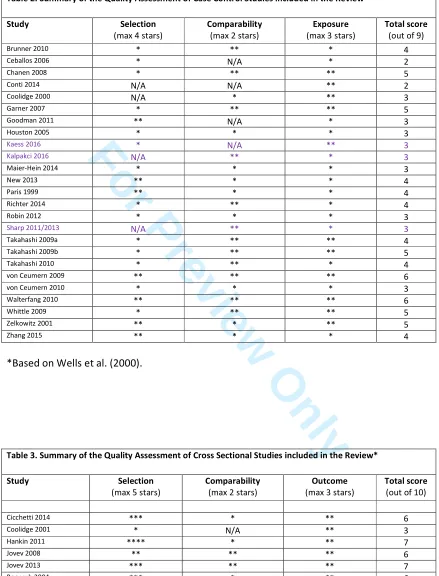
assessment form was also produced based on the Newcastle-Ottawa Scale (NOS) (Wells et
al., 2000), which can be adapted for the assessment of non-randomised cross-sectional and
case control studies. For case control studies we assessed the quality domains of selection
(maximum of 4 stars), comparability (maximum of 2 stars) and exposure (maximum of 3
stars). For cross-sectional studies which did not use a case control design we used the
adapted scale by Herzog et al. (2013), covering the domains of selection (maximum of 5
stars), comparability (maximum of 2 stars), and outcome (maximum of 3 stars).
Results
Search results
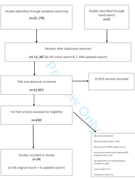
Of the original 8,195 abstracts scanned, 209 text articles were selected for full text retrieval
(Figure 1). There was a high level of agreement between raters (Kappa = 0.82). The authors
met to discuss discrepancies regarding selected articles, which were largely due to
uncertainty regarding sample characteristics (e.g., the sample was primarily defined
according to another mental illness) or age (the age was not reported in the abstract). If
there was doubt over whether an abstract should be included for full text retrieval, the
decision was made to include. Of the 209 full text articles reviewed, we identified 25 studies
providing information on the neurobiological correlates of BPD. We identified a further
three relevant studies via hand search (Coolidge, Thede, & Jang, 2001; Houston, Ceballos,
Hesselbrock, & Bauer, 2005; Jovev et al., 2008). The 50% full text reliability check indicated a
high level of agreement between raters (Kappa=0.80). The cross-referenced updated search 3
For Preview Only
conducted on June 4th 2016 yielded a total of 12, 367 abstracts (i.e., when the original and
updated searches were combined and all duplicates removed). We identified a further 6
articles from the updated search (Cicchetti, Rogosch, Hecht, Crick, & Hetzel, 2014; Conti et
al., 2013; Kaess, Parzer, Koenig, Resch, & Brunner, 2016; Kalpakci, Vanwoerden, Elhai, &
Sharp, 2016; Richter et al., 2014; Zhang et al., 2015). Therefore, a total of 34 studies are
included in the review. Please see Table 1 for a description of studies and summary of main
findings.
Studies comprised a mix of clinical, high-risk and non-clinical populations.
Distribution of gender within samples varied across studies, with most studies having a
preponderance of female participants (with the exception of two early studies which had a
majority of male participants (Paris, Zelkowitz, Guzder, Joseph, & Feldman, 1999; Zelkowitz,
Paris, Guzder, & Feldman, 2001)). All studies, with the exception of two longitudinal studies
(Belsky et al., 2012; Bornovalova, Hicks, Iacono, & McGue, 2009) were cross-sectional in
design. Cross-sectional studies were a mix of case control studies and those assessing
associations between neurobiological features and continuous BPD outcome measures (i.e.,
scales of BPD symptoms).
Twenty six studies utilised adolescent samples (i.e., youth aged 12 years or older),
and eight child samples (or a mixture of children and adolescents) ranging from 4 to 17
years of age (Cicchetti et al., 2014; Coolidge, Segal, Stewart, & Ellett, 2000; Coolidge et al.,
2001; Hankin et al., 2011; Jovev et al., 2013; Paris et al., 1999; Rogosch & Cicchetti, 2005;
Zelkowitz et al., 2001). The majority of the adolescent studies assessed BPD features with
the Structured Clinical Interview for DSM-IV Axis II Personality Disorders (SCID-II), commonly
used in adult BPD studies (Maffei et al., 1997). Studies with children used a range of
validated BPD assessment tools, some of which had been adapted from widely used adult 3
For Preview Only
diagnostic tools, i.e., the Child version of the Diagnostic Interview for Borderlines (C-DIB),
which has five sub-scales incorporating social adaptation, impulsivity, affect, psychosis and
interpersonal relations (Paris et al., 1999; Zelkowitz et al., 2001). Others were adapted from
dimensional assessments used in adult BPD populations. The Borderline Personality
Features Scale for Children (BPFS-C) developed by Crick, Murray–Close, and Woods (2005)
covered the four domains of: affective instability, identity problems, self-harm, and negative
relationships. The Children in the Community-Self Report described in Crawford et al. (2005)
was based on the DSM-IV conceptualisation of BPD.
We organised the studies into three main types:
(1) Those reporting on the genetic underpinnings (i.e., heritability, molecular genetic
studies; gene-environment interactions) of BPD;
(2) Those exploring neurophysiological correlates (i.e., electrophysiological measures,
physiological measures) and brain structures of BPD;
(3) Those examining performance on neuropsychological (i.e., cognition, emotion
recognition, mentalisation) tasks.
Quality assessment of studies
Table 2 presents a summary of the quality assessment for case control studies, and Table 3
a summary of cross-sectional studies (e.g., those assessing associations with continuous BPD
scales). Studies varied widely in quality according to the Newcastle Ottawa Scale (NOS), with
scores ranging from 2 to 7 (out of a possible 9/10). Most studies demonstrated some degree
of selection bias, usually in terms of issues with the representativeness of cases (e.g., self-3
For Preview Only
selection bias). A number of studies also demonstrated comparability bias by not sufficiently controlling for pertinent confounding variables (e.g., whole brain volume).
The genetic underpinnings of BPD in childhood and adolescence
Family studies
We identified three studies examining the heritability of BPD in young twin populations.
Coolidge et al. (2001) reported that the monozygotic (MZ) correlation for BPD symptoms in
a sample of 4 to 15 years olds was significantly greater than the dizygotic (DZ) correlation
(rMZ = 0.70; rDZ =0.39). Structural equation modelling confirmed a substantial genetic
component, with a heritability figure of 0.76. In the first of two prospective studies,
Bornovalova et al. (2009) examined heritability rates at 4 discrete periods from 14 to 24
years of age. MZ correlations were higher than DZ correlations: 14 years (rMZ = 0.48; rDZ
=0.38); 17 years (rMZ = 0.50; rDZ =0.30); 20 years (rMZ = 0.43; rDZ =0.35) and 24 years (rMZ =
0.48; rDZ =0.22). Heritability figures ranged from a moderate 0.3 to 0.5 across the four time
points, with a trend towards increased heritability from age 14 to 24. Finally, in a twin
sample of 12 year olds, Belsky and colleagues (2012) found a higher correlation of BPD
symptoms between MZ (0.66) than DZ twins (0.29). Biometric modelling indicated that
genetic factors accounted for 66% of the variance in BPD symptoms.
Molecular genetic studies
We identified two molecular genetic studies examining associations between candidate
genes and BPD symptoms in childhood and adolescence (Cicchetti et al., 2014; Hankin et al.,
2011). Hankin and colleagues (2011) used two independent samples of 9 to 15 year olds to
assess the association between the 5-HTTLPR serotonin transporter gene and BPD
symptoms. Participants were divided into groups according to variants of the 5-HTTLPR gene 3
For Preview Only
(2 short alleles; 2 long alleles; or 1 short and 1 long allele). In both samples, mean BPD traits
significantly differed as a function of 5-HTTLPR polymorphism (Sample 1, F2, 239 =4.33, p = .01; Sample 2, F2, 144 =4.97, p= .008). Participants with two short alleles exhibited
significantly higher BPD trait scores than participants with two long alleles. Participants
carrying one short and one long allele (S/L) exhibited intermediate BPD traits.
Cicchetti et al. (2014) examined associations between two candidate genes (oxytocin
receptor gene and FKBP5) and BPD symptoms in a sample of 8 to 12-year-old children. The
authors selected the oxytocin receptor gene (OXTR) due to its relation to variation in social
behaviour, attachment, affiliation and aggression; and the FKBP5 gene due to its role in the
pathogenesis of stress-related psychopathology. There were no significant main effects of
OXTR or FKBP5 on BPD symptoms in childhood.
Gene-environment interactions
Belsky et al. (2012) prospectively demonstrated the impact of genetic vulnerability in
combination with environmental risk on the development of BPD in early adolescence.
Young adolescents with a genetic risk (i.e., a family history of psychiatric disorder) and
exposed to physical maltreatment had a 13-fold increased risk of being in the extreme (>95th
percentile of symptoms) BPD group. In contrast, those without a genetic risk but exposed to
harsh parenting had only a two-fold increased risk of being in the extreme BPD group. A
similar effect was observed for high maternal negative expressed emotion, with a 15-fold
increased risk for adolescents with genetic risk and exposure to negative expressed emotion
compared to a five-fold increased risk for those just exposed to high expressed emotion.
Cicchetti et al. (2014) tested three-way interactions between variations in genotype
(OXTR and FK506), environmental risk, and gender on the development of BPD symptoms in
late childhood. Results indicated differential effects for males and females. For girls, effects 3
For Preview Only
were most consistent with a stress-diathesis effect, i.e., genotype (OXTR: AG-AA genotype;
FK506: 1 to 2 copies of the CATT haplotype) was associated with BPD symptoms in the
presence of maltreatment only. For boys, observed effects were most consistent with a
differential susceptibility effect (i.e., genetic predisposition increased susceptibility for both
better and worse outcomes). Boys exposed to maltreatment had significantly higher BPD
scores than non-maltreated boys if they had the GG genotype of OXTR (there was no
difference between maltreatment groups for those with the AG-AA genotype). For FK506,
maltreated boys had significantly higher BPD scores than non-maltreated boys if they had
the zero copy CATT haplotype (maltreatment groups did not differ for boys with the one to
two copies of the CATT haplotype).
Neurophysiological correlates and brain structures
Neurophysiological correlates
We identified 2 studies using P300 Event Related Potential (ERP) measurements to examine
differences in brain maturation between adolescents with and without BPD (Ceballos,
Houston, Hesselbrock, & Bauer, 2006; Houston et al., 2005). In a sample of 14 to 19 year old
girls, Houston et al. (2005) used a visual oddball task to compare P300 amplitudes between
4 groups (BPD <16.5 years; BPD>16.5 years; no BPD <16.5 years; no BPD>16.5 years).
ANCOVAs, adjusting for comorbid conduct disorder and depression symptoms,
demonstrated a significant interaction. Girls with BPD features did not evidence the
expected age-related reductions in P300 amplitude, suggesting impairment in brain
maturation. Ceballos et al. (2006) failed to find similar neurophysiological abnormalities in
BPD adolescents in the absence of co-morbid conduct disorder symptoms. Again using the
visual oddball paradigm in a sample of 14 to 19 year olds, P300 amplitudes were compared 3
For Preview Only
across 4 groups (BPD only, Conduct Disorder only, BPD plus CD, no BPD or CD). With
increasing age, abnormal brain maturation (i.e., lack of age related reductions) was only
observed in the BPD plus CD and CD groups. The authors attributed the discrepancy in
results (i.e., lack of impairment in BPD only subjects) to sex. Indeed, when they reanalysed
their data with the females only (Houston’s study used females only) they observed the
expected impairment in brain maturation in the BPD only group.
We identified one study examining dysfunction of the neurosteroid system in
adolescents with BPD (Conti et al., 2013). The authors compared BPD patients (Mage=15.5;
SD=1.2) to healthy controls on diazepam binding inhibitor (DBI) and
dehydroepiandrosterone sulphate (DHEA-S) plasma levels, and also cortisol to DHEA-S molar
ratio (CDR). There was no difference between groups in DBI plasma levels; however, BPD
patients had significantly increased (approx. 70%) DHEA-S levels (t=3.023; p=.0054) and decreased CDR (t=2.401; p=.0235). The authors hypothesised that DHEA-S may represent a trait marker for the altered stress response observed in BPD.
Brain structures
We identified fourteen structural neuroimaging studies examining whether adolescents with
BPD demonstrate brain abnormalities. Eight studies were derived from the Orygen Youth
Health Research Centre in Melbourne (Chanen, Velakoulis, et al., 2008; Garner et al., 2007;
Jovev et al., 2008; Takahashi, Chanen, Wood, Walterfang, et al., 2009; Takahashi et al.,
2010; Takahashi, Chanen, Wood, Yücel, et al., 2009; Walterfang et al., 2010; Whittle et al.,
2009) and three from the University of Heidelberg (Brunner et al., 2010; Maier-Hein et al.,
2014; Richter et al., 2014). These two study groups used the same respective cohort of
patients for each study, but examined different brain structures or utilised varying imaging
technologies. Two studies were derived from the Mount Sinai Hospital in New York 3
For Preview Only
(Goodman et al., 2011; New et al., 2013). Of note, the adolescents in these two studies were
all (with the exception of one) co-morbid for Major Depressive Disorder. In view of the very
high levels of comorbidity observed between BPD and depression in adolescence (Glenn &
Klonsky, 2013), and because these studies adjusted for depression symptoms within the
analysis, they were included in the review. Results from these two studies should be
interpreted with caution, however, as they are not directly generalisable to all adolescents
with BPD features (i.e., the patients in these studies likely represent the severe end of the
spectrum of BPD psychopathology (Goodman et al., 2011)). The final study utilised a high
risk sample selected from sixth grade students in Melbourne, Australia (Jovev et al., 2013).
Grey matter structures of the frontolimbic network
Two studies reported reductions in Orbitofrontal Cortex (OFC) volume in BPD compared to
control groups (Brunner et al., 2010; Chanen, Velakoulis, et al., 2008), while one study
reported no difference in OFC between groups (Goodman et al., 2011).
Using Region of Interest (ROI) methodology, Chanen, Velakoulis, et al. (2008) found
that 15-19 year old BPD patients demonstrated significant OFC grey matter loss in
comparison to healthy controls (HCs): F1.35=8.62, p=.006. Inspection of the data indicated a reversal of the normal asymmetry associated with BPD, with a reduction in the right OFC. Brunner et al. (2010), using Voxel-Based Morphometry (VBM) techniques, found that BPD
patients aged 14-18 years exhibited significant volume reductions in the left OFC in comparison to healthy (but not clinical) controls: t =6.11, p=.002. Conversely, Goodman et al. (2011) found no difference in OFC grey matter volume between BPD patients with
co-morbid major depressive disorder (Mage=15.8, SD=1.1) and HCs using ROI methodology.
For Preview Only
Two studies reported anterior cingulate (AC) volume reductions (Goodman et al.,
2011; Whittle et al., 2009) in adolescents with BPD in comparison to controls, while one
study did not find any significant difference between groups (Brunner et al., 2010).
Using a subsample of female patients from the Melbourne group, Whittle et al. (2009)
reported a decrease in left AC cortex volume (across limbic and paralimbic regions) in
patients with BPD compared to HCs: t29 =5.82, p=.023. Post hoc partial correlations controlling for age and whole brain volume indicated that volumetric change was
significantly correlated with parasuicidal behaviour: rs = -.675 and impulsivity: rs=.575. Goodman et al. (2011) found that BPD/MDD patients had smaller relative volume (averaged
across grey and white matter) in Brodmann area 24 (i.e., part of the anterior cingulate) in
comparison to HCs: F4, 96 =3.43, p=.03. Of note, smaller BA 24 volume was associated with BPD (r= -.45, p=.022) but not MDD indices. Conversely, Brunner et al. (2010) did not report any ACC abnormalities in BPD patients in comparison to healthy or clinical controls (CCs).
Two studies assessed dorsolateral cortex (DLPFC) volume in adolescents with BPD.
Brunner et al. (2010) reported bilateral volume reduction of the DLPFC in adolescents
compared to healthy, but not clinical, controls. In contrast, Goodman et al. (2011) did not
find any difference in DLPFC volume in BPD/MDD patients compared to HCs.
In another Orygen research group study, Takahashi, Chanen, Wood, Yücel, et al.
(2009) found no significant difference in insular cortex volume (a frontolimbic integration
cortex) between BPD patients and HCs. BPD patients reporting violent episodes during the
previous 6 months, however, had a smaller insular volume bilaterally than those who had
not been violent: F1, 16 = 5.56, p=.031.
Only one (Richter et al., 2014) of three studies comparing amygdala volume in
adolescents with BPD to controls reported a significant group difference. Chanen, 3
For Preview Only
Velakoulis, et al. (2008) did not find any differences in amygdala volume between patients
with BPD and HCs (p>.05). Sub-analysis with female BPD patients only, however,
demonstrated a significant negative correlation between right amygdala volume and BPD
total symptom score: r= -.613, p=.026. Similarly, Brunner et al. (2010) found no significant difference in amygdala volume between BPD patients, CCs and HCs. In a follow-up to
Brunner et al. utilising FreeSurfer software to reanalyse the data, Richter et al. (2014) found volumetric reductions in the right amygdala of BPD patients compared to healthy and
clinical controls. These differences only reached significance for comparison with the HC
group: BPD=1613 (49.58) mm3; HC=1777 (38.16) mm3; p=.024. There was no significant difference between CC and HC groups in amygdala volume: CC=1712.45 (33.78) mm.3
Four studies assessed hippocampal volume in adolescents with BPD; two reported
significant differences between BPD patients and controls (Jovev et al., 2013; Richter et al.,
2014) and two reported no difference (Brunner et al., 2010; Chanen, Velakoulis, et al.,
2008). Chanen, Velakoulis, et al. (2008) found no difference in hippocampal volume
between patients with BPD and HCs (p<.05) using ROI methodology. Similarly, Brunner et al. (2010) reported no hippocampal volume differences between BPD, CC and HC groups using
VBM methodology. In re-analysis with the same sample, but using FreeSurfer technology, Richter et al. (2014) demonstrated group (i.e., BPD, CC, HC) differences in right and left
hippocampal volumes, with BPD patients evincing the smallest hippocampal volume. Post
hoc tests revealed significant group differences between patients with BPD and HCs in both
the right (BPD=3977.65 [70.49] mm3 versus HC=4339. 8 [74.66] mm3; p=.003) and left (BPD=
3748.75 [82.26] mm3 versus HC=4167.5 [81.87] mm3; p=.008) hippocampus, as well as
group differences between the CC (4066.35[66.47] mm3; p=.033) and HC in the right hippocampus. Finally, Jovev et al. (2013) reported an association between atypical 3
For Preview Only
rightward hippocampal asymmetry and BPD symptoms in 11-13 year olds, but only via the
moderating effects of temperament (i.e., there was no significant main effect of
hippocampal asymmetry on BPD symptoms). Two significant three-way interactions (i.e.,
sex, temperament and hippocampal asymmetry) were observed. Boys were more likely to
have BPD symptoms if they were high on affiliation (representing a desire for closeness with
others) and had atypical rightward hippocampal asymmetry. Girls were more likely to have
elevated BPD symptoms if they were low in effortful control (representing poor
self-regulation) and had atypical rightward hippocampal asymmetry.
White matter structures of the frontolimbic network
Brunner et al. (2010) failed to find any differences in white matter structures between BPD,
CC, and HC groups using VBM. In a follow-up study, Maier-Hein et al. (2014) analysed the
same data using Diffusion Tensor Imaging (DTI). Tractography methods were used to
explore group differences in the fornix (white matter tract of the limbic system), cingulum (a
major frontolimbic tract) and uncinate fasciculus (a major frontotemporal tract).
Tract-Based Spatial Statistics (TBSS) analysis was used for a global (exploratory) assessment. The
BPD group demonstrated significantly lower fractional anisotropy (reflecting lower
myelination and organised directionality of white matter tracts) in the bilateral fornices in
comparison to clinical (x2 =13.11, p=.009) and healthy (x2=4.52, p=.097) controls. TBSS indicated disorder specific white matter alterations in the long association bundles
interconnecting the heteromodal association cortex, and in connections between the
thalamus and hippocampus. The authors concluded that a large-scale network of emotion
processing is disrupted in adolescents with BPD. In a second DTI study examining
adolescents with BPD, New et al. (2013) reported bilateral tract specific decreased
fractional anisotropy (FA) in the inferior longitudinal fasciculus (ILF) (i.e., a fibre bundle 3
For Preview Only
connecting the temporal lobe and occipital lobe) of BPD adolescents (Mage=15.8 [1.1]) in
comparison to HCs (left ILF: t=3.13; p<.005; right ILF: t=2.92; p<.008). Follow-up TBSS analysis indicated a lower FA in BPD adolescents in comparison to HCs in the uncinate and
occipitofrontal fasciculi (i.e., the white matter tracts connecting parts of the limbic system
to the OFC among other frontal regions). The authors hypothesised that these findings
indicate a possible neural substrate for the previously reported OFC-amygdala disconnect in
adults with BPD (New et al., 2007).
Using the Orygen sample, Walterfang et al. (2010) failed to find a significant difference in
corpus callosum size or shape between BPD and HC groups.
Other brain regions
Again using the sample from the Orygen research group, Takahashi, Chanen, Wood,
Walterfang, et al. (2009) examined several midline brain structures, including the adhesio
interthalamica (AI), the cavum septum pellucidum (CSP), and the third ventricular.
Compared to the HCs, the length of the AI was significantly shorter (F1, 34 = 11.45, p=.002)
and the third ventricle significantly larger (F1, 34 = 4.56, p=.040) in the BPD group. In a
subsequent study led by Takahashi (Takahashi et al., 2010), BPD patients and healthy
controls did not significantly differ in superior temporal gyrus (STG) volumes (p>.05). BPD patients with a history of violent episodes, however, had a smaller left caudal STG volume
than those without violent histories (F4, 72= 2.81, p=.032). Walterfang et al. (2010) found no group differences in lateral ventricular volume between BPD and HC groups.
Indicators of neuroendocrine functioning
Only two studies have considered potential markers of Hypothalamic-Pituitary-Adrenal
(HPA) axis functioning. In the first from the Orygen research group, Garner et al. (2007)
examined whether adolescent patients with BPD differed from HCs in pituitary gland 3
For Preview Only
volume (PGV). There were no significant differences in PGV between BPD patients and HCs
(F1, 39 = 0.5, p=.5). In an extension to this study with just the BPD patient group, Jovev et al. (2008) found that lifetime parasuicidal events significantly predicted increased PGV (β =
71.76 [29.78], p=.029) following adjustment for age, sex and internalising problems.
The neuropsychological correlates of BPD in childhood and adolescence
Neurological soft signs/executive function
Seven studies examined neuropsychological soft signs (NSS) in youth with BPD (Belsky et al.,
2012; Coolidge et al., 2000; Kaess et al., 2016; Paris et al., 1999; Rogosch & Cicchetti, 2005;
Zelkowitz et al., 2001; Zhang et al., 2015).
Belsky et al. (2012) examined the prospective association between executive
functioning (maze task, non-verbal Stroop task, and sentence working memory task) in early
childhood and BPD symptoms. Executive functioning at 5 years was significantly negatively
(though weakly) correlated with BPD symptoms at 12 years (r= - .06, p < .05).
In two related studies (Paris et al., 1999; Zelkowitz et al., 2001), Paris and colleagues
examined deficits in executive function using the Wisconsin Card Sorting Task (WCST) and
Continuous Performance Test (CPT). Children with BPD aged 7 to 12 years of age
significantly differed from clinical controls on a number of WCST (i.e., poorer learning
efficiency, more perseverative responses and more errors) and CPT (i.e., more risk taking,
slower and more inconsistent responses) tasks (Paris et al., 1999). Extending these findings,
Zelkowitz et al. (2001) reported that the CPT index (OR=1.12; 95% CI=1.01, 1.23) and WCST
learning efficiency (OR=7.08; 95% CI=1.98, 25.35) remained significant predictors of
borderline pathology after adjustment for psychosocial risk factors (i.e., sexual abuse and
witnessing violence). 3
For Preview Only
Coolidge et al. (2000) compared the parent-reported neurocognitive skills of 5 to 17
year olds with BPD to those with other personality disorders. Children and adolescents with
BPD demonstrated significantly higher scores on the executive function deficits (M=63.1
[SD=10.6] vs. M=52.3 [SD=9.8], p=.001) and mild neurocognitive disorder (M=66.3 [SD=14.7] vs. M=54.4 [SD=10.7], p=.005) scales than controls with other personality disorders.
Rogosch and Cicchetti (2005) compared low income children aged 6-12 high in BPD traits to
those low in BPD traits on alerting, orienting and conflict attention network tasks. There
were no group differences for alerting and orienting; however, children with BPD had
significantly higher conflict network scores (F1, 359 = 10.66, p =.001) interpreted as less efficient processing of the executive attention network.
Zhang and colleagues (Zhang et al., 2015) examined the prevalence and severity of
neurological soft signs (NSS) in adolescents aged 14 to 18 with BPD. Using the soft sign
subscales of the Cambridge Neurological Inventory (i.e., motor coordination - MC, sensory
integration - SI, and disinhibition - DI), they examined group differences between 14 to 18
years olds’ with BPD traits versus those without any personality disorder. Five NSS (i.e.,
go-no go-test, mirror movements [left], finger aggo-nosia [right; left], left-right orientation) were
significantly more frequent in adolescents with BPD traits. In total, 59.6% of adolescents
with BPD traits exhibited at least one neurological soft sign, while 42.7% exhibited at least
two. In comparison, 34.8% of adolescents in the control group exhibited one soft sign and
just 16.9% exhibited at least two soft signs.
In a recent study, Kaess and colleagues (2016) presented adolescent females with a
dual-task paradigm to examine functioning of the central executive system within stress and
non-stress conditions. There were no group differences in task performance between
adolescents with BPD and healthy controls (HCs). Under stress conditions, performance on 3
For Preview Only
the auditory (but not visual) task decreased for both groups, but there were no significant
group differences. HCs (but not the BPD group) showed an increase in heart rate following
stress induction. The authors hypothesised that this finding may contradict current theories
suggesting that the affective hyper-responsivity in BPD is biologically based.
Social cognition
Facial emotion recognition
Three studies assessed emotion processing as a likely attentional bias in BPD in adolescence
(Robin et al., 2012; von Ceumern-Lindenstjerna et al., 2009, 2010). Using a visual dot
paradigm and emotional face stimuli, von Ceumern-Lindenstjerna et al. (2009) reported an
interaction between current mood and hypervigilance towards negative emotional stimuli in
13 to 19 year olds with BPD (i.e., an attentional bias towards negative emotional stimuli was
observed when BPD patients were in a negative mood). In a second study with the same
sample, groups were compared on attentional orienting to negative emotional faces (von
Ceumern-Lindenstjerna et al., 2010). Adolescents with BPD perceived more negative faces
than healthy controls; however, adolescents with mixed psychiatric diagnoses also
demonstrated this bias. Finally, Robin et al. (2012) used a dynamic paradigm in which
neutral faces were morphed into fully expressed emotions (i.e., sadness, anger, happiness,
disgust, surprise and fear) to examine whether 15 to 19 year olds with BPD process facial
expressions differently to healthy matched controls. There were no significant differences in
the accuracy of responses between groups; however, adolescents with BPD were less
sensitive to facial expressions of anger and happiness (i.e., they required more intense
expressions to be able to accurately label emotions).
Mentalisation
For Preview Only
Four studies reported disturbances in aspects of mentalisation (i.e., understanding others
behaviour in mental state terms, also referred to as “theory of mind”) in child or adolescent
BPD populations.
Belsky et al. (2012) examined the prospective association between theory of mind
(ToM) at 5 years and BPD symptoms at 12 years. ToM, measured with a battery of tests to
determine the child’s ability to attribute first and second order false beliefs, was significantly
negatively correlated with BPD features (r= - .11, p<.001).
Sharp and colleagues conducted a series of studies to examine mentalisation abilities
in adolescents with BPD (Kalpakci et al., 2016; Sharp et al., 2013; Sharp et al., 2011). In the
first, the authors examined associations between mentalisation, emotion regulation, and
BPD traits in adolescent inpatients. Mentalisation (i.e., undermentalising, no mentalisation,
and excessive or hypermentalising reflecting an over-interpretation of mental states) was
assessed with the Movie Assessment for Social Cognition (MASC) task. Emotion regulation
and psychopathology were assessed via self-report. The authors found that
hypermentalising (but not undermentalising) was independently associated with BPD traits
(B=0.91, p=.002) and diagnosis (B=0.17, p=.04) following adjustment for age, sex,
externalising, internalising, and psychopathy symptoms. In cross-sectional analysis (thus not
indicative of temporal ordering), the association between hypermentalising and BPD was
significantly mediated (i.e., partly explained) by difficulties in emotion regulation.
In a subsequent study, Sharp et al. (2013) investigated whether a reduction in
hypermentalisation may be achieved between the admission and discharge of adolescent
inpatients. They found that hypermentalisation (but not other forms of social-cognitive
reasoning) was responsive to milieu-based inpatient treatment (i.e., treatment placing an
emphasis on forming close relationships with mental health workers to provide structure 3
For Preview Only
and discipline). The effect was significantly more pronounced for patients with BPD in
comparison to psychiatric controls (interaction effect for BPD and hypermentalising: F=5.30,
p=.02, partial eta squared = .03).
Finally, Kalpakci et al. (2016) examined associations between emotion regulation,
hypermentalisation (assessed with the MASC), and cognitive and affective empathy
(assessed with the Basic Empathy Scale: BES) in female adolescent inpatients. Adolescents
with BPD had greater affective (but not cognitive) empathy than non-BPD adolescents
(Mean = 3.70, SD =0.70 vs Mean = 3.48, SD =0.65, p = .01). Emotional dysregulation was
associated with increased affective empathy in BPD patients (β= 0.01, SE: 0.00, p=.01), while
hypermentalisation was related to decreased cognitive empathy (β= - 0.03, SE: 0.01, p=.01).
There was no relation between hypermentalisation and either type of empathy for the
psychiatric controls.
Discussion
As far as we are aware this is the first systematic review of studies examining the
neurobiological correlates of BPD features in child and adolescent populations. Before we
evaluate individual study findings, compare them with the adult literature, and
contextualise within a neurodevelopmental framework, a consideration of methodological
limitations observed across studies is warranted.
First, as observed within the adult literature (van Zutphen et al., 2015), findings
regarding brain abnormalities (e.g., structural volumes and emotional processing) were
somewhat inconsistent. This is unsurprising given the small sample sizes (meaning that
some studies could have been underpowered); variance in sample characteristics;
divergence in imaging techniques (potentially varying in sensitivity); and variations in BPD 3
For Preview Only
assessment tools (though the majority of imaging studies with adolescent patients tended
to use the SCID-II for BPD diagnosis). A number of studies used exclusively female
participants (e.g., von Ceumern et al., 2010) or a majority of female participants (e.g.,
Walterfang et al., 2010) making generalisations to males difficult (Grant et al., 2008). Some
study samples encompassed a wide age range (e.g., 5 to 17 years) spanning both childhood
and adolescence (Coolidge et al., 2000), which is problematic in view of likely developmental
differences in neurobiological features across development (e.g., changes in gray and white
matter, executive functioning, and social cognition (Blakemore & Choudhury, 2006)). In two
of the studies we included in our review, all participants had co-morbid depression, limiting the comparability (and generalisability) of the findings. Our formal quality assessment using
the NOS indicated variations in risk of bias across studies, with some studies scoring low in
the domains of selection (impacting on the generalisability of findings), and comparability
(failure to control for important confounding factors, such as whole brain volume). This,
along with the observation that a number of studies used healthy controls only, indicates
that future studies should focus on corroborating the specificity of findings for BPD by
carefully controlling for co-morbid symptomatology (Goodman et al., 2013) and selecting
appropriate clinical control groups.
Second, although samples comprised young individuals in the very early stages of the
disorder, the confounding effects of treatment cannot be totally ruled out; a substantial
proportion of young participants in some of the identified studies were taking a variety of
psychotropic medications. Furthermore, as nearly all of the studies were cross-sectional, it
was not possible to ascertain whether neurobiological features predated the development
of the disorder, or elucidate the progression of neurobiological perturbations across
development (e.g., whether alterations in some brain structures or biological processes had 3
For Preview Only
a cascade effect, see Selby and Joiner Jr (2009)). Cross-sectional studies do not allow us to
disentangle the core pathophysiological processes of BPD from the effects of pre-existing
illness and adverse life experiences on brain development (Mazzone & Curatolo, 2010), or
allow for the study of intra-individual change over time (Crone & Elzinga, 2015). Just two of
the included studies were prospective, and only one examined prospective pathways to BPD
involving gene-environment interactions, and neuropsychological dysfunction (Belsky et al.,
2012).
Third (and related to the previous point), although there is growing evidence for the
validity of BPD in adolescence (Ensink, Biberdzic, Normandin, & Clarkin, 2015; Kaess et al.,
2014; Winsper, Marwaha, et al., 2015), it is recognised that a proportion of youths
demonstrating the BPD phenotype will not be diagnosed with BPD in adulthood. Thus,
findings from the cross-sectional literature (although suggestive) will require further
elaboration from longitudinal studies to identify the neurobiological underpinnings of
chronic BPD symptom trajectories.
Fourth, neuroimaging findings were based on a limited number of independent
cohorts utilising the same patient groups (i.e., the 14 neuroimaging studies drew on only
four independent cohorts), thus limiting replication of specific findings. Further, all of these
studies utilised structural neuroimaging techniques. While we can speculate regarding
associations between alterations to frontolimbic structures and BPD pathology,
functional imaging studies are needed to more explicitly determine links between brain
activity and the clinical features of BPD (Weber & Thompson-Schill, 2010).
Overview of main findings and comparison with the adult literature 3
For Preview Only
Twin studies indicated a moderate to high level of heritability for BPD symptoms in
adolescent and child populations ranging from.30/.50 to.76 (Belsky et al., 2012; Bornovalova
et al., 2009; Coolidge et al., 2001). These figures are largely similar to those reported in
adult BPD (.40) and overlap with bipolar (.79) populations (Amad et al., 2014; Cardno et al.,
1999; Kendler, Myers, & Reichborn-Kjennerud, 2011; Torgersen, 2000). Also congruent with
some of the adult literature (Lis, Greenfield, Henry, Guilé, & Dougherty, 2007; Lynch et al.,
2006), a significant association between the serotonin transporter gene 5-HTTLPR
(specifically the short allele) and BPD traits in 9 to 15 year olds was reported (Hankin et al.,
2011). Previous adult studies have reported that risk-allele carriers with a history of
childhood abuse show increased probability of BPD diagnosis (Wilson et al., 2012).Two
studies included in this review support that gene-environment (i.e., childhood
maltreatment) interactions may play a role in the early development of BPD (Belsky et al.,
2012; Cicchetti et al., 2014), though there may be complex variations in effects according to
gender (Cicchetti et al., 2014).
Both structural and functional neuroimaging studies in adult populations suggest
that the frontolimbic network (encompassing the anterior cingulate cortex [ACC],
orbitofrontal cortex [OFC], dorsolateral prefrontal cortex [DLPFC], amygdala, and
hippocampus) is dysfunctional in individuals with BPD and that this dysfunction mediates
most BPD symptomatology (Ensink et al., 2015; Krause-Utz et al., 2014). The neuroimaging
studies we identified in adolescent populations examined only structural aspects of this
network, including both grey (i.e., cells involved in processing and cognition) and white (i.e.,
neural substrates of connectivity) matter structures. Congruent with findings from adult
studies (Hazlett et al., 2005; Tebartz van Elst et al., 2003) there was some evidence of
volumetric reduction in grey matter structures of the frontolimbic network, including the 3
For Preview Only
OFC, ACC, hippocampus and to a lesser degree, amygdala. Two studies (utilising different
methodologies) reported reductions in OFC volume in comparison to healthy, but not
clinical, controls (Brunner et al., 2010; Chanen, Velakoulis, et al., 2008). Findings regarding
ACC alterations followed a similar pattern, with two studies reporting reductions in ACC
volume in comparison to HCs (Goodman et al., 2011; Whittle et al., 2009). In contrast to the
adult literature (Driessen et al., 2000; C. Schmahl, Vermetten, Elzinga, & Douglas Bremner,
2003; Tebartz van Elst et al., 2003), initial studies using Voxel Based Morphology and Region
Of Interest methodologies found no difference between hippocampal or amygdala volumes
in BPD and control groups (Brunner et al., 2010; Chanen, Velakoulis, et al., 2008). These
findings led the authors to conjecture that alterations in these structures may be acquired
later on in development as a consequence of changes to the OFC (Chanen, Velakoulis, et al.,
2008). More recent studies utilising different methodologies or considering interactional
effects (with temperament) indicate that hippocampal and amygdala abnormalities may be
present early on in the course of the disorder (Jovev et al., 2013; Richter et al., 2014),
suggesting that previous null findings could potentially reflect insufficient sensitivity in
neuroimaging techniques (Dewey et al., 2010). Whether discrepancies in findings between
adolescent and adult studies reflect methodological artefacts or developmentally sensitive
alterations in amygdala and hippocampus regions requires further explication (e.g.,
prospective studies with repeated assessments).
While there were no functional brain imaging studies in adolescent populations,
studies denoting alterations in white matter structures were compatible with adult
functional studies (Silbersweig et al., 2007) in suggesting possible regions of disconnect
between brain structures in the frontolimbic system. New et al. (2013), for example,
reported decreased fractional anisotropy in white matter tracts between the limbic system 3
For Preview Only
and frontal brain regions, which may manifest as diminished top-down control of affective
and aggression responses (Leichsenring, Leibing, Kruse, New, & Leweke, 2011). Similarly,
Maier-Hein et al. (2014) reported white matter alterations in a large-scale network (i.e.,
limbic system, bilateral fornices) of emotion processing in adolescents with BPD.
Findings from the neuropsychological studies are somewhat consistent with those
from adult studies. Studies utilising a variety of methodologies (e.g., questionnaires,
behavioural tasks) demonstrated that children and adolescents (like adults) with BPD evince
problems in executive functioning (Belsky et al., 2012; Coolidge et al., 2000; Paris et al.,
1999; Rogosch & Cicchetti, 2005; Zelkowitz et al., 2001; Zhang et al., 2015). Comparisons
with adult studies (Bazanis et al., 2002; Posner et al., 2003; Ruocco, 2005); however, should
be considered through a developmental lens, as executive processes develop throughout
childhood to adolescence (Ensink et al., 2015).
Results regarding social cognition suggested a tendency towards hypervigilance in
adolescents with BPD. Two studies indicated a hypervigilance towards negative emotional
faces at both the initial (von Ceumern-Lindenstjerna et al., 2010) and later stages (von
Ceumern-Lindenstjerna et al., 2009) of emotional processing. There was an indication that
this tendency may be mood dependent (von Ceumern-Lindenstjerna et al., 2010).
Congruent with these findings, Sharp and colleagues (2011; 2013) found that adolescents
with BPD tended to “hypermentalise” or over-interpret the actions of others (i.e., make
negative assumptions about other people’s mental states). Studies with BPD adults have
suggested atypical or superior mentalisation ability (Arntz & Veen, 2001; Fertuck et al.,
2009; Franzen et al., 2011), though superior awareness appears to relate to explicit, external
features (e.g., face, behaviour) rather than internal (e.g., putative thoughts and feelings)
features (Sharp et al., 2013). 3
For Preview Only
Study findings contextualised within a neurodevelopmental frameworkSeveral pathways to BPD incorporating various neurobiological markers (i.e., genetic,
structural, neuropsychological) are indicated by the included studies (see Figure 2), though
these hypotheses remain tentative pending future prospective research. Indeed, the current
literature does not provide information on the temporality of the neurobiological
underpinnings of adolescent BPD (with the exception of Belsky et al., 2012), thus our
interpretations are guided by current neurodevelopmental models (Ensink et al., 2015;
Hughes et al., 2012).
Contemporary theories for the development of BPD assert that an inborn biological
vulnerability is potentiated across development by environmental risk factors giving rise to
more extreme emotional, behavioural, interpersonal, and cognitive dysregulation until
these precursors eventuate in clinically relevant BPD (Crowell et al., 2009; Selby & Joiner Jr,
2009). Pathways to adolescent BPD are likely overlapping, encompassing genetic, biological,
and environmental influences which make reciprocal contributions to the development of
the disorder (Judd, 2005).
At the genetic level, polymorphisms, such as the short allele of the 5-HTTLPR
genotype (Hankin et al., 2011) and the OXTR genotype (Cicchetti et al., 2014), may underpin
problems with self-and interpersonal-regulation, both of which may be exacerbated across
development by environmental risk factors (e.g., childhood maltreatment, Belsky et al.,
2012; Cicchetti et al., 2014). Epigenetic mechanisms, in turn, may impact on gene
expression. Prenatal maternal depression, for example, may modulate infant stress
responsiveness through the methylation of glucocorticoid receptors (Steele & Siever, 2010)
increasing risk of adolescent BPD (Winsper, Wolke, & Lereya, 2015). Of note, individuals 3
For Preview Only
who inherit a genetic predisposition to BPD are also at heightened risk of environmental
adversity (e.g., insecure attachment), as observed in the children of mothers with BPD
(Eyden, Winsper, Wolke, Broome, & MacCallum, 2016).
At the structural level, alterations to frontolimbic structures (associated with BPD)
may begin as early infancy within the context of poor mother-child attachment experiences
(Schore, 2000). We cannot glean from the current literature when in the developmental
trajectory (or in what order) the observed alterations to frontolimbic structures occurred.
Overall, however, findings of grey and white matter alterations are consistent with
diminished top-down control of the limbic system. At the neuropsychological level,
frontolimbic dysfunction may impact on attentional control, executive function, and
mentalisation domains (Ensink et al., 2015). Consistent with this theory, studies in our
review indicated child or adolescent markers of diminished executive function (e.g.,
Zelkowitz et al., 2001), impaired mentalisation (e.g., Sharp et al., 2011), and biases in
emotion recognition (e.g., von Ceumern-Lindenstjerna, et al., 2009).
At the phenotypic (or BPD precursor) level, frontolimbic dysregulation may
contribute to dysregulation of the interpersonal, emotional, behavioural, and cognitive
domains, via an exacerbation of “cascades of emotion” in a self-amplifying positive feedback
loop of rumination, negative emotions, and dysregulation during the day (Selby & Joiner Jr,
2009) and night via increased risk of nightmares (Lereya, Winsper, Tang, & Wolke, 2016).
As mentioned, neurobiological features (e.g., at the neuropsychological and
phenotypic level) are believed to interact with one another in complex (reciprocal) ways on
the pathway to BPD (Lenzenweger & Castro, 2005), and a number of possible routes are
suggested by the reviewed literature. Diminished executive function, for example, could
increase the risk of impulsive behaviours (Pharo, Sim, Graham, Gross, & Hayne, 2011), and 3
For Preview Only
thus subsequent BPD. Hyper-mentalisation could increase risk of BPD by exacerbating levels
of emotional dysregulation (Sharp et al., 2011). Attentional bias to negative faces may be
moderated by mood states (von Ceumern-Lindenstjerna et al., 2010), and further
exacerbate a tendency to hyper-mentalise.
Conclusions and future research directions
The main aims of this review were to ascertain whether the neurobiological abnormalities
associated with BPD in adulthood are also observed in child and adolescent populations,
and to contextualise findings within a neurodevelopmental framework to highlight areas for
future research.
Accepting limitations of the extant studies, we found that BPD symptoms in
adolescence are associated with similar neurobiological features (e.g., structural
frontolimbic abnormalities, neurocognitive deficits) to BPD in adulthood. This suggests that
these features are not simply a non-specific consequence of chronic illness, nosological
overlap, or prolonged medication use.
The cross-sectional findings summarised in this review provided an important
platform from which we could hypothesise about the neurodevelopment of adolescent BPD.
However, there remain a number of gaps in our knowledge particularly regarding the
temporal unfurling of neurobiological features. Thus, an important next step is the use of
longitudinal studies with repeated, prospective assessments of biological and environmental
risk indicators for adolescent BPD. These studies could help clarify how, and in what order,
various life experiences impact on neurobiological factors (e.g., child maltreatment may
produce “limbic scars” on brain functioning and structure) across development. Further,
they would facilitate the statistical examination of complex reciprocal effects between 3
For Preview Only
environment and biology. For a fuller understanding of BPD pathology, the effects of
epigenetic programming (e.g., the impact via methylation of child maltreatment on the
expression of stress-related genes) should also be considered (Perroud et al., 2013).
Genome wide studies are indicated as multiple genes (e.g., MAOA, BDNF, DRD2, and COMT)
are thought to play a role in moderating the impact of early life stress on the development
of BPD (Prados et al., 2015).
There has been debate regarding the conceptualisation of BPD. The DSM-5 (Section III:
Emerging Models and Measures) now presents an alternative hybrid
dimensional-categorical model for further research. The new model emphasises dimensional functional
impairment criteria and personality traits mapping onto six categorical personality
disorders, including BPD (Anderson & Sellbom, 2015). This, and emerging data on the
meta-structure of psychopathology (Ofrat & Krueger, 2012), challenge the notion of BPD as a
categorical construct. A neurodevelopmental approach to the aetiology of BPD may sit well
with these developments, as it seeks to elucidate the neurobiological correlates of
dysfunction (associated with BPD) at varying levels of explanation (e.g., endophenotypic:
executive dysfunction; phenotypic: emotional dysregulation), which may underlie multiple
disorders.
A neurobiological understanding of adolescent BPD offers promise for the
development of refined treatment programmes, which can be implemented early on when
traits may be more malleable (Lenzenweger & Cicchetti, 2005). For example, findings from
neurocognitive studies may directly inform social-cognitive therapies, i.e., adolescents may
be taught to replace emotionally driven interpretations (Sharp et al., 2013), while
neurochemical studies could underpin pharmacological regimens (Conti et al., 2013). 3