REVIEW
Adaptations to deep and prolonged diving in phocid seals
Arnoldus Schytte Blix1,2,*ABSTRACT
This Review focuses on the original papers that have made a difference to our thinking and were first in describing an adaptation to diving, and less on those that later repeated the findings with better equipment. It describes some important anatomical peculiarities of phocid seals, as well as their many physiological responses to diving. In so doing, it is argued that the persistent discussions on the relevance and differences between responses seen in forced dives in the laboratory and those during free diving in the wild are futile. In fact, both are two sides of the same coin, aimed at protecting the body against asphyxic insult and extending diving performance.
KEY WORDS: Hypoxia, Asphyxia, Blood flow, Bradycardia, Deep diving, Anaerobic metabolism, Pinnipeds
Introduction
‘One day they may do it better, but, remember boys: we did it first!’
P. F. Scholander
It has been known for some time that northern elephant seals (Mirounga angustirostris) can dive to a depth of 1500 m and stay submerged for 77 min (De Long and Stewart, 1991), and that
southern elephant seals (Mirounga leonina) can occasionally dive to
2000 m (McIntyre et al., 2010) and stay submerged for a staggering 120 min (Hindell et al., 1991, 1992). Even the much smaller hooded
seal (Cystophora cristata) can, at least, dive to 1000 m and stay
submerged for 60 min (Folkow and Blix, 1999). Such deep diving implies that the animals are exposed to hydrostatic pressures in excess of 100 atmospheres, and hence many have wondered how these animals can withstand such pressures. However, hydrostatic pressure is hardly the problem: when the seal reaches a depth of about 100 m, depending on the species, the chest and the lungs will collapse and the animal will then, for practical purposes, be reduced
to a sack of water, which is incompressible, and after that depthper
seno longer matters.
It is, however, unclear how, for example, the elephant seals, at great depth avoid the narcotic effects of extreme tissue nitrogen tension, oxygen poisoning and other such calamities. There is also the danger of rupturing the tympanic membranes, but Stenfors et al. (2001) have shown that the tympanic membranes of the deep-diving hooded seal are protected by the presence of cavernous (erectile) tissue in the middle ear.
The collapse of the lungs at depth has, in fact, a real advantage: Scholander (1940) suggested, and it was later shown by Kooyman and associates (for references, see Kooyman, 1963; Fahlman et al., 2017), that because the airways are reinforced with an unusual amount of cartilage that extends to the openings of the
alveolar sacs, it is the alveoli that collapse first under pressure. The alveolar air is thereby shifted into the airways, in which gas
exchange is no longer possible; hence accumulation of N2, which
causes decompression sickness in humans, is mitigated, but
marine mammals may still have to manage high N2loads (Hooker
et al., 2012).
The problem
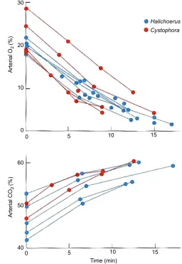
The real problem facing air-breathing animals is, of course, that they have to stop breathing, and, in consequence, the arterial oxygen content is ever-decreasing and the arterial carbon dioxide content is ever-increasing, as first shown by Scholander (1940) in hooded
seals and grey seals (Halichoerus grypus) (Fig. 1).
The solution
One obvious way to mitigate this problem is for habitually diving animals to be able to suppress breathing far better than non-divers. This is reflected in their ventilatory response to increased carbon dioxide, which is clearly less pronounced in seals than in terrestrial mammals (Irving et al., 1935b; Robin et al., 1963).
More important, however, if you want to stay submersed for an extended period, is to bring with you as much oxygen as you can and to economize with it to the best of your ability from the very start of the dive.
Oxygen stores
The lungs of phocid seals (see Glossary) are not particularly big and they will normally expire before a dive, to reduce buoyancy
and avoid divers’disease (Scholander, 1940). The size of the lung
oxygen stores was first estimated by Packer et al. (1969) and Lenfant et al. (1970) in juvenile seals and later by Burns et al. (2007) in adult hooded seals. Assuming 15% oxygen in the lung air and that 50% of lung volume is expired before the dive, the lung
oxygen store in hooded seals amounts to 6 ml O2kg−1, compared
with some 9 ml O2 kg−1 in the fully expanded lungs of man.
However, at depth, as the alveoli of the lungs collapse because of the hydrostatic pressure (Kooyman et al., 1970; Falke et al., 1985; Moore et al., 2011), the blood is shunted through the lung with minimal opportunity for gas exchange (Sinett et al., 1978; Kooyman and Sinnett, 1982; Falke et al., 1985, Fahlman et al., 2017). Accordingly, Miller et al. (2006) have shown that in seals the surfactants in the lungs are not primarily there to reduce surface tension to very low values, as in terrestrial mammals, but also have an anti-adhesive function, enabling the lungs to reopen following collapse during deep diving.
Hunter (1787) was probably the first to notice that the blood volume of seals was larger than in quadrupeds, and Irving et al. (1935a) found it more than twice as large as in humans. This was later confirmed by Lenfant et al. (1970), and subsequently by others (Box 1). Not only is the blood volume large: the hematocrit value (see Glossary), and thereby the hemoglobin content of the blood is also very high, amounting to about 60% compared with 45% in 1Department of Arctic and Marine Biology, UiT–The Arctic University of Norway,
9037 Tromsø, Norway.2St Catharine’s College, Cambridge CB2 1RL, UK. *Author for correspondence ([email protected])
A.S.B., 0000-0002-7824-0464
Journal
of
Experimental
humans (Lenfant et al., 1969; Packer et al., 1969; Lenfant et al., 1970; Burns et al., 2007) (Fig. 2). The oxygen-carrying capacity of the blood and hence the blood oxygen store in hooded seals may
therefore be as high as 45 ml O2kg−1, whereas in humans it is about
11 ml O2kg−1(Packer et al., 1969; Burns et al., 2007), but this is not
to say that the entire blood oxygen store is available at all times. Kooyman et al. (1980) noticed a rise in aortic hemoglobin
concentration in diving Weddell seals (Leptonychotes weddellii)
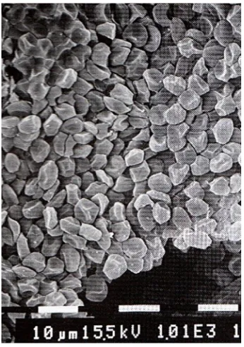
and assumed that it was caused by release of previously sequestered red blood cells in the venous sinuses. Qvist et al. (1986) and Hurford et al. (1996) observed the same, but suggested that the increase was caused by release of large amounts of red blood cells from the spleen. The spleen is large in phocid seals, in particular in deep-diving species such as Weddell seals and hooded seals, in which it
may amount to 2–4% of body mass. In a 200 kg hooded seal it may
release in excess of 3 litres of blood with a hematocrit of about 90% (Fig. 3) into the circulation (Cabanac et al., 1997). Cabanac et al. (1999) have described the structure and the filling and release mechanism of the spleen, and it is now generally accepted that the spleen functions as an important oxygen reservoir, which is utilized when the seal starts to dive. Moreover, Elsner and Meiselman
(1995) pointed out the advantage of being able to reduce the viscosity of the blood, which is mainly determined by the hematocrit, by withdrawal of a substantial fraction of the
30
Halichoerus Cystophora
20
Arterial O
2
(%)
Arterial CO
2
(%)
10
0
60
50
40
0 5 10
Time (min)
15
[image:2.612.304.560.59.430.2]0 5 10 15
Fig. 1. Arterial oxygen and carbon dioxide content in hooded and grey seals during forced dives.The forced dives were of up to 17 min duration in a bathtub; upper panel: oxygen; lower panel: carbon dioxide; red symbols, hooded seals; blue symbols, grey seals. Redrawn from Scholander (1940). GLOSSARY
ADL
Aerobic dive limit; the amount of time a seal can dive without releasing lactate into the circulation after the dive.
Aerobic respiration
Respiration in the presence of oxygen. Anaerobic respiration
Respiration in the absence of oxygen. Apnea
Cessation of breathing. Asphyxia
A combination of severe hypoxia and hypercapnia. Bradycardia
Reduced heart rate. Cardiac output
The amount of blood the left ventricle of the heart pumps out in one minute.
Coronary blood vessels
Blood vessels providing circulation of the heart muscle. Hematocrit
Volume percentage of red blood cells in blood. HIF-1α
Hypoxia inducible factor 1α.
Hypercapnia
Increased partial pressure of carbon dioxide. Hypoxia
Reduced partial pressure of oxygen. Ischemia
Obstruction of blood supply. Myoglobin
Iron- and oxygen-binding protein of vertebrate muscle. Oxygen tension
Partial pressure of oxygen (in fluids). Phocid seals
True earless seals. Q10
Increased rate of an activity caused by a 10°C increase in temperature. Tachycardia
Heart rate above resting value. Vasa vasorum
Network of small blood vessels that supply the walls of large blood vessels.
Box 1. Historical perspective
The first student of adaptations to diving habit was probably Robert Boyle,
who, in 1670, was introduced to the Royal Society in London as ‘the
indefatigable benefactor to philosophy’. A benefactor to philosophy
perhaps, but hardly to animals, Boyle (1670) exposed a great number of
diving and non-diving animals to evacuated chambers and found‘to his
wonder’that they were‘all recoverably gone’after a very short time. Without
apparent wonder, however, he also noticed that ducks can endure much longer underwater exposure than hens. About a hundred years later, Hunter (1787) found that seals and whales have much larger blood volumes and hence larger oxygen stores than terrestrial animals, and this was rediscovered another hundred years later by Bert (1870), who also found that ducks have much larger blood volumes than hens. This impressed him and his successors as the ultimate answer, to the extent that the interest in the problem vanished for more than 60 years. Then, however, Irving and Scholander, within a few years, delineated the basic problems, performed a number of crucial experiments, and predicted the answers to the most pertinent questions regarding adaptations to diving. The development up until that time is outlined in detail in Andersen (1966).
Journal
of
Experimental
circulating red blood cells when they are not needed between diving bouts.
The colour of seal muscle is conspicuously dark, sometimes almost black, owing to the fact that it is very rich in myoglobin (see Glossary). The myoglobin concentration in seals was probably first determined by Robinson (1939) and Scholander (1940). Since then, a number of determinations in a wide variety of age groups of a number of pinniped species, with a great variety of accuracy, have been made (Fig. 2), but for the sake of comparison, we will continue to focus on the hooded seal. In this species the myoglobin content
varies in different muscles, but in total it has a capacity to bind and
store oxygen to the amount of 37 ml O2kg−1(Burns et al., 2007),
which is about six times that of humans. Thus, the grand total of
oxygen stores of the hooded seal is a staggering 90 ml O2 kg−1,
whereas in humans it amounts to approximately 25 ml O2kg−1, of
which about 30% is contained in the lungs.
It is intriguing that the hooded seal is born with fully developed hemoglobin stores, whereas their myoglobin stores are only about 25% of that of the adult (Geiseler et al., 2013). The reason for this is probably that a large myoglobin concentration in the muscles would be counterproductive in that the myoglobin would hold large amounts of oxygen, never to be released during fetal life. It appears, however, that the myoglobin stores are established shortly after birth thanks to iron stores in the liver (Geiseler et al., 2013). This build-up coincides with a remarkably rapid development in diving performance by the pups, which may dive to 100 m, and stay submerged for 15 min, within 3 weeks of age, and reach below 700 m within less than a year, with a body mass of less than 100 kg (Folkow et al., 2010). A similar, albeit slower, development has been observed in Weddell seal pups (Burns, 1999) and southern elephant seal pups (Hindell et al., 1999).
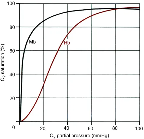
There is, however, an important peculiarity with regard to myoglobin, which has frequently been overlooked when the so-called total available oxygen stores and aerobic dive limit (ADL) (see Glossary) have been estimated. This is that myoglobin has a drastically higher affinity for oxygen than hemoglobin (Fig. 4), which implies that if the muscles are circulated during a dive then the oxygen on the myoglobin will only be available for transport elsewhere at a time when the oxygen content of the blood and tissues has reached precipitously low values. This problem and its implications will be the topic of discussion repeatedly throughout this Review.
Simulated diving in the laboratory Economy with oxygen stores
The fact remains that although the mass-specific oxygen stores (lung air included) of the hooded seal are only about four times those of humans (Fig. 2), the hooded seal can dive 20 times longer 100
Muscle
Blood Lung 80
Oxygen stores (ml O
2
kg
–1
)
60
40
20
0
Crabeater Leopard Harbour
Ringed
Grey Harp Ribbon Baikal
N. elephant
W
eddell
Hooded
Steller SL
New Zealand SL
California SL Australian SL Northern FS
W
[image:3.612.52.363.57.305.2]alrus Man
Fig. 2. Mass-specific total available oxygen stores for a variety of adult seals and man, with relative distribution in muscle, blood and lungs.Oxygen
measured in ml O2kg−1; SL, sea lion; FS, fur seal.
Redrawn from Burns et al. (2007).
Fig. 3. Scanning electron micrograph of the dense aggregate of red blood cells in the dilated spleen of the hooded seal.Scale: each division is
10 µm. From Cabanac et al. (1999).
Journal
of
Experimental
[image:3.612.88.261.460.706.2](Folkow and Blix, 1999). This begs the question: how is this possible?
As early as 1877, the eminent Danish physiologist Christian Bohr suggested that anaerobic processes (see Glossary) must cover important parts of the energy needs when diving animals are submerged (Bohr, 1877). His paper was, however, written in Danish and therefore did not reach the international scientific community. The breakthrough came instead with Lawrence Irving, who, studied changes in blood flow through brain and muscle in diving muskrats (Ondatra zibethicus) and beavers (Castor canadensis) (Irving, 1938).
In a seminal review he (Irving, 1939) concluded:‘The storage of
oxygen is inadequate to provide for its use by all tissues, but differential control of the distribution of oxygen might reasonably serve to maintain the brain, allowing the less sensitive tissues or those
with fair capacity for anaerobic metabolism to do without oxygen.’
Peripheral vasoconstriction
It did not take long to prove Irving right. At the very same time, P. F. Scholander, who worked in Oslo, was investigating responses to
diving in hooded seals and grey seals and found that arterial lactate did not increase much as long as the seal was submerged, whereas he recorded a surge as soon as the animal surfaced to breathe (Fig. 5). From this, he drew the conclusion that the muscles were excluded from circulation and that they first used the oxygen held by the muscle myoglobin, whereafter they metabolized anaerobically, during the dive. The muscles were subsequently recirculated and re-oxygenated when breathing recommenced (Scholander, 1940). This selective peripheral vasoconstriction virtually transforms the animal
into a‘heart–brain–lung preparation’, reserving the blood oxygen
store for the brain, whereas the rest of the body has to rely on local stores of oxy-myoglobin and subsequently anaerobic metabolism. This was later nicely illustrated by Bron et al. (1966) by use of angiography (Fig. 6).
Decisive as the above concept was, it raised a host of questions, which have kept physiologists well occupied ever since. For a start, how is it possible for the arteries to stay constricted with a steadily increasing tissue pH, following the rise in intracellular lactate? The solution to this problem was first found in ducks by Folkow et al. (1966), and later confirmed in seals by White et al. (1973): expert divers have the ability to constrict the arteries, leading to, but outside, the organs, unlike terrestrial forms that control the resistance to flow at the arteriolar level inside the organ. This is also apparent in Fig. 6. However, regardless of where a vascular constriction of the magnitude seen in diving seals occurs, it will imply a dramatic increase in total peripheral resistance to flow, and, unless compensated, should result in a disastrous increase in blood pressure! The solution to this imminent problem is bradycardia (see Glossary).
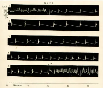
Bradycardia
Scholander (1940) was probably first to record an electrocardiogram (ECG) from seals, but the phenomenon was not put into context until Irving et al. (1942) recorded unchanged arterial blood pressure in diving seals (Fig. 7). From that, it was fully understood that bradycardia provided the reduction of cardiac output (see Glossary) necessary to maintain central arterial pressure in response to the increased resistance caused by the peripheral vasoconstriction. Later, several studies showed that cardiac output, by and large, is reduced in proportion with the slowing of the heart (Murdaugh et al., 1966; Blix et al., 1976; Zapol et al., 1979; Blix et al., 1983). However, a reduction in stroke volume (Blix et al., 1983) caused by
a reduction of ventricular contractility (dP/dtmax; rate of left
ventricle pressure rise in early systole) (Kjekshus et al., 1982) and myocardial wall tension (Elsner et al., 1985) contribute to the same end. This is not to say that the systolic ejection of the stroke volume 100
Mb Hb
80
60
40
20
0 20 40 60 80
O2 partial pressure (mmHg)
O2
saturation (%)
[image:4.612.56.294.57.287.2]100
Fig. 4. Myoglobin has a much higher affinity for oxygen than hemoglobin. Hemoglobin (Hb) with four heme groups has sigmoidal oxygen dissociation curves, whereas myoglobin (Mb) with only one has a hyperbolic dissociation curve and a much higher affinity for oxygen, which implies a one-way flow of oxygen from the blood to the skeletal muscles.
Lactic acid (mg %)
O2 CO2
(vol. %) 160
60
40
20 120
25
20
15
10
5
0 Dive
mmol l
−
1
80
40
0
CO2 %
O2 %
LA
Fig. 5. Arterial oxygen, carbon dioxide and lactate content in a grey seal before, during and after an 18 min dive.Red trace, oxygen; blue trace, carbon dioxide; yellow trace, lactate (LA). Redrawn from Scholander (1940).
Journal
of
Experimental
[image:4.612.49.377.595.737.2]against a vastly increased peripheral resistance is without potential problems for central arterial blood pressure. Previously, Burow (1838) had described the presence of a bulbous enlargement of the ascending aorta in seals, and Drabek (1975) later described its morphology in some detail. However, its significance as a windkessel (elastic chamber) that accommodates the systolic ejection and through elastic recoil contributes to the maintenance of blood pressure during the extended diastole was not fully
appreciated until Rhode et al. (1986) studied its pressure–volume
characteristics. Recently, Blix et al. (2016) have shown that the wall of the bulb in hooded seals is so thick that an extensive vasa vasorum (see Glossary) is required for its maintenance.
Myocardial workload and metabolism
The importance of the profound bradycardia, which may reach
values as low as 4–6 beats min–1, is unquestionably, first and
foremost, to balance central arterial blood pressure against the dramatic increase in peripheral vascular resistance during diving. However, the bradycardia is in itself a great benefit, in that reduced heart rate, myocardial contractility and wall tension all also contribute to a profound reduction of the workload of the heart. It
was earlier assumed that the perfusion of the heart was much increased to compensate for the reduction in arterial oxygen content during diving (Johansen, 1964) and this was even emphasized in a much-used textbook (Schmidt-Nielsen, 1975). Blix et al. (1976) demonstrated, however, that the myocardial blood flow in diving seals is instead reduced to about 10% of the pre-dive level, as would be expected from the reduction of cardiac output. In fact, the reduction is such that zero coronary blood flow (see Glossary) is
regularly sustained for 10–45 s (Elsner et al., 1985). The great
reduction in myocardial energy demand also allows the heart to shift from aerobic to anaerobic metabolism (see Glossary), probably mainly based on rich local glycogen deposits (Kerem et al., 1973) from the very beginning of the dive, without any evidence of myocardial dysfunction (Kjekshus et al., 1982).
Brain circulation and metabolism
Having dealt with the heart and peripheral vasculature, what about the brain? Elsner et al. (1970b) demonstrated by encephalographic (EEG) recordings in Weddell seals that cerebral integrity is maintained down to an arterial oxygen tension (see Glossary) of
10 mmHg. This is much lower than the critical arterial O2tensions
A
B
Fig. 6. Angiogram of peripheral (abdominal) arteries of aharbour seal.(A) During breathing at the surface, arteries of flanks (thin arrow) and hindflippers (thick arrow) are well filled with blood. (B) During diving, the same arteries become profoundly constricted.
Also shown is the bladder (‘B’) in which the contrast medium ends up.
[image:5.612.49.333.59.173.2]Figure from Bron et al. (1966).
Fig. 7. First recording of blood pressure in the femoral artery of a seal, at intervals, during an 8 min dive demonstrating dramatic bradycardia and a fairly well maintained central arterial blood pressure.Blood pressure measured in mmHg. Figure from Irving et al. (1942).
Journal
of
Experimental
[image:5.612.49.396.441.736.2]of 25–40 mmHg at which impairments from limitations in ATP production are first seen in brains of terrestrial mammals (Erecinska and Silver, 2001). So, how does the seal brain cope with repeated extreme hypoxia (see Glossary)?
The blood flow to the brain is, as suggested by Irving (1939) and Scholander (1940), pretty well maintained at the end of a long dive (Blix et al., 1983; Fig. 8), and blood glucose is fairly well maintained (Scholander, 1940; Guppy et al., 1986). Endogenous stores of glycogen are two to three times as large as in terrestrial mammals, but still quite small (Kerem et al., 1973; Czech-Damal et al., 2014). Cerebral capillary density is somewhat higher in seals than in non-diving mammals (Kerem and Elsner, 1973) and one would have assumed that neuroglobin (Burmester et al., 2000),
which is further assumed to facilitate O2diffusion, would be more
abundant in seal brains. In the deep-diving hooded seal, cerebral neuroglobin levels are no higher than in mouse or human (Mitz et al., 2009), but have an unusual distribution, with a higher concentration in glial cells (astrocytes) than in neurons. This suggests that glial cells are more involved in aerobic metabolism than neurons, which may further imply that seal brain neurons depend more on anaerobic metabolic pathways, whereas glial cells may remove and metabolize the lactate that is thereby produced (Larson et al., 2014).
In terrestrial mammals, the tetrameric lactate dehydrogenase (LDH) enzyme consists of two types of subunits, designated H and M. A random combination of these two subunits results in
five possible isozymes, LDH-1 (H4) to LDH-5 (M4) (Appella
and Markert, 1961). The isozymes consisting of M-subunits preferentially convert pyruvate to lactate, whereas those with H-subunits preferentially convert lactate to pyruvate. The LDH-1 isozyme is predominantly found in brain and heart tissue, whereas LDH-5 dominates in skeletal muscle. However, Blix and From (1971) found that all the isozymes were represented in the brain of the hooded seal, and Murphy et al. (1980) found that LDH activity was twice that of the ox in the Weddell seal brain, whereas the activities of oxidative enzymes were similar, or somewhat lower. Recently, Hoff et al. (2016) rediscovered the data by Blix and From (1971) and also found that maximum LDH activity was significantly higher in the brain of hooded seals than in mice, but
less than that of the ferret (Mustela putorius furo). Murphy et al.
(1980) also found that glucose uptake by the brain was only slightly
increased during dives and that a relatively large fraction (20–25%)
of the glucose taken up was released as lactate. Hochachka (1981) therefore suggested that brain metabolism is not oxygen limited during diving in the Weddell seal, but this was based on a number of assumptions of brain blood flow (Zapol et al., 1979), brain venous drainage (Harrison and Tomlinson, 1956), and a host of other factors that might not be correct (Hol et al., 1975; Blix et al., 1983).
Ramirez et al. (2011) have demonstratedin vitrothat neocortical
slices from hooded seals may maintain high spiking activity for up to 60 min in severe hypoxia. Hooded seal cerebellar slices may even
maintain high spontaneous activity for durations of 10–15 min in
complete anoxia (L. P. Folkow, S. Ludvigsen and S. Geiseler, unpublished observations). Folkow et al. (2008) have further demonstrated in cortical pyramidal neurons of brain slices from hooded seals that resting membrane potential and ability to generate action potentials are maintained under severe hypoxic conditions. Geiseler et al. (2016) have shown that the synaptic activity in hooded seal hippocampal slices decreased, but unlike terrestrial mammals, it remained at more than 30% of normoxic amplitude throughout 3 h of severe hypoxia, and upon re-oxygenation, the signal recovered to 50% of the amplitude under normoxic conditions. Finally, Fabrizius et al. (2016) used transcriptome analysis to study the stress tolerance of the seal brain and found that it has a lower aerobic capacity than a terrestrial animal, the ferret. Hoff et al. (2017) with the same approach found up-regulation of genes related to inflammation and a down-regulation of genes involved in ion transport and other neuronal processes, indicative of neuronal shutdown in response to hypoxia in brain slices from the visual cortex of hooded seals.
Hochachka (1986) suggested that seals are able to overcome the challenges of extreme hypoxia by what he termed metabolic arrest, to be achieved by means of a reversed Pasteur effect, in combination with a maintenance of membranes of low permeability, probably by reduced densities of ion-specific channels. Evidence for such ion
channel arrest was later obtained in liver cellsin vitrofrom Antarctic
seals by Hochachka et al. (1988). This concept, now termed neuronal shut-down and nervous network reconfiguration, in which some neurons shut down to save energy, whereas others of eminent importance maintain activity in response to hypoxic insult, has been further developed by Ramirez et al. (2007).
Having a small brain would, at least as far as diving capacity is concerned, be an advantage, as the brain is the major consumer of oxygen during a dive, but it does not appear that seals have brain sizes relative to body mass that differ from the overall mammalian line (Worthy and Hickie, 1986). In fact, given that as much as 50% of body mass may be made up of almost metabolically inert blubber (Aarseth et al., 1999), brain size relative to lean body mass is large and would put seals at a disadvantage.
0
% Pre-dive blood flow –20
–40
–60
–80
–100
Cortex
Cerebellum
Lung Heart Liver
Kidney
Stomach Muscle
[image:6.612.64.289.365.664.2]20 40 60 80 100 120
Fig. 8. Tissue blood flow in the organs indicated after 5 and 10 min of submersion in spotted and grey seals.These two species are not significantly different; brain blood flow is increased, whereas most other organs are more or less excluded from circulation at the end of long experimental dives. Blood flow was measured by use of radioactive microspheres as % of pre-dive flow; red bars, 5 min of submersion; blue bars, 10 min of submersion.
Redrawn from Blix et al. (1983).
Journal
of
Experimental
Selective brain cooling
Regardless of brain size and mode of ATP production, reduced oxygen consumption by the brain would be an advantage. Many decades ago, Scholander et al. (1942a) recorded a drop in brain temperature in diving seals, but they used mercury thermometers that were stuck into the brain through holes that were drilled through the skull. Probably owing to their not-so-modern approach, their finding was lost to science until confirmed by Odden et al. (1999), who found a 3°C drop in brain temperature during 15 min dives in a juvenile hooded seal. These results were later confirmed by Blix et al. (2010) who also demonstrated that the brain is cooled in a controlled manner by cold blood returning through the large superficial veins
from the front flippers. Assuming that theQ10effect (see Glossary)
on cerebral metabolism is the same in seals as in piglets (Busija and Leffler, 1987; Laptook et al., 1995), a reduction of brain temperature of 3°C in a seal should result in a reduction of brain oxygen demand
of 15–20%. That would extend the diving capacity substantially, and
in addition provide neuroprotection against hypoxic injury (e.g. Laptook et al., 1995). However, any reduction of brain temperature will normally lead to vigorous shivering in mammals (Simon et al., 1986) and, if so, compromise brain cooling and hence in that context be rather counterproductive. Accordingly, Kvadsheim et al. (2005) have shown that the normal shivering response to brain cooling is itself inhibited as part of the response package that is elicited upon diving. Several other aspects of thermoregulation in seals are reviewed in Blix (2016).
Kidney function during diving
Selective arterial vasoconstriction also affects the kidneys, and renal blood flow is reduced to less than 10%, and sometimes ceases altogether, during prolonged dives (Elsner et al., 1966; Blix et al., 1976; Zapol et al., 1979). This implies that the kidneys can be exposed to warm ischemia (see Glossary) for periods up to 1 h, a stress that kidneys of terrestrial mammals cannot tolerate. Bradley and Bing (1942) showed that diving results in a profound reduction of glomerular filtration and urine production in seals, and their results were confirmed by Murdaugh et al. (1961b) who reported that glomerular filtration and urine flow in harbour seals (Phoca vitulina) ceased completely during 10 min dives. Halasz et al. (1974) compared the renal function of isolated fresh kidneys from harbour seals and dogs and found that urine production recovered promptly after 1 h of warm ischemia in seals, whereas dog kidneys remained anuric after the insult. This shows that the seal kidneys have an amazing tolerance to warm ischemia that deserves further study.
Skeletal muscle metabolism and buffering capacity
In 1939, Scholander, in the spirit of the times, cut a hole in the back of a seal and demonstrated that the bleeding from the wound stopped completely while the seal was under water, but bled profusely as soon as the animal resumed breathing (Scholander, 1940). Scholander (1940) and Scholander et al. (1942b) further showed that the complete cessation of circulation in the muscle resulted in a massive build-up of lactic acid (Fig. 5), which started at the time when the muscle myoglobin had reached complete reduction. This implies that the skeletal muscles depend on anaerobic metabolism after the oxy-myoglobin stores have been exhausted during dives of long duration. Accordingly, Kanatous et al. (1999) found that short-duration divers, such as the harbour seal, have a well-developed capacity for aerobic metabolism, particularly in the typical swimming muscles. Even so, such seals still have a smaller capillary-to-fibre interface and capillary supply per fibre mitochondrial volume than dogs
(Kanatous et al., 2001). Weddell seals, with an ability for dives of very long duration, by contrast, do not have enhanced aerobic capacities compared with those of terrestrial mammals and short-duration divers (Kanatous et al., 2002).
The enormous build-up of lactate in the skeletal muscles of diving seals requires some sort of buffering if undue changes in blood and tissue pH are to be avoided. It took quite some time before this important issue attracted interest, but Castelini and Somero (1981) found that marine mammals have higher muscle buffering capacity on average than terrestrial mammals. They also noticed strong correlations between buffering capacity and myoglobin concentration, and between buffering capacity and muscle LDH activity. Much later, Lestyk et al. (2009) studied the
development of the buffering capacity in harp seals (Pagophilus
groenlandicus) and hooded seals and found that it was very high in the adult seal and that the buffering capacity of neonate muscle was as high as 75% of the adult value in spite of their much lower myoglobin concentration. This suggests, as one would expect, that other muscle proteins might contribute significantly to the buffering capacity in the muscles during the dive. At the end of the dive, when breathing and the circulation of the muscles are resumed, however, the lactate that has accumulated during the dive is washed into the general circulation and has the potential to cause a disastrous decrease in arterial blood pH. This is, at least in part, mitigated in that the muscles are not perfused wholesale, but rather brought back in circulation gradually (Blix et al., 1983). Even so, pH values as low as 6.8 may be reached in arterial blood after long dives (Scholander, 1940; Kooyman et al., 1980).
It is also possible that the anticipatory tachycardia (see Glossary), which is often observed when seals are on return to the surface, reflects
reperfusion of the previously ischemic blubber in which N2is perhaps
five times more soluble than in other tissues, and that the rise in arterial
N2 which would otherwise ensue is mitigated. The fact that seals
habitually exhale before diving also contributes to the same end. However, it is not only nitrogen and lactate that may be a problem after long dives. The danger of reperfusion injury is also considerable. Elsner et al. (1998) and Vázquez-Medina et al. (2007) suggested that
this may be mitigated with enhanced O2 free radical scavenging,
especially through elevated glutathione levels and increased activities of enzymes involved in glutathione recycling. Moreover, Vázquez-Medina et al. (2011) have suggested that repeated apneas (see Glossary) stimulate adaptive responses in elephant seal pups by
up-regulation of their anti-oxidant system, HIF-1α(see Glossary) and
myoglobin.
Selective distribution of cardiac output
Although Elsner et al. (1966) made the first direct measurements of renal blood flow, it was not until the introduction of radioactive microspheres (Blix et al., 1976) that the overall distribution of cardiac output could be assessed. This method was further employed in two similar studies (Elsner et al., 1978), later published as Blix et al. (1983) and Zapol et al. (1979). All three studies confirmed the original assumption by Irving and Scholander that all the major organs, such as kidneys, liver, gut, skeletal muscle, and even the heart, are rendered almost without circulation, whereas the brain and to some extent the adrenals receive most of the cardiac output during diving (Fig. 8). However, there were differences: Zapol et al. (1979) studied Weddell seals with a diving capacity of 60 min (Kooyman et al., 1980) that were submerged for only
8–12 min, whereas Blix et al. (1983) studied spotted seals (Phoca
vitulina largha) and grey seals, with much inferior diving capacity,
which dived for 10 min. Thus, Zapol et al. (1979) found that
Journal
of
Experimental
cerebral blood flow was maintained at the end of the dive, whereas Blix et al. (1983) found that it was reduced to only 35 and 42% of the pre-dive value after 2 and 5 min diving, respectively, whereas it had more than doubled in this part of the brain after 10 min of diving.
Moreover, Zapol et al. (1979) found that lung (tissue) perfusion was reduced to about 60% of the pre-dive value during the dive, whereas Blix et al. (1983) found that it was reduced to only 12%. The reason for this discrepancy is most likely that the lungs in the former study received microspheres both from the bronchial artery, and from the systemic circulation, as a result of recirculation through arteriovenous shunts, known to be numerous in seals (Molyneux and Bryden, 1975).
Venous circulation in seals
With the massive and widespread arterial constriction and the collapse of the lungs at depth, a question arises concerning where all the blood goes. Houston (1835) believed that the blood in peripheral tissues was displaced by the hydrostatic pressure into what he had found to be a large vena cava and hepatic sinuses (in which he surmised that the pressure would be lower) in seals. Shortly after, Burow (1838) made a detailed description of the inferior caval veins that communicate with hepatic sinuses of unusual size. He found that the inferior vena cava passes through a sphincter of striated muscle located immediately anterior to the diaphragm, later shown to be operated by a branch of the phrenic nerve (Burne, 1910; Harrison and Tomlinson, 1956). Paramore (1910) did not believe that the thorax could collapse in deep diving and suggested that the sphincter prevented the central venous pool of blood from engorging the heart when the seal was put under pressure. It was not until Elsner et al. (1971) and Hol et al. (1975) studied the action
of the sphincterin vivoby use of angiography that its true function
was revealed. They demonstrated that the sphincter prevents engorgement of the heart, not because of pressure from outside, but as a result of the constriction of the arteries whereby the blood is shifted to the central veins. This creates a major reservoir of oxygen-rich blood in the vena cava, which is metered into circulation in proportion with the cardiac output. In the latter part of a dive the blood in this reservoir may even have an oxygen content that is higher than in the arterial blood (Elsner et al., 1964).
It was previously suggested that mixing of the oxygen-rich blood in the vena cava with the oxygen-depleted venous blood from the brain was avoided, as the venous drainage from the brain was assumed to go in a retrograde direction through the extradural intravertebral vein and reach the vena cava from behind by way of numerous venous plexuses during diving (Ronald et al., 1977). However, this much-cited suggestion was not supported by Nordgarden et al. (2000), who found that the blood in the extradural intravertebral vein does not flow in a retrograde direction during diving. Instead, Blix (2011) has suggested that the unusually large intravertebral vein of phocid seals (see Glossary) provides the animal with an overflow shunt through which retrograde central venous flow can be routed back to the anterior caval vein when the animals squeeze themselves through blowholes in the ice or slide over edges of ice floes to get into water. The importance of the very many and spectacular venous plexuses in phocid seals (Houston, 1835; Burow, 1838) is not fully understood, but Blix et al. (1975) have shown that they are an important component of a brown fat thermogenic tissue complex in the newborn pup.
Integration of cardiovascular and respiratory responses Huxley (1913), Andersen (1963) and Djojosugito et al. (1969) demonstrated that ducks lacking the cerebrum developed the normal
responses to diving, which implies that at least some very basic components in the adjustments to diving are integrated at the non-cerebral (medullary) level of the brain. The integration of the diving responses was under intense study from the late 1960s throughout the 1970s and this research has been thoroughly reviewed by Blix and Folkow (1983) and Butler and Jones (1997). In summary, apnea is induced by stimulation of trigeminal and glossopharyngeal receptors in the mouth, and the following asphyxia (see Glossary) stimulates the peripheral chemoreceptors, which causes a gradually increasing peripheral vasoconstriction and bradycardia. However, if the peripheral chemoreceptors are stimulated (by hypoxia) while the animal is breathing it causes increased ventilation, heart rate (cardiac output) and vasodilatation of the skeletal muscles. This is known as the primary chemoreceptor response, whereas when they are stimulated in the apneic animal the secondary chemoreceptor response is elicited, and is then characterized by peripheral vasoconstriction and bradycardia (Daly and Scott, 1963). There is no known study of the responses in decerebrate seals, but Elsner et al. (1977) have shown that simultaneous activation of the chemoreceptors and stimulation of the superior laryngeal nerve reinforced the bradycardia component of the diving responses. Moreover, Daly et al. (1977) demonstrated that withdrawal of chemoreceptor stimulation in diving seals suppressed the bradycardia component. It appears therefore, that whereas the chemoreceptors are a prerequisite for the full development of the responses in ducks, they are only necessary for the maintenance of the responses, at least in anesthetized seals, where the modulating influences of cortico-hypothalamic centres are more or less blocked. In seals it also appears that the responses of the carotid baroreceptors are augmented during diving (Angell-James et al., 1978). It also seems safe to say that the vasoconstrictor and bradycardia components of the diving responses emanate as tightly coupled efferent responses initiated via reflex suppression of the respiratory centres and are subsequently reinforced and stabilized by the chemoreceptor reflex response. In this context, the carotid baroreceptors no doubt have important modulating reflex influences and contribute to adjusting the vasoconstriction and bradycardia components towards oxygen saving, while properly maintaining blood pressure.
It is in this respect interesting to view the reactions of the seal fetus. Kooyman (1966) reported that the pregnant seal continues her normal diving behaviour until shortly before delivery and the fetus is therefore exposed to severely hypoxic conditions. Lenfant et al. (1969) found that the fetal hemoglobin of the Weddell seals has a much higher affinity for oxygen than that of its mother. Elsner et al. (1970a) studied the responses to clamping of the endotracheal tube in anesthetized pregnant Weddell seals, and Liggins et al. (1980) exposed them to simulated diving of 20 min duration. Although the latter authors claim that the responses are similar in both mother and fetus, it is quite clear from both studies that the bradycardia response develops much more slowly in the fetus than in the mother. This is to be expected in a fetus that is unable to know what is going on outside, and, without the cortico-hypothalamic input of the mother, its responses will have to rely on chemoreceptor input.
Metabolism during diving
Scholander (1940) compared the oxygen debt after dives with pre-dive oxygen consumption and concluded that the oxygen consumption during a dive was reduced by about 70%. He attributed the reduction to the shift to anaerobic metabolism, but cooling of the body (Kooyman et al., 1980; Hill et al., 1987; Blix et al., 2010) probably also contributed to the hypo-metabolism, as
previously suggested by Scholander et al. (1942a). Castellini et al.
Journal
of
Experimental
(1992) measured the metabolic rate of freely diving Weddell seals and found it to be about 20% higher than the resting rate in short dives, whereas it was about 20% lower than resting metabolic rate in long dives. However, their value for oxygen uptake during diving was an average of the value while the animal was under water plus that at the subsequent period at the surface, and it is therefore likely that the diving value is even lower. Le Boeuf et al. (1989) found that northern elephant seals dived fairly continuously, staying submerged for up to 62 min and reaching depths below 1000 m, and in this species the recovery time at the surface (<3.5 min) was independent of diving time. This would imply either that metabolism is conspicuously depressed, possibly as a result of body cooling, to an extent to allow it to be aerobic for such an extended period, or that lactate is somehow utilized during dives. However, the total available oxygen stores in these animals are calculated based on body mass, and in elephant seals with sometimes more than 50% of body mass in metabolically inert blubber the amount of oxygen available for the metabolizing lean body mass will be much greater. However, we have also learnt that seals may minimize their energy expenditure while under water by behavioural means, such as the use of prolonged gliding, allowed by the buoyancy reduction following lung compression during descent (Williams et al., 2000; Miller et al., 2012; Maresh et al., 2015).
Unrestrained diving at sea: facts and fiction
The story outlined above was pretty much the gospel of adaptations to diving habit until the 1970s, when time-depth recorders and radio-telemetry were introduced. Kooyman (1965) was the first to deploy a time-depth recorder on Weddell seals, whereas Harrison et al. (1972) recorded heart rate by radio-telemetry in grey seals, and others did the same in ducks (see Butler and Jones, 1997, for references). Subsequently, as more studies were conducted, it became clear that the bradycardia response was usually much smaller, and in some cases absent, in voluntary diving animals. This caused a state of insecurity in the diving community, and the feeling grew that the reactions observed during experimental diving were somehow false. This notion was further developed when Kanwisher
et al. (1981) published a paper in Science venturing the bold
suggestion that the reactions recorded in experimental dives were mainly caused by emotional stress and had little to do with natural responses to diving. This caused so much further emotion that the International Union of Physiological Sciences at its World Congress in Vancouver in 1986 dedicated a session to discuss the topic.
My view, then as now, was that the reactions that are elicited upon forced submersion expose the basic defence mechanisms against asphyxic insult, which albeit expressed to a different extent, are common to all mammals. As a seal that is forced under water is unable to know how long the dive will last, it will turn on its defences in full, from the very beginning, to be able to expand its diving capacity to its full extent. This has one crucial implication: seals must be able to control their cardiovascular responses by cortical (conscious) input. This paradigm was pitched hard in the reviews of Blix and Folkow (1983) and Blix (1987), but unfortunately, we termed it suprabulbar influences, a term that was lost on most actors in the field.
Besides the fact that the reactions seen during forced diving are common reactions to asphyxic insult in most animals from man to fish, our argument was based on a series of previous observations. Scholander (1940) reported that a number of psychological stimuli could elicit apnea and a variable bradycardia in seals, and Murdaugh et al. (1961a) and Jones et al. (1973) recorded prompt bradycardia
and heart rates as low as 7 beats min−1in freely diving seals, and
also anticipatory bradycardia before dives. Moreover, both Jones et al. (1973) and Casson and Ronald (1975) recorded anticipatory tachycardia in seals before surfacing. Thus, all these results reflect how higher central nervous system centres can turn on full oxygen-conserving responses through modulating descending pathways allowing them to adjust their cardiovascular responses to submersion to the current situation, as perceived by the cortical-limbic handling of tele-receptor information and earlier experiences. Even so, this rather absurd controversy prevailed until Thompson and Fedak (1993) at the meeting of the Society for Marine Mammals in Galveston presented a paper showing that heart rate declined as a function of dive duration in grey seals freely diving at sea. Even more convincing, they also reported that, in long dives, the heart rate could drop to as low as
4 beats min−1 for extended periods, whereas in short dives the
bradycardia was only moderate.
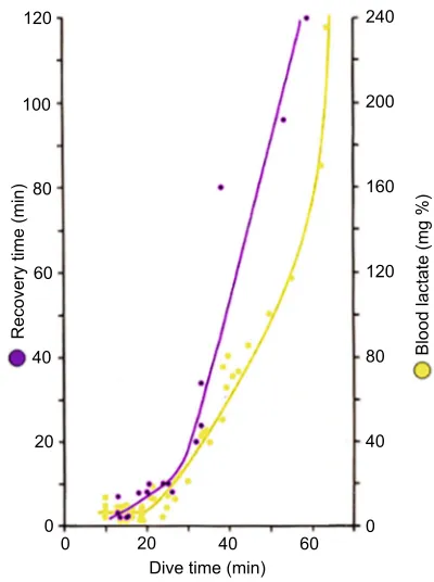
Aerobic dive limit: use and abuse
So, what else came out of the technological‘revolution’? First and
foremost, in a seminal study of Weddell seals in Antarctica, Kooyman et al. (1980) pioneered a technique by which the rather tame Weddell seals were brought to a breathing hole in the ice, far from other holes, and over which they put a laboratory hut (without a floor) into which the seal had to return after each dive. First, they found that the seals usually did a series of dives lasting up to
20–25 min, and only occasionally dived for extended periods up to
60 min. Even more important, they found that when dives were
shorter than 20–25 min, there was no post-dive lactic acid in the
arterial blood, whereas if the dives were longer, the arterial lactate concentration increased in a roughly exponential manner (Fig. 9). Moreover, after the short dives a very short recovery period was needed, whereas after dives of 60 min a recovery period of 2 h ensued. The advantage of performing a series of short dives is that
120
200 240
160
120
Blood lactate (mg %)
Recovery time (min)
80
40
0 100
80
60
40
20
0 20 40
[image:9.612.337.537.427.695.2]Dive time (min) 60 0
Fig. 9. Voluntary diving in Weddell seals.Peak arterial lactate
concentrations upon emergence from dives of various durations (yellow), and the recovery time required at the surface after dives of various durations (blue).
Redrawn from Kooyman et al. (1980).
Journal
of
Experimental
the animal can spend about 80% of the time under water, whereas the total time spent under water when the dives are long is much reduced. Based on these results, Kooyman et al. (1983) later coined the term aerobic dive limit (ADL) for the time a seal could dive
without a post-dive increase in arterial lactate concentration–that is
the time a seal could metabolize aerobically under water. This term is very useful when it is actually measured. The problem soon arose, however, that instead of being measured a great number of papers appeared in which the ADL was calculated on the basis of assumed field metabolic rates and determinations of total oxygen stores, in which the determinations of the myoglobin stores were of variable quality. Even worse, some started to use heart rate and calculated ADLs, which (of course) often was exceeded, to reach behavioural and ecological conclusions of a sometimes dubious nature. This made
Butler (2006) raise the crucial question:‘Aerobic dive limit. What is it
and is it always used appropriately?’In so doing, he emphasized the
problems with estimating field metabolic rate, and the fact that the blood oxygen stores can never be depleted completely if the animal is to survive the dive. He also emphasized the complications which may come from the possible role of phosphocreatine as a source of phosphorus for the production of ATP, but Blix (1971) found no difference in creatine concentration in brain, heart or skeletal muscle
between seals and sheep (Ovis aries). Butler (2006), however, did not
mention the most crucial link in the chain of arguments: the great difference in the affinity for oxygen between hemoglobin and myoglobin (Fig. 4) that makes it impossible to transfer oxygen from myoglobin to the blood for use elsewhere, whereas in Butler (2004) he did allude to this problem.
Thus, for dives to be truly aerobic, the blood flow to working muscles has to be shut off so that the oxygen on the myoglobin can
be utilized in situ, which requires some degree of bradycardia.
Alternatively, there is no peripheral vasoconstriction and therefore no bradycardia, whereby the blood oxygen store can be depleted, whereas the muscle oxygen store will remain fully loaded at the end of the dive, which consequently will have to be much shorter than any calculated ADL.
Alternatively, at the other extreme, if the animal decides to go for a long dive, it will shut off circulation to all the muscles and visceral organs from the very beginning, accompanied by profound bradycardia. In this case the blood oxygen store is reserved for the brain, whereas the muscles first use up the oxygen on the myoglobin and thereafter metabolize anaerobically, which is exactly the same as happens when a trained seal is forced under water in a bathtub (Scholander, 1940).
In most cases, however, the seals appear to go for an intermediate strategy, in which the most active muscles, which would otherwise consume a significant amount of the blood oxygen, are shut off from circulation and use the local oxy-myoglobin for aerobic metabolism, without compromising the blood oxygen store. However, this cannot be done without a certain degree of bradycardia to compensate for the ensuing increase in peripheral vascular resistance, and that is what is observed in most voluntary dives (e.g. Kooyman and Campbell, 1972; Hill et al., 1987). Thus, without knowing how the animal is managing its muscle oxygen stores, any discussion of the ecological implications of whether the animal is exceeding its calculated ADL (e.g. Costa et al., 2001), even when in rare cases the size of the total oxygen stores is determined correctly, will, in my opinion, be a futile exercise.
Concluding remarks
So what else have we learned from the study of free-swimming seals? We know that lactate accumulates during long dives
(Kooyman et al., 1980), and that heart rate is variable and related to expected duration, with extremely low values in very long dives (Kooyman and Campbell, 1972; Hill et al., 1987; Thompson and Fedak, 1993). We know that the myoglobin-bound oxygen stores are not utilized in correlation with overall swimming activity, and are even sometimes re-oxygenated from the blood during dives (Guyton et al., 1995), and that (renal) glomerular filtration rate is maintained in short dives, but is reduced over 90% during dives longer than the ADL (Davis et al., 1983). Furthermore, we know that blood cells are released from the spleen at the beginning of dives (Hurford et al., 1996), that body temperature is reduced (Kooyman et al., 1980; Hill et al., 1987; Meir and Ponganis, 2010), that the adrenals are perfused with blood, albeit at a reduced rate and that the concentration of catecholamines (noradrenaline and adrenaline) in the blood is correlated with increasing reliance on anaerobic metabolism (Hochachka et al., 1995). We know
that arterial and venous oxygen tensions as low as 12–23 and
2–10 mmHg, respectively, can be experienced and well tolerated
(Meir et al., 2009). Moreover, Meir et al. (2013) have shown in juvenile elephant seals that the blood oxygen stores are depleted to the same extent irrespective of dive function, and we know that the development of the diving responses of the fetus are slow in their onset (Hill et al., 1987).
Thus, the responses seen in seals diving freely at sea are physiologically the same as those seen during forced dives in the laboratory. These responses are, moreover, not specific and unique
to water immersionper se, but instead reflect protective mechanisms
against asphyxia, which, to a variable degree, are common to all mammals. To what extent these basically medullary mechanisms are facilitated or depressed by cortico-hypothalamic centres depends greatly on how the seal judges the situation. It follows that when a seal is forced under water not knowing the duration of the dive, it will immediately turn on the full oxygen-conserving responses, whereas it can suppress them to a variable extent, if the dive at sea is anticipated to be brief (Blix and Folkow, 1983). In a recent review by Davis (2014) of the work of his associates, this
concept is still not fully adopted and it is emphasized that ‘to
understand the significance of biological adaptations (e.g. the dive
response), one must study animals in their natural environment’.
Our understanding of how seals can dive to extreme depths is now well advanced, but there are still questions that remain to be answered. The apparent ability of certain cells to shut down to save energy when oxygen is in short supply, and in particular how neuron metabolism is organized, is one. In this context, use of modern technology such as computed tomography (CT) scan, magnetic resonance imaging (MRI) and positron emission tomography (PET) scans (e.g. Wehrl et al., 2013) has so far not been exploited as it should. The finding that the surface intervals do not vary with the duration of preceding or succeeding dives in northern elephant seals (Le Boeuf et al., 1989) is another. There are also several other aspects of the intermediary metabolism, in particular how lactate is eliminated and how the toxic effects of high partial pressures of nitrogen and oxygen at depth are avoided, that remain to be understood. There is also great promise in further development of blood sampling devices (Takei et al., 2016) for use on seals at sea.
Acknowledgements
I have benefited from discussions with Lars P. Folkow, Philip Oliver and Lars Walløe during the preparation of this Review.
Competing interests
The author declares no competing or financial interests.
Journal
of
Experimental
References
Aarseth, J. J., Nordøy, E. S. and Blix, A. S.(1999). The effect of body fat on basal metabolic rate in adult harp seals (Phoca groenlandica).Comp. Biochem. Physiol.
124, 69-72.
Andersen, H. T.(1963). The reflex nature of the physiological adjustments to diving and their afferent pathway.Acta Physiol. Scand.58, 263-273.
Andersen, H. T.(1966). Physiological adaptations in diving vertebrates.Physiol. Rev.46, 212-243.
Angell-James, J. E., Daly, M. de B. and Elsner, R.(1978). Arterial baroreceptor reflexes in the seal and their modification during experimental dives. Am. J. Physiol.3, H730-H739.
Appella, E. and Markert, C. L.(1961). Dissociation of lactate dehydrogenase into subunits with guanidine hydrochloride. Biochem. Biophys. Res. Commun.6, 171-176.
Bert, P.(1870).LeÇons sur la physiologie comparée de la respiration. pp. 526-553. Paris: Baillière.
Blix, A. S. (1971). Creatine in diving animals – a comparative study. Comp. Biochem. Physiol.40A, 805-807.
Blix, A. S.(1987). Diving responses: fact or fiction?NIPS2, 64-66.
Blix, A. S.(2011). The venous system of seals, with new ideas on the significance of the extradural intravertebral vein.J. Exp. Biol.214, 3507-3510.
Blix, A. S.(2016). Adaptations to polar life in mammals and birds.J. Exp. Biol.219, 1093-1105.
Blix, A. S. and Folkow, B. (1983). Cardiovascular adjustments to diving in mammals and birds. InHandbook of Physiology. The Cardiovascular System III. Peripheral Circulation and Organ Blood Flow (ed. J. T. Shepherd and F. M. Abboud). pp. 917-945. Bethesda: American Physiological Society.
Blix, A. S. and From, S. H.(1971). Lactate dehydrogenase in diving animals–a comparative study with special reference to the eider (Somateria mollissima). Comp. Biochem. Physiol.40 B, 579-584.
Blix, A. S., Grav, H. J. and Ronald, K.(1975). Brown adipose tissue and the significance of the venous plexuses in pinnipeds. Acta Physiol. Scand. 94, 133-135.
Blix, A. S., Kjekshus, J. K., Enge, I. and Bergan, A.(1976). Myocardial blood flow in the diving seal.Acta Physiol. Scand.96, 277-280.
Blix, A. S., Elsner, R. and Kjekshus, J. K. (1983). Cardiac output and its distribution through capillaries and A-V shunts in diving seals. Acta Physiol. Scand.118, 109-116.
Blix, A. S., Walløe, L., Messelt, E. B. and Folkow, L. P.(2010). Selective brain cooling and its vascular basis in diving seals. J. Exp. Biol.213, 2610-2616.
Blix, A. S., Kuttner, S. and Messelt, E. B.(2016). Ascending aorta of hooded seals with particular emphasis on its vasa vasorum.Am. J. Physiol.311, R144-R149.
Bohr, C. (1877). Bidrag til svømmefuglernes fysiologi. (Contribution to the physiology of swimming birds).K. Dan. Vidensk. Selsk. 2.
Boyle, R. (1670). New pneumatical experiments about respiration. Philos. Trans. R. Soc. London5, 2011-2034.
Bradley, S. E. and Bing, R. J.(1942). Renal function in the harbor seal (Phoca vitulinaL.) during asphyxia ischemia and pyrogenic hyperemia.J. Cell. Comp. Physiol.19, 229-237.
Bron, K. M., Murdaugh, H. V., Milen, J. E., Lenthall, R., Raskin, P. and Robin, E. D.(1966). Arterial constrictor response in a diving mammal.Science152, 540-543.
Burmester, T., Weich, B., Reinhardt, S. and Hankeln, T.(2000). A vertebrate globin expressed in the brain.Nature407, 520-523.
Burne, R. H.(1910). Note on the veins of a seal.Proc. Zool. Soc. London385-387.
Burns, J. M.(1999). The development of diving behavior in juvenile Weddell seals: pushing physiological limits in order to survive.Can. J. Zool.77, 737-747.
Burns, J. M., Lestyk, K. C., Folkow, L. P., Hammill, M. O. and Blix, A. S.(2007). Size and distribution of oxygen stores in harp and hooded seals from birth to maturity.J. Comp. Physiol.177, 687-700.
Burow, (1838). Ueber das Gefässsystem der Robben. Arch. Anat., Physiol. (Müller’s Arch.). 230-258. Berlin: Verlag von Veit et Comp.
Busija, D. W. and Leffler, C. W.(1987). Hypothermia reduces cerebral metabolic rate and cerebral blood flow in newborn pigs.Am. J. Physiol.253, H869-H873.
Butler, P. J.(2004). Metabolic regulation in diving birds and mammals.Resp. Physiol. Neurobiol.141, 297-315.
Butler, P. J.(2006). Aerobic dive limit. What is it and is it always used appropriately? Comp. Biochem. Physiol.145, 1-6.
Butler, P. J. and Jones, D. R.(1997). Physiology of diving of birds and mammals. Physiol. Rev.77, 837-899.
Cabanac, A., Folkow, L. P. and Blix, A. S.(1997). Volume capacity and contraction control of the seal spleen.J. Appl. Physiol.82, 1989-1994.
Cabanac, A. J., Messelt, E. B., Folkow, L. P. and Blix, A. S.(1999). The structure and blood-storing function of the spleen of the hooded seal (Cystophora cristata). J. Zool., Lond.248, 75-81.
Casson, D. M. and Ronald, K.(1975). The harp seal,Pagophilus groenlandicus (Erxleben, 1777)-XIV. Cardiac arrhythmias. Comp. Biochem. Physiol.50 A, 307-314.
Castellini, M. A. and Somero, G. N.(1981). Buffering capacity of vertebrate muscle: correlations with potentials for anaerobic function. J. Comp. Physiol. 143, 191-198.
Castellini, M. A., Kooyman, G. L. and Ponganis, P. J.(1992). Metabolic rates of freely diving Weddell seals: correlations with oxygen stores, swim velocity and diving duration.J. Exp. Biol.165, 181-194.
Costa, D. P., Gales, N. J. and Goebel, M. E.(2001). Aerobic dive limit: how often does it occur in nature?Comp. Biochem. Physiol.129 A, 771-783.
Czech-Damal, N. U., Geiseler, S. J., Hoff, M. L. M., Schlieb, R., Ramirez, J.-M., Folkow, L. P. and Burmester, T.(2014). The role of glycogen, glucose and lactate in neuronal activity during hypoxia in the hooded seal (Cystophora cristata) brain.Neuroscience275, 374-383.
Daly, M. de B. and Scott, M. J.(1963). The cardiovascular responses to stimulation of the carotid body chemoreceptors in the dog. J. Physiol. London165, 179-197.
Daly, M. de B., Elsner, R. and Angell-James, J. E.(1977). Cardiorespiratory control by carotid chemoreceptors during experimental dives in the seal. Am. J. Physiol.232, H508-H516.
Davis, R. W.(2014). A review of the multi-level adaptations for maximizing aerobic dive duration in marine mammals: from biochemistry to behavior.J. Comp. Physiol.184 B, 23-53.
Davis, R. W., Castellini, M. A., Kooyman, G. L. and Maue, R.(1983). Renal glomerular filtration rate and hepatic blood flow during voluntary diving in Weddell seals.Am. J. Physiol.245, R743- R748.
De Long, R. L. and Stewart, B. S.(1991). Diving patterns of northern elephant seal bulls.Mar. Mamm. Sci.7, 369-384.
Djojosugito, A. M., Folkow, B. and Yonce, L. R.(1969). Neurogenic adjustments of muscle blood flow, cutaneous A-V shunt flow and venous tone during‘diving’in ducks.Acta Physiol. Scand.75, 377-386.
Drabek, C. M.(1975). Some anatomical aspects of the cardiovascular system of Antarctic seals and their possible functional significance in diving.J. Morphol.
145, 85-105.
Elsner, R. and Meiselman, H. J.(1995). Splenic oxygen storage and blood viscosity in seals.Mar. Mamm. Sci.11, 93-96.
Elsner, R., Scholander, P. F., Craig, A. b., Dimond, E. G., Irving, L., Pilson, M., Johansen, K. and Bradstreet, E.(1964). A venous oxygen reservoir in the diving elephant seal.Physiologist7, 124.
Elsner, R., Franklin, D. L., Van Citters, R. V. and Kenney, D. W. (1966). Cardiovascular defense against asphyxia.Science153, 941-949.
Elsner, R., Hammond, D. D. and Parker, H. R.(1970a). Circulatory responses to asphyxia in pregnant and fetal animals: a comparative study of Weddell seals and sheep.Yale J. Biol. Med.42, 202-217.
Elsner, R., Shurley, J. T., Hammond, D. D. and Brooks, R. E.(1970b). Cerebral tolerance to hypoxemia in asphyxiated Weddell seals.Resp. Physiol.9, 287-297.
Elsner, R., Hanafee, W. N. and Hammond, D. D.(1971). Angiography of the inferior vena cava of the harbor seal during diving.Am. J. Physiol.220, 1155-1157.
Elsner, R., Angell-James, J. E. and Daly, M. de B. (1977). Carotid body chemoreceptor reflexes and their interactions in the seal.Am. J. Physiol.232, H517-H525.
Elsner, R., Blix, A. S. and Kjekshus, J. K.(1978). Tissue perfusion and ischemia in diving seals.Physiologist21, 33.
Elsner, R., Millard, R. W., Kjekshus, J. K., White, F., Blix, A. S. and Kemper, W. S.(1985). Coronary blood flow and myocardial segment dimensions during simulated dives in seals.Am. J. Physiol.249, H1119-H1126.
Elsner, R., Øyasæter, S., Almås, R. and Saugstad, O. D.(1998). Diving seals, ischemia-reperfusion, and oxygen radicals. Comp. Biochem. Physiol. 119, 975-980.
Erecinska, M. and Silver, I. A.(2001). Tissue oxygen tension and brain sensitivity to hypoxia.Respir. Physiol.128, 263-276.
Fabrizius, A., Hoff, M. L. M., Engler, G., Folkow, L. P. and Burmester, T.(2016). When the brain goes diving: transcriptome analysis reveals a reduced aerobic energy metabolism and increased stress proteins in the seal brain. BMC Genomics17, 583.
Fahlman, A., Moore, M. J. and Garcia-Parraga, D.(2017). Respiratory function and mechanics in pinnipeds and cetaceans.J. Exp. Biol.220, 1761-1773.
Falke, K. J., Hill, R. D., Quist, J., Schneider, R. C., Guppy, M., Liggins, G. C., Hochachka, P. W., Elliott, R. E. and Zapol, W. M.(1985). Seal lungs collapse during free diving: evidence from arterial nitrogen tensions.Science229, 556-558.
Folkow, L. P. and Blix, A. S.(1999). Diving behavior of hooded seals (Cystophora cristata) in the Greenland and Norwegian Seas.Polar Biol.22, 61-74.
Folkow, B., Fuxe, K. and Sonnenschein, R. R.(1966). Responses of skeletal musculature in its vasculature during‘diving’in the duck: peculiarities of the adrenergic vasoconstrictor innervation.Acta Physiol. Scand.67, 327-342.
Folkow, L. P., Ramirez, J.-M., Ludvigsen, S., Ramirez, N. and Blix, A. S.(2008). Remarkable neuronal tolerance in the deep-diving adult hooded seal (Cystophora cristata).Neurosci. Lett.446, 147-150.
Folkow, L. P., Nordøy, E. S. and Blix, A. S.(2010). Remarkable development of diving performance and migrations of hooded seals (Cystophora cristata) during their first year of life.Polar Biol.33, 433-441.