Original Article
Development and performance test of biodegradable
polymer porous composite membrane
micro-coil skeleton system
Qihong Wang1, Ming Wang1, Wei Zhong2, Sheng Meng2, Yunzhao Jiang3
1Department of Neurosurgery, Ruijin Hospital, Shanghai Jiao Tong University School of Medicine, Shanghai City,
China; 2Key Laboratory of Molecular Engineering of Polymers and Department of Macromolecular Science, Fudan
University, Shanghai City, China; 3Department of Neurosurgery, Wuxi Third People’s Hospital, Wuxi, Jiangsu
Prov-ince, China
Received February 4, 2018; Accepted March 14, 2018; Epub April 15, 2018; Published April 30, 2018
Abstract: Objective: To make biodegradable porous composite membrane micro-coil (MC) skeleton system with phosphorylchline-grafted chitosan (PC-Chi) and to analyze its mechanical properties. Methods: Based on the re-striction of the thickness and pore size of porous membrane, the interfacial bonding tightness of biodegradable polymer materials (chitosan, poly-ε-caprolactone and its phosphorylchline grafted polymer) and tungsten, platinum, stainless steel materials was studied by using scratch tester and X-ray diffraction technique. Step-by-step casting, dipping and phase separation were adopted to combine the biodegradable polymer material on MC so as to prepare porous composite membrane MC skeleton system. Scanning electron microscopy was used to observe the adhe-sion morphology of porous composite membrane on MC. The biosensor was used to measure the tensile strength and elastic modulus of porous composite membrane MC skeleton system. Results: In comparison, the compressive stress of PC-Chi film was the largest; the minimum tensile stress was -243.1 MPa; the peel adhesion-critical load Lc was 5.9N. In this experiment, the binding ability of tungsten was the best. PC-Chi could well cover the surface of tungsten MC, but its elastic stress-strain had no significant difference comparing with common MC. Conclusion: PC-Chi can adhere well to metal surfaces. Within the elastic range, the porous composite membrane MC skeleton system with PC-Chi as coating material has favorable elastic stress and is suitable for practical application.
Keywords: Phosphorylchline-grafted chitosan, X-ray diffraction, thin film, bond stress, scratch method, elastic stress-strain, micro-coil
Introduction
Micro-coil (MC) skeleton system is a kind of embolic material widely used in interventional therapy [1]. It mainly occludes lesion vessels and interrupts their blood supply to effectively control hemorrhage or treat diseases such as vascular lesions and tumors [2]. However, at present, many MC materials cannot meet clini-cal application standards [3, 4]. Relevant schol-ars have shown that rapid coagulation near the MC and good biological properties require MC materials with better structural performance and surface design [5]. With the rise of surface
modification technology as well as the applica -tion and promo-tion of coagula-tion-degradable polymer materials, the coils with metal
materi-als containing biodegradable macromolecule will replace the bare coils to become a new
direction in intervention field [6, 7].
Biodegradable macromolecule material is a polymer that can be degradable into micromol-ecule after a certain period of time and under certain conditions; the biodegradable polymer materials used for human body implantation can be divided into natural polymers (chitosan and collagen, etc.) and synthetic polymers (polylactic acid, poly-ε-caprolactone (PCL)) [8]. Chitosan (Chi) is a very abundant natural poly-saccharide made from the shell of some
shell-fish and chitin deacetylation of some fungi’s
indus-tries [9-11]. By graft modification and other
chemical methods, the advantages of chitosan and other substances can be effectively com-bined to obtain excellent properties of poly-mers [12-14]. PCL has favorable biocompatibil-ity, and its degradation products are harmless to the human body. Related studies have shown that phosphorylchline (PC) grafted polymer has good blood compatibility [15, 16]. Coating the
graft-modified polymer can achieve the required properties of the material, but sufficient adhe -sive strength is the precondition for any coating to perform its functions [17-19]. Moreover, the
coating film has an influence on the original
mechanical properties of practical materials [20-22]. So, it is necessary to analyze the
mechanical properties of the modified materi -als. In this paper, we used biodegradable
poly-mer material (modifier of natural macromole -cule degradable material chitosan: phosphory- lchline-grafted chitosan (PC-Chi)), which was suitable for autologous vascular endothelial cells culture, to coating (with solution) different metal surfaces, to prepare metal-based coated test specimens and porous composite mem-brane MC skeleton system test samples. Its structural properties were analyzed to obtain an ideal porous composite membrane MC. Materials and methods
Equipment
Parameters of X350A X-ray stress meter: tube voltage 30 kV, tube current 10 mA, focal size 0.5*0.5 mm2, integrated stability ±0.1%,
scan-ning angle range 120°≤2θ≤170° and angular
accuracy 0.002°.
Parameters of STRA-1 surface profiler: probe radius 2 μm, sampling interval 2.5 μm. During
the experiment, the scanning length was 17.5 mm, so that each contour had a total of 7,000 data, which were stored in the computer for morphology analysis.
Parameters of WS-2000 film adhesion scratch
tester: radius of diamond indenter tip 0.2 mm, scratch speed 2 mm/min, loading speed 2 N/ min, scratch range 20-30 mm and ending load 100 N.
Parameters of AGS2500ND strong tensile machine (from Shimadzu, Japan): clip distance 80 mm, pretension 0.05 N.
Automatic displacement booster: self-develo- ped.
Sample preparation
Metal substrate and coating test samples: Tungsten, platinum and stainless steel were processed to sheet samples with thickness of 3 mm and diameter of 20 mm. After polishing
and cleaning samples’ surface, 1%-3% aque -ous solutions containing Chi, PC-Chi, PCL, phosphorylchline grafted poly-ε-caprolactone (PC-PCL) polymers were spin-coated on the sur-face of materials respectively, after natural air-ing for 12 hours then dryair-ing in a vacuum oven at 60°C.
Porous composite membrane MC skeleton sys-tem test samples: The Chi, PC-Chi polymers
were configured as aqueous solutions and dip -ping on the surface of the tungsten MC by trolling the treated time, temperature and con-centration, after natural airing for 12 hours then drying in a vacuum oven at 60°C. The sur-face of MC substrate formed relatively com-plete porous polymer membrane with pore size 1-50 μm (standby application).
Analysis of structural performance
Adhesion analysis of metal substrate and coat-ing: Internal stress was a common problem in
thin film materials. The causes of internal stress in thin film were complicated. Its mea -surement methods were divided into measur-ing the lattice distortion and the substrate deformation, while X-ray diffraction was a gen-eral method to measure lattice distortion. The X350A X-ray stress meter was used to measure the phase structure of coating and
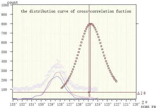
the internal stress in the film. The instrument was using roll-fixed Ψ scan mode, and
cross-correlation function was used to determine the peak.
determined by measuring the interplanar spac-ing correspondspac-ing to different azimuth angles
(Ψ).
When X-ray was incident on the material, the diffraction phenomenon occurred inevitably. The interplanar spacing of the material (d), the
X-ray diffraction angle θ and wavelength λ fol
-lows the Bragg equation: 2d sinθ=λ.
According to the theory of elastic mechanics
and the Bragg equation, the residual stress σ
could be deduced as follows: (K as the X-ray
stress constant of the material, 2θ as the dif
-fraction angle and ρ as crystalline density of the sample): σ=K (ә (2θ)/ә (sin2ψ)).
The film adhesion was measured by a scratch tester, and the film thickness of each sample was measured by a surface profilometer with
computer data acquisition system.
WS-2002 coating adhesion scratch tester was used in this experiment (Figure 2). Acoustic emission detection technology, tangential force detection technology and microcomputer-con-trolled technology were used. Load was contin-uously added to the stylus (diamond indenter) through the automatic loading mechanism while moving the samples, so the stylus could
contact the coating (0.5-20 μm) surface.
Acoustic emission signals, load variations, and tangential force variations were obtained by
each sensor while scratching. After amplifica -tion, they were input into a computer, and the measurement results were drawn into graphs through A/D conversion. Thereout, bonding strength (critical load) Lc of the substrate and
the film (with a thickness of 3-5 microns) could
be obtained.
Tensile strength and elastic modulus test of porous composite membrane MC skeleton sys-tem: There were 6 groups of samples, including
[image:3.612.92.523.75.274.2]φ6 mm MC, Chi-MC and PC-Chi-MC, as well as φ10 mm MC, Chi-MC and PC-Chi-MC respec -Figure 1. Interplanar spacing in different azimuths.
Figure 2. Sketch of scratch measurement.
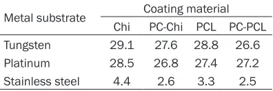
Table 1. Scratch test of metal substrate and
coating film (critical load, N)
Metal substrate Coating material Chi PC-Chi PCL PC-PCL
Tungsten 29.1 27.6 28.8 26.6
Platinum 28.5 26.8 27.4 27.2
[image:3.612.91.287.309.454.2] [image:3.612.90.288.516.582.2]tively. The mechanical properties were mea-sured by a Shimadzu AGS2500ND strong ten-sile machine [23]. Scanning electron microscopy was used to determine the structure of PC-Chi
film and MC multiple layers.
Test methods of mechanical properties: auto-matic displacement booster was used for sam-ple gripping; the center of the straightened coil was the base point; displacement of 10 mm, 15 mm, 20 mm and 25 mm was another point
of action; the lifting speed of the fixture was
250 mm/min. After completion, the change of level two spiral was observed; meanwhile, elec-tron microscopy was used for the observation of porous composite membrane MC system after stretching.
the use of under-film substrate diffraction
me-thod. Corresponding measurements are shown in Figures 3, 4, Tables 2 and 3.
Different substrate materials resulted in differ-ent positions of diffraction peaks; differdiffer-ent coatings on the surface caused to different
changes of diffraction peak’s position while the
diffraction angle was changing, so that the cor-responding bonding stress between the sub-strate and the material could be calculated. The measurement results of each sample are shown in Table 4.
[image:4.612.91.374.72.257.2]The surface of the metal substrate had com-pressive stress (negative stress) itself. It was more meaningful to compare the adhesion of Figure 3. Binding force of phosphorylchline-grafted chitosan coating and
tungsten.
Figure 4. Binding force of chitosan coating and platinum.
Statistical method
SPSS19.0 statistical software was used for statistical analy-sis. Enumeration data were expressed as rate and tested by chi-square test. P<0.05 for the difference was
statistical-ly significant.
Results
Binding ability of film
While coating the stainless steel surface, coating materi-als were clustered as ball shapes and not easy to form a
film. Measurement showed that the thickness of the film
was uneven, and scratch test showed that its load was extremely low, which proved that the adhesion was not good, so, we did not use stain-less steel for further phase structure and internal stress tests. Coating on the surface of tungsten and platinum had good adhesion. See Table 1. Phase structure of coating
and internal stress in film
measured by X-ray stress meter
The stress of the film system
was indirectly characterized by measuring the stress of
[image:4.612.90.364.306.497.2]the films on the same metal substrate material
surface. For example, the tungsten samples had a gradually decreased order of PCL > Chi > PC-PCL > PC-Chi about substrate compressive stress (negative stress). Moreover, taking into account that the change trend of substrate
stress was the opposite of the film, the above
gradual decrease order of substrate
compres-sive stress reflected the gradual increase of film compressive stress or the gradual decrease of film tensile stress, which was helpful for improving the bonding force between the film
and the substrate. Therefore, in comparison,
PC-Chi film had the largest compressive stress,
the least tensile stress and the best binding ability with tungsten.
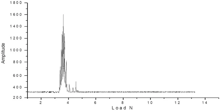
Film adhesion determined by scratch test The bonding strength tests of the metal
sub-strate and coating film were shown in Figures 5 and 6.
The experiment showed that both PC-Chi and Chi materials were able to adhere well to metal surfaces compared with other materials. Detection of porous composite membrane MC skeleton system
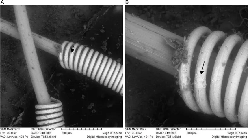
After Chi and PC-Chi were coated on the sur-face of the tungsten MC, there was no obvious change in the appearance of the coil. Scanning electron microscopy showed that the membra-nous material uniformly covered on the surface
of the MC with a thickness of 10 μm or less. Where there was a film shedding, white tung -sten MC body could be seen (indicated by arrow in Figure 7).
Tensile strength and elastic modulus test of porous composite membrane MC skeleton system
As can be seen from the stress-strain curve shown in Figure 8, the shapes of the various MC tensile curves on the basis of the original metallic material were similar and naturally retracted to near the original baseline after stretching, indicating that the materials were good elastomers. However, after several long-distance stretching, their self-contrast showed a decrease in baseline and kurtosis, indicating a decrease of elasticity.
The values recorded in Table 5 were statisti-cally analyzed. Comparing the stress and strain
of the MC with different diameters (φ6 mm and φ10 mm) but with the same composite mem
-brane, it was found that the strain of φ6 mm
coil had greater response to stress (better
elas-ticity), while the diameter of φ10 mm coil
showed poor elasticity. The difference between
the two was statistically significant (P<0.01).
Within the tested elongation range, the
chang-es of level two spiral were not significant in both diameter φ6 mm and φ10 mm MC, and were
considered to be within their elastic limits.
Comparing MC with the same diameter (φ6
[image:5.612.91.288.86.191.2]mm) but with different materials, it was found Table 2. Binding force of pc-chi and tungsten
Measurement results
ψ 0.0° 45.0°
2θρ 124.766° 124.969° Peak count 442 364 Width of half peak 1.87° 2.07° Integral intensity 885 816 Integral width 2.00° 2.24°
Stress σ -243.1 Mpa
[image:5.612.92.289.257.362.2]Note: Ψ as azimuth angle, 2θ as diffraction angle, ρ as crystalline density of the sample, stress σ as residual stress.
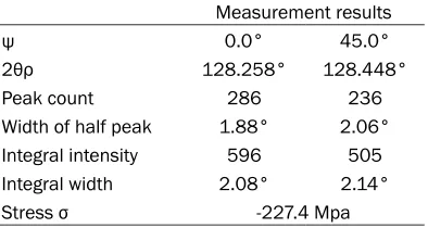
Table 3.Binding force of Chi and platinum
Measurement results
ψ 0.0° 45.0°
2θρ 128.258° 128.448° Peak count 286 236 Width of half peak 1.88° 2.06° Integral intensity 596 505 Integral width 2.08° 2.14°
Stress σ -227.4 Mpa
Note: Ψ as azimuth angle, 2θ as diffraction angle, ρ as crystalline density of the sample, stress σ as residual stress.
Table 4. Measurement results of stress (base stress, MPa)
[image:5.612.90.292.439.503.2]that there was a significant difference (P<0.01)
between Chi-MC and MC; the comparison between PC-Chi-MC and Chi-MC was also with
significant difference (P<0.01). However, stress change showed no significant difference among three materials MC with larger diameter of φ10
mm when the tensile length was less than 20 mm. When the tensile length was longer than
20 mm, there was significant difference
(P<0.01). Discussion
[image:6.612.93.520.78.300.2]At present, MC for embolization therapy is the main method of vascular intervention therapy. MC mainly includes bare coil and biologically-Figure 5. Bonding strength of film and substrate. Changes of phosphorylchline-grafted chitosan and tungsten film-substrate binding force, the critical load Lc was 5.9 N.
[image:6.612.91.521.354.573.2]Figure 7. Scanning electron microscopy graphs of porous composite membrane MC skeleton system. A: X97, B: X293.
modified coil. Due to the bio-inertness of bare coil, inflammation occurs easily with slow scar
formation, which leads to recanalization of
aneurysm. However, the biologically-modified
coil, with good biocompatibility, can accelerate the organization of tissues in aneurysm, and it is the future direction of development. Its
sur-face is modified with extracellular matrix pro -teins, biodegradable polymers, cationic im-
plants and fiber forming cells that can secrete
meter, scratch tester, tensile strength and elas-tic modulus, system performance of different MC was tested to provide some reference for MC study. Experimental study on metal-based coatings revealed that the biodegradable poly-mers including Chi, PC-Chi, PCL and PC-PCL had poor adhesion and film-forming ability on
stainless steel surface, but their binding force with tungsten and platinum was good [27]. The
results of under-film substrate diffraction test
Figure 8. Stress-strain curve of micro-coils with different diameters and materi-als.
growth factors. For example, lactic-co-glycolic
acid-modi-fied platinum coils are used
to increase the postopera-tive recanalization rate [24]; hydrolysable and water-ex- pansion MC is used to in-
crease the filling rate of
embolized vessels [25]; vas-cular endothelial growth
factor-modified MC has bet -ter occlusion for aneurysm [26].
In this paper, MC modified
[image:7.612.92.382.373.551.2]showed that the PC-Chi combined with
tung-sten had the highest under-film compressive
stress, the highest critical load Lc and better binding force, while Chi combined with plati-num had the highest under-film compressive
stress, better critical load Lc and better binding force.
Tensile strength and elastic modulus test of porous composite membrane MC skeleton sys-tem showed that PC-Chi could adhere well to tungsten surface compared with other materi-als. This may be related to the presence of a large number of free amino groups in chitosan, for they increased the adhesion to the metal surface. Within the elastic range, the porous composite membrane MC skeleton system with PC-Chi as coating material had favorable elas-tic stress. Different diameters of MC varied widely in their stress. The smaller the diameter, the larger their tensile stress. Coils with the same diameter, but with different materials had different stress, with the increase of ten-sile length, the difference of stress between different materials also increased.
In summary, PC-Chi can be well attached to the tungsten coil surface. Within the elastic range,
the porous composite membrane MC skeleton system with PC-Chi as coating material has favorable elastic stress and is suitable for prac-tical application. However, there are some shortcomings in this study. For example, based on the binding force between the
biodegrad-able polymer film and the metal substrate, we
only chose tungsten (with better binding force) as coil skeleton system for performance study. We did not carry out research about porous composite membrane platinum coil skeleton system, neither did we compare that with PC-Chi composite membrane tungsten coil skeleton system. The shortcomings will be sup-plemented in subsequent studies.
Acknowledgements
This work was supported by National Natural Science Foundation of China (No. 81271304). Disclosure of conflict of interest
None.
[image:8.612.93.522.83.349.2]Address correspondence to: Yunzhao Jiang, De- partment of Neurosurgery, Wuxi Third People’s Hospital, No. 585 Xingyuan North Road, Wuxi Table 5. Elastic stress changes and baseline shift of different materials MC with different elongation
Diameter Coating Stress change (N) Baseline shift (N)
10 mm 15 mm 20 mm 25 mm 10 mm 15 mm 20 mm 25 mm
6 mm MC 1.50 2.60 4.30 0.05 0.25
1.43 2.55 4.20 0.05 0.32
1.45 2.55 4.00 0.02 0.05 0.35
Chi-MC 0.70 1.75 2.95 4.18 0.05 0.20 0.58
0.73 1.85 3.00 4.13 0.07 0.25 0.63
0.70 1.85 2.85 3.95 0.08 0.28 0.65
PC-Chi-MC 1.40 2.55 3.65 > 4.25 0.06 0.20 0.45 0.80
1.45 2.56 3.58 > 4.25 0.06 0.20 0.48 0.80
1.40 2.45 3.50 4.15 0.08 0.20 0.50 0.80
10 mm MC 0.10 0.20 0.30 0.41 0.04 0.08
0.10 0.21 0.32 0.39 0.06 0.09
0.10 0.20 0.33 0.39 0.06 0.10
Chi-MC 0.13 0.20 0.40 0.50 0.03 0.05 0.08
0.12 0.23 0.39 0.45 0.02 0.03 0.06 0.08
0.12 0.23 0.38 0.48 0.03 0.03 0.08 0.08
PC-Chi-MC 0.10 0.18 0.40 0.50 0.05 0.05 0.07
0.10 0.17 0.40 0.48 0.02 0.07 0.07 0.08
0.11 0.14 0.35 0.48 0.03 0.08 0.08 0.10
214041, Jiangsu Province, China. Tel: +86-021-62108620; Fax: +86-021-+86-021-62108620; E-mail: jiang- [email protected]
References
[1] Poursaid A, Jensen MM, Huo E and Ghande-hari H. Polymeric materials for embolic and chemoembolic applications. J Control Release 2016; 240: 414-433.
[2] Zidan M, Gawlitza M, Metaxas G, Foussier C, Soize S and Pierot L. Endovascular treatment of intracranial aneurysms with barricade coils: feasibility, procedural safety, and immediate postoperative anatomical results. J Neuroradi-ol 2016; 43: 353-357.
[3] Gruber A, Dorfer C and Knosp E. Recurrent and incompletely treated aneurysms. Acta Neuro-chir Suppl 2014; 119: 13-20.
[4] Spiotta AM, Fargen KM, Lena J, Chaudry I, Turner RD, Turk AS, Huddle D, Loy D, Bellon R and Frei D. Initial technical experience with the SMART coil for the embolization of intracranial aneurysms. World Neurosurg 2017; 97: 80-85. [5] Poncyljusz W, Zarzycki A, Zwarzany L and
Burke TH. Bare platinum coils vs. HydroCoil in the treatment of unruptured intracranial aneu-rysms-A single center randomized controlled study. Eur J Radiol 2015; 84: 261-265. [6] Miura Y, Tanemura H, Fujimoto M, Hamada K,
Miyamoto K, Toma N, Imanaka-Yoshida K, Mat-sushima S, Yoshida T, Taki W and Suzuki H. Aneurysm organization effects of gellan sul-fate core platinum coil with Tenascin-C in a simulated clinical setting and the possible mechanism. J Stroke Cerebrovasc Dis 2016; 25: 771-780.
[7] Mitome-Mishima Y, Oishi H, Yamamoto M, Ya-tomi K, Nonaka S, Miyamoto N, Urabe T and Arai H. Differences in tissue proliferation and maturation between Matrix2 and bare plati-num coil embolization in experimental swine aneurysms. J Neuroradiol 2016; 43: 43-50. [8] Witecka A, Yamamoto A, Idaszek J, Chlanda A
and Swieszkowski W. Influence of biodegrad -able polymer coatings on corrosion, cytocom-patibility and cell functionality of Mg-2.0Zn-0.98Mn magnesium alloy. Colloids Surf B Biointerfaces 2016; 144: 284-292.
[9] Zou P, Yang X, Wang J, Li Y, Yu H, Zhang Y and Liu G. Advances in characterisation and bio-logical activities of chitosan and chitosan oli-gosaccharides. Food Chem 2016; 190: 1174-1181.
[10] Ryu JH, Hong S and Lee H. Bio-inspired adhe-sive catechol-conjugated chitosan for biomedi-cal applications: a mini review. Acta Biomater 2015; 27: 101-115.
[11] Patrulea V, Ostafe V, Borchard G and Jordan O. Chitosan as a starting material for wound heal-ing applications. Eur J Pharm Biopharm 2015; 97: 417-426.
[12] Pradhan AK, Rana PK and Sahoo PK. Biode-gradability and swelling capacity of kaolin based chitosan-g-PHEMA nanocomposite hy-drogel. Int J Biol Macromol 2015; 74: 620-626. [13] Yong SK, Shrivastava M, Srivastava P, Kunhi-krishnan A and Bolan N. Environmental appli-cations of chitosan and its derivatives. Rev Environ Contam Toxicol 2015; 233: 1-43. [14] de Y Pozzo L, da Conceição TF, Spinelli A,
Scharnagl N, Pires ATN. Chitosan coatings crosslinked with genipin for corrosion protec-tion of AZ31 magnesium alloy sheets. Carbo-hydr Polym 2018; 181: 71-77.
[15] Schlenoff JB. Zwitteration: coating surfaces with zwitterionic functionality to reduce non-specific adsorption. Langmuir 2014; 30: 9625-9636.
[16] He L, Huang L, Zhang S, Chen Y and Luo X. Poly(epsilon-caprolactone) modification via surface initiated atom transfer radical polym-erization with bio-inspired phosphorylcholine. Mater Sci Eng C Mater Biol Appl 2017; 77: 45-51.
[17] Bedair TM, Cho Y, Kim TJ, Kim YD, Park BJ, Joung YK and Han DK. Reinforcement of inter-facial adhesion of a coated polymer layer on a cobalt-chromium surface for drug-eluting stents. Langmuir 2014; 30: 8020-8028. [18] Lekjing S. A chitosan-based coating with or
without clove oil extends the shelf life of cooked pork sausages in refrigerated storage. Meat Sci 2016; 111: 192-197.
[19] Bellucci D, Bianchi M, Graziani G, Gambardella A, Berni M, Russo A and Cannillo V. Pulsed electron deposition of nanostructured bioac-tive glass coatings for biomedical applications. Ceramics International 2017; 43: 62-67. [20] Seuss S, Lehmann M and Boccaccini AR.
Alter-nating current electrophoretic deposition of antibacterial bioactive glass-chitosan compos-ite coatings. Int J Mol Sci 2014; 15: 12231-12242.
[21] Wang TJ, Wang IJ, Lu JN and Young TH. Novel chitosan-polycaprolactone blends as potential scaffold and carrier for corneal endothelial transplantation. Mol Vis 2012; 18: 255-264. [22] Huang D, Zuo Y, Zou Q, Zhang L, Li J, Cheng L,
Shen J and Li Y. Antibacterial chitosan coating on nano-hydroxyapatite/polyamide66 porous bone scaffold for drug delivery. J Biomater Sci Polym Ed 2011; 22: 931-944.
behavior of tubular collagen type I templates. Acta Biomater 2017; 59: 234-242.
[24] Piotin M, Pistocchi S, Bartolini B and Blanc R. Intracranial aneurysm coiling with PGLA-coat-ed coils versus bare platinum coils: long-term anatomic follow-up. Neuroradiology 2012; 54: 345-348.
[25] Maleux G, Deroose C, Fieuws S, Van Cutsem E, Heye S, Bosmans H and Verslype C. Prospec-tive comparison of hydrogel-coated microcoils versus fibered platinum microcoils in the pro -phylactic embolization of the gastroduodenal artery before yttrium-90 radioembolization. J Vasc Interv Radiol 2013; 24: 797-803; quiz 804.
[26] Wang Q, Gao Y, Sun X, Ji B, Cui X, Liu Y, Zheng T, Chen C, Jiang X, Zhu A and Quan D. Accelera-tion of aneurysm healing by P(DLLA-co-TMC)-coated coils enabling the controlled release of vascular endothelial growth factor. Biomed Mater 2014; 9: 045004.