Original article
Haploinsufficiency of A20 impairs
protein–protein interactome and leads
into caspase-8-dependent enhancement
of NLRP3 inflammasome activation
Kristiina Rajamäki,1 Salla Keskitalo,2 Mikko Seppänen,3 Outi Kuismin,4
Paula Vähäsalo,5 Luca Trotta,6 Antti Väänänen,7 Virpi Glumoff,8 Paula Keskitalo,5
Riitta Kaarteenaho,9,10 Airi Jartti,11 Nina Hautala,12 Päivi Jackson,12
Dan C Nordström,13 Janna Saarela,6 Timo Hautala,14 Kari K Eklund,15,16,17
Markku Varjosalo2,18
To cite: rajamäki K, Keskitalo S, Seppänen M, et al. Haploinsufficiency of a20 impairs protein– protein interactome and leads into caspase-8-dependent enhancement of nlrP3 inflammasome activation. RMD Open 2018;4:e000740. doi:10.1136/ rmdopen-2018-000740
►Prepublication history and additional material for this paper are available online. to view these files, please visit the journal online (http:// dx. doi. org/ 10. 1136/ rmdopen- 2018- 000740).
Kr and SK contributed equally. tH, KKe and MV contributed equally.
received 11 June 2018 revised 3 august 2018 accepted 7 September 2018
For numbered affiliations see end of article.
Correspondence to
Dr Markku Varjosalo; markku. varjosalo@ helsinki. fi © author(s) (or their employer(s)) 2018. re-use permitted under cc BY-nc. no commercial re-use. See rights and permissions. Published by BMJ.
AbstrAct
Objectives TNFAIP3 encodes a20 that negatively regulates nuclear factor kappa light chain enhancer of activated B cells (nF-κB), the major transcription factor coordinating inflammatory gene expression. TNFAIP3
polymorphisms have been linked with a spectrum of inflammatory and autoimmune diseases and, recently, loss-of-function mutations in a20 were found to cause a novel inflammatory disease ‘haploinsufficiency of a20’ (Ha20). Here we describe a family with Ha20 caused by a novel TNFAIP3 loss-of-function mutation and elucidate the upstream molecular mechanisms linking Ha20 to dysregulation of nF-κB and the related inflammasome pathway.
Methods nF-κB activation was studied in a mutation-expressing cell line using luciferase reporter assay. Physical and close-proximity protein–protein interactions of wild-type and TNFAIP3 p.(lys91*) mutant a20 were analysed using mass spectrometry. nF-κB -dependent transcription, cytokine secretion and inflammasome activation were compared in immune cells of the Ha20 patients and control subjects.
Results the protein–protein interactome of p.(lys91*) mutant a20 was severely impaired, including interactions with proteins regulating nF-κB activation, Dna repair responses and the nlr family pyrin domain containing 3 (nlrP3) inflammasome. the p.(lys91*) mutant a20 failed to suppress nF-κB signalling, which led to increased nF-κB -dependent proinflammatory cytokine transcription. Functional experiments in the Ha20 patients’ immune cells uncovered a novel caspase-8-dependent mechanism of nlrP3 inflammasome hyperresponsiveness that mediated the excessive secretion of interleukin-1β and interleukin-18.
Conclusions the current findings significantly deepen our understanding of the molecular mechanisms underlying Ha20 and other diseases associated with reduced a20 expression or function, paving the way for future therapeutic targeting of the pathway.
InTROduCTIOn
Tumour necrosis factor alpha-induced protein 3 (TNAP3, also known as A20), encoded by the TNFAIP3 gene, is a key negative regulator of nuclear factor kappa B (NF-κB) activation downstream a large number of immune receptors, including the TNF and Toll/interleukin(IL)-1 receptor families and the T and B cell antigen recep-tors.1–5 A20 achieves this via its dual function as a ubiquitin-editing enzyme: an N-terminal ovarian tumour (OTU) domain removes activating K63-linked polyubiquitin from key
Key messages
What is already known about this subject? ► a20 is a negative regulator of nuclear factor kappa B
(nF-κB) having a key role in immune activation and autoimmunity.
► loss-of-function mutations in a20 are reported to lead to an inflammatory disease termed haploinsuf-ficiency of a20 (Ha20).
What does this study add?
► We report a novel p.(lys91*) a20 mutation and use it as a model to uncover molecular mechanisms relat-ed to the role of a20 in Ha20 development. ► the p.(lys91*) fails to suppress nF-κB activity and
has severely impaired protein interactome com-pared with the wild-type a20 in HeK cells, and caus-es aberrant nlrP3 inflammasome rcaus-esponsivencaus-ess in patient peripheral blood mononuclear cells.
How might this impact on clinical practice? ► this study significantly deepens our understanding
on the role of a20 in rheumatic diseases and may provide novel approaches for the development of therapeutic approaches.
on May 7, 2020 by guest. Protected by copyright.
http://rmdopen.bmj.com/
protein–protein interactions, whereas a C-terminal zinc finger domain catalyses the attachment of K48-linked polyubiquitin to induce proteasomal degradation.3 6 7 In addition, A20 inhibits NF-κB activation via a non-cata-lytic mechanism involving its binding to linear ubiquitin on NF-κB essential modulator (NEMO).8 9 The mRNA expression of A20 is NF-κB-inducible,10 thus generating a negative feedback loop to attenuate NF-κB responses.
Recently Zhou et al reported six families with hetero-zygous loss-of-function mutations in the TNFAIP3 gene that led to haploinsufficiency of A20 (HA20) and caused an early-onset Behçet-like disease.11 Studies in mouse models have previously demonstrated a crucial role for A20 in autoinflammation and autoimmunity as mice with a germline deletion of Tnfaip3 spontaneously develop severe multiorgan inflammation and tissue damage resulting in perinatal death,12 and cell-specific ablation of Tnfaip3 results in diverse symptoms of immune dysreg-ulation.13 14 Similar to Tnfaip3 deficiency in mice,12–15 human HA20 led to exaggerated NF-κB responses and increased secretion of NLR family pyrin domain containing 3 (NLRP3) inflammasome target cytokines.11 Additional reports from these16 and further identified patients17–21 have expanded the clinical spectrum of HA20 to comprise diseases such as autoimmune lymph-oproliferative syndrome, systemic juvenile arthritis, psori-atic arthritis, Crohn’s disease and Hashimoto’s thyroiditis. The increasing number of diseases associated with A20 has emphasised its role in inflammatory diseases and in autoimmunity and raised the possibility that mutations in
TNFAIP3 may be an unexpectedly common underlying factor in the pathogenesis of rheumatic and other auto-immune diseases.
We describe here a family with polyautoimmunity and autoinflammatory symptoms caused by a novel hetero-zygous TNFAIP3 p.(Lys91*) loss-of-function mutation resulting in haploinsufficiency. Our aim was to further elucidate the upstream molecular mechanisms linking HA20 to dysregulated NF-κB activation and NLRP3 inflammasome responses. Thus, we analysed protein– protein interactions of wild-type (wt) and p.(Lys91*) A20 in HEK cells and performed functional experiments in patients’ immune cells to study NLRP3 inflammasome responses. As A20 is rapidly emerging as a key regulator of immune activation and autoimmunity in humans, the detailed characterisation of A20 effector mechanisms paves the way for future therapeutic targeting of the pathway.
MaTeRIals and MeTHOds Genetic analyses
DNA samples were extracted from total peripheral blood using standard methods. Whole-exome sequencing and data analysis were performed as previously described.22 23 All the common (frequencies >0.01 in the general popu-lation) and non-coding variants were discarded. We
the inferred autosomal-dominant inheritance pattern (figure 1A). The variant identified in the TNFAIP3
gene (Ensembl ENSG00000118503:ENST00000433680: exon2:c.A271T: p.(Lys91*) was confirmed using PCR and capillary electrophoresis.
Creating a20 expressing Flp-In 293 T-Rex cell lines
A20 mutant plasmids were created using QuickChange Site-Directed Mutagenesis kit (Agilent Technologies) to wt TNFAIP3 obtained from the human Orfeome collec-tion (Horizon Discovery). TNFAIP3 constructs were further subcloned into N-terminal MAC-tag Gateway destination vector.24 All generated constructs were confirmed with direct sequencing. The MAC-tagged expression constructs were transfected into Flp-In 293 T-REx cells (Invitrogen, Life Technologies) with FuGENE HD (Promega). The cells were grown according to manufacturer’s instructions under selec-tion with Hygromycin B (Thermo Fisher Scientific) to create stable cell lines. Expression of stable trans-genes was confirmed by western blotting using anti-he-magglutin (HA) primary (Biolegend, MMS-101R) and horseradish peroxidase-conjugated secondary anti-body.
luciferase assay
For reporter assays, HEK293 cells were transfected with in total 50 ng A20 mutant or wt MAC-tagged construct and GFP or empty MAC-tag-vector,24 40 ng of NF-κB lucif-erase reporter plasmid (Cignal 45), and 1.4 ng Renilla luciferase reporter plasmid (pRL-SV40). Transfected cells were stimulated with indicated amounts of tumour necrosis factor alpha TNF-α (R&D Systems) for 16 hours, lysed with 1× passive lysis buffer (Promega) and luciferase assays performed with Dual-Glo Luciferase Assay System according to manufacturer’s instructions (Promega).
affinity purification and mass spectrometry
For each pull-down approximately 5×10⁷ cells (5×15 cm dishes) were induced with 2 µg/mL tetracycline (Thermo Fisher Scientific) for 24 hours (for BioID additional 50 µM biotin was added). After induction, cells were washed, harvested, pelleted by centrifugation, snap-frozen and stored at −80°C until analysis. Affinity purification of AP-MS and BioID was performed as described in Turunen
et al 25 and Heikkinen et al,26 respectively. Sample prepa-ration for mass spectrometry and LC-MS/MS analysis on Orbitrap Elite ETD mass spectrometer was performed as in Heikkinen et al.26 After peptide reconstitution, 4 µL was analysed from both Strep-Tag and BioID-samples.
Culture and stimulation of peripheral blood mononuclear cells (PBMCs)
PBMCs were isolated by density gradient centrifugation in Ficoll-Paque PLUS (GE Healthcare) and seeded at 1.5×10⁶ cells/mL in Macrophage-SFM medium supplemented with penicillin-streptomycin or in RPMI 1640 supplemented with GlutaMAX, penicillin-streptomycin and 10% FBS (all
on May 7, 2020 by guest. Protected by copyright.
Beta strand Helix Turn
92 263
OTU rep.1 rep.2
286 317 324 356 790
K91X
A
1 2
1 2
K91X -/- K91X NA
K91X
NA K91X -/- K91X -/+ K91X
-/+
K91X -/+
3 4
1
Fatal liver failure
Autoimmune thyroiditis
Autoimmune thyroiditis juvenile idiopathic arthritis
Autoimmune thyroiditis Psoriasis, psoriatic arthropathy
Atrophic gastritis Lung disease
G
ener
ations
I
II
III
B
C
A20
A20 A20-K91X
OTU domain -N
-C
-N zf
1 zf2 zf3 zf4 zf5 zf6 zf7 381 – 416
472 – 507 515 – 548 601 – 636 651 – 686
[image:3.595.146.441.47.452.2]710 – 745 756 – 790
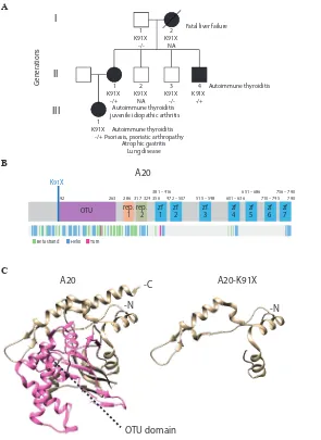
Figure 1 Phenotypic and protein-level characteristics of a family with newly identified TNFAIP3 c.A271T: p.(Lys91*) mutation.
(A) A pedigree showing the TNFAIP3 p.(Lys91*) mutation carriers (–/+, solid symbols) in three generations (I–III). Autoimmune thyroiditis was diagnosed in all carriers (I-2, II-1, II-4, III-1) at an early age. In addition, the index patient (II-1) suffered from psoriasis, articular symptoms, atrophic gastritis, severe autoinflammatory lung reaction, anaemia, genital papillomatosis and repeated genital HSV infections. Daughter of the index (III-1) was diagnosed with polyarticular juvenile idiopathic arthritis at the age of 4 years, and mother of the index (I-2) died of liver failure at the age of 46 years. (B) Schematic illustration of A20 structure showing the protein domains (upper panel) and the folding (lower panel). Position of the novel p.(Lys91*) mutation is indicated in blue. (C) 3D structure models of wild-type and mutant p.(Lys91*) A20 (based on PDB accession 5LRX). OTU domain is highlighted with pink. HSV, herpes simplex virus; OTU, ovarian tumour.
from Gibco). PBMCs were allowed to rest for a minimum of 3 hours before starting the stimulations with 1 µg/mL lipopolysaccharides (LPS) from Escherichia coli O111:B4 (Sigma), 1 µg/mL Pam3Cys-SKKKK (Pam3Cys; EMC micr-ocollections), 10 µg/mL polyinosinic-polycytidylic acid (poly(I:C); InvivoGen) and 5 mM adenosine 5′ -triphos-phate (ATP; Sigma) for the indicated times. Inhibitors Z-YVAD-FMK (20 µM; R&D Systems), Z-IETD-FMK (5 µM; BD Bioscience) and necrostatin-1 (30 µM; Enzo Life Sciences) were added to the cells simultaneously with LPS in the 16-hour stimulations or 30 min before LPS in the LPS 6 hour+ATP 45 min stimulations.
Measurement of cytokine secretion
TNF-α and the mature, cleaved forms of interleukin (IL)-1β and IL-18 were detected from PBMC culture media supernatants using Human TNF-α DuoSet ELISA, Human IL-1β/IL-1F2 DuoSet ELISA and Human Total IL-18 DuoSet ELISA (all from R&D Systems).
Quantitative real-time PCR
RNA was isolated from PBMCs using RNeasy Plus Mini Kit (Qiagen) and cDNA synthesised with iScript kit (Bio-Rad). Quantitative real-time PCR was performed from 10 ng of cDNA per reaction using LightCycler480 SYBR Green I
on May 7, 2020 by guest. Protected by copyright.
http://rmdopen.bmj.com/
primer sequences are listed in online supplementary table 1. Relative gene expression was calculated using the 2(-ΔΔCt) method, normalising to the geometric mean of expression of two housekeeping genes, ribosomal protein lateral stalk subunit P0 and beta-2 microglobulin.
Monocyte caspase-1 activity measurement
Aliquots of EDTA-anticoagulated blood were incubated for 6 hours at +37°C under gentle rocking with or without 10 ng/mL LPS (Sigma). ATP at 5 mM (Sigma) was added for 20 min, followed by 2-hour incubation at +37°C with a cell-permeable, irreversibly binding FAM-FLICA fluores-cent caspase-1 substrate (ImmunoChemistry Technolo-gies). After red blood cell lysis and staining with the mono-cyte marker anti-human CD14-APC (Miltenyi Biotec) for 15 min at +4°C, the cells were analysed using BD Accuri C6 flow cytometer (BD Biosciences; instrument maintained by the Biomedicum Flow Cytometry Unit).
ResulTs
Identification of the mutation and the clinical manifestations of the patients
Exome sequencing of the index patient (II-1) identi-fied a novel nonsense mutation in the exon 2 of the
TNFAIP3 gene: c.A271T: p.(Lys91*) (ENSG000001185 03:ENST00000433680) (figure 1A,B). The mutation is not listed in Genome Aggregation Database (gnomAD, Cambridge, Massachusetts, USA) and was predicted to be pathogenic according to the ACMG Standards and Guide-lines.27 The TNFAIP3 p.(Lys91*) mutation was confirmed in the patient’s genomic DNA and screened in the other members of the family. Two other affected family members (II-4, III-1) were observed to also harbour the mutation, (I-2 is an obligate carrier). The clinical manifestations of the carriers included autoimmune thyroiditis, which was diagnosed in all carriers (I-2, II-1, II-4, III-1) at an early age. The index patient suffered from psoriasis, articular symp-toms, atrophic gastritis, severe inflammatory lung reaction, anaemia and repeated genital HSV infections. Daughter of the index was diagnosed with polyarticular juvenile idiopathic arthritis at the age of 4. See also the legend of
figure 1A.
effect of the TnFaIP3 p.(lys91*) mutation on protein structure and expression
The 3D protein structures of wt and p.(Lys91*) A20 were visualised by processing the PDB (http://www. rcsb. org28) accession number 5LRX (chain A29 with Chimera,30 which yielded a severely truncated protein lacking the complete OTU domain (figure 1C). To further analyse the expres-sion and possible physical and functional changes caused by the TNFAIP3 p.(Lys91*) mutation, stable Flp-In 293 T-REx cell lines expressing wt and p.(Lys91*) A20 were created. The wt HA-tagged A20 was expressed in western blot, but the p.(Lys91*) mutant was detected only with an N-terminal tag (figure 2A), suggesting that only an N-ter-minal fragment is produced and no alternative start codons
confirmed that only the N-terminal region was detectable in the p.(Lys91*) mutant (figure 2B). The expression levels of the N-terminal peptides/protein of the wt and p.(Lys91*) mutant A20 were highly comparable (figure 2B).
a20 p.(lys91*) mutant fails to suppress nF-κB activation and shows severely impaired protein–protein interactome
To analyse the possible functional consequences of the p.(Lys91*) mutation and the loss of OTU domain on the A20 functions, we monitored the NF-κB pathway activity using a luciferase-based reporter assay. The wt A20 dose-de-pendently inhibited TNF-α induced NF-κB pathway activa-tion, whereas the p.(Lys91*) mutant failed to do so, clearly suggesting a haploinsufficiency (figure 2C).
The physical effects of the p.(Lys91*) mutation were further analysed by proteome-wide affinity purification mass spectrometry (AP-MS) (see online supplementary figure 1 for details). These analyses revealed that the p.(Lys91*) mutant completely loses physical interactions with BRCC3, TRAD1, WRIP1, TRAF2, BIRC2, F175B, USP9X and HUWE1, with several additional weakened interactions (figure 2D). To obtain more insight into the molecular mechanisms of A20 and especially on the p.(Lys91*) muta-tion, we further quantitatively analysed the functional and close-proximity interactions using BioID MS24 (see online supplementary figure 1 for details). This analysis revealed complete loss of interactions with AZI2, TNIP1, TBK1, CYLD, CCD50, SLK, ITSN1, HAUS6, OGA, SH3K1, WDCP, TRIM9, PDLI1, FHL3, KPYM, COPE, UBA1, CE152 and PGK1. Many more interactions were severely diminished (figure 2D). Majority of the detected interactions of wt A20 were novel (figure 3). Functional annotation revealed extensive interactions with ubiquitinylation-related, prote-asome-related and NF-κB-related proteins, as well as with proteins involved, for example, in cell fate decisions (14-3-3 proteins), DNA repair (53BP1 complex) and glycolysis (figure 3). Of these, the interactions with ubiquitinyla-tion-related and NF-κB-related proteins were most severely affected in the A20 p.(Lys91*) mutant.
Blood immune cells of TnFaIP3 p.(lys91*) carriers display strongly increased secretion of inflammasome-controlled cytokines Il-1β and Il-18
The NF-κB pathway and A20 have been shown to play regu-latory roles in the activation of NLRP3 inflammasome, a pathway triggering caspase-1-mediated proteolytic matu-ration and secretion of proinflammatory cytokines IL-1β
and IL-18.15 31 32 Excessive NLRP3-dependent cytokine secretion was demonstrated in HA20,11 yet the mecha-nism(s) linking reduced A20 to NLRP3 inflammasome acti-vation in patient cells were not elucidated. We stimulated PBMCs of the HA20 patients with Toll-like receptor (TLR) 4 agonist LPS, with or without a subsequent ATP pulse; both stimuli trigger the NLRP3 inflammasome in mono-cytes via distinct mechanisms and with different kinetics.31 We found elevated secretion of IL-1β and IL-18 in PBMCs of the TNFAIP3 p.(Lys91*) carriers in response to LPS +ATP-induced ‘canonical’ (figure 4A,B; left panels) as well
on May 7, 2020 by guest. Protected by copyright.
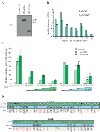
Figure 2 The p.(Lys91*) mutant A20 fragment is expressed but shows severe impairment of protein–protein interactions and of inhibitory activity on nuclear factor (NF)-κB signaling. (A) Expression profile analysis of wild type (wt) A20 and the p.(Lys91*) mutant in HEK293 cells. The wt protein is detected with both as N- and C-terminally tagged constructs, whereas the p.(Lys91*) mutant is only detected as N-terminally tagged illustrating that no alternative start site is used and only short N-terminal fragment is produced. (B) Quantitative mass spectrometry-based proteomics analysis shows that the N-terminal part of the wt A20 and p.(Lys91*) mutant are expressed in similar levels as shown by the quantitative abundance of the peptides derived from the N-termini. (C) wt A20 dose-dependently inhibits the NF-κB signaling induced by tumour necrosis factor alpha (TNF-α) (light green=10 ng/mL and dark green=25 ng/mL), whereas the p.(Lys91*) mutant shows clearly diminished inhibitory activity suggesting haploinsufficiency. The asterisks represent the significance values; *<0.05, ** <0.01, *** <0.001. (D) Affinity purification mass spectrometry (AP-MS) (upper panel) and BioID (lower panel) analysis of wt and p.(Lys91*) mutant A20 show clear differences in their stable protein–protein interactions and BioID analysis displays differential molecular context of the p.(Lys91*) mutant compared with the wt A20. (The size and the colour gradient shows the relative abundance of the interacting prey protein detected in association with A20, red typeface designates the complete loss of the corresponding interactions and yellow >50% decrease compared with the wt A20).
as LPS-induced ‘alternative’ pathway of NLRP3 inflam-masome activation (figure 4A,B; right panels). To control for potential confounding factors in FBS preparations, PBMC cytokine secretion was analysed both in commer-cial serum-free monocyte-macrophage medium (figure 4) and in RPMI containing 10% FBS (online supplementary figure 2), yielding similar results. Also, TNF-α secretion in
response to LPS, Pam3Cys and poly(I:C) was increased in the patients (figure 4C, online supplementary figure 2C. We further studied the levels of IL-1β and IL-18 in patients’ plasma by ELISA and found increased levels of circulating IL-18 in TNFAIP3 p.(Lys91*) carriers compared with the age-matched and sex-matched controls (online supple-mentary figure 3A), whereas IL-1β was not detectable.
on May 7, 2020 by guest. Protected by copyright.
http://rmdopen.bmj.com/
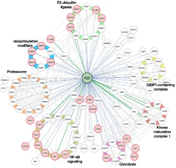
Figure 3 Interactome analysis reveals known and novel interactions for A20. Affinity purification mass spectrometry and BioID analysis of A20 identified 98 high-confidence protein–protein interactions (known interactions are shown with green lines, novel interaction detected on this study with blue and known prey–prey interactions with a dashed line). The interacting proteins are grouped based on their molecular functions/complexes. Interactions, which are lost with the p.(Lys91*) mutation, are illustrated with red node fill colour.
The increased nlRP3 inflammasome response in TnFaIP3 p.(lys91*) carriers is dependent on enhanced pro-Il-1β transcription and caspase-8 activity, but not on RIPK1 To elucidate the mechanism of altered NLRP3 inflam-masome activation in the HA20 patients’ PBMCs, we analysed the mRNA expression of inflammasome pathway components and cytokines and the effect of inflammasome-related inhibitors on cytokine secretion. PBMCs of patient III-1 displayed strongly increased baseline expression of pro-IL-1β and NLRP3 receptor, whereas patient II-1 cells showed only moderate changes (online supplementary figure 3B). After LPS stimulation, PBMCs from both patients showed slightly elevated levels of pro-IL-1β mRNA and NLRP3 receptor expression remained moderately elevated only in patient III-1 (figure 5A). LPS-induced expression of NF-κB target cytokines IL-6, TNF-α and IL-8 was elevated in patient III-1, and IL-6 also in patient II-1 (figure 5B). Receptor interacting serine/threonine-protein kinase 1 (RIPK1) inhibitor necrostatin-1 did not affect IL-1β or IL-18 secretion in the patients’ PBMCs (figure 4A,B), although it normalised the excessive TNF-α response to 16-hour LPS treatment (figure 4C, right panel).
As expected, both canonical and alternative NLRP3 inflammasome responses were efficiently blocked by the caspase-1 inhibitor Z-YVAD-FMK (figure 4A,B). However, levels of active caspase-1 in monocytes after whole blood inflammasome stimulation were increased only in patient II-1 (figure 5C), supporting a more indirect enhancement of inflammasome complex func-tion. Remarkably, caspase-8 inhibitor Z-IETD-FMK effi-ciently suppressed IL-1β and IL-18 secretion in PBMCs of the TNFAIP3 p.(Lys91*)-positive patients, particu-larly after canonical NLRP3 inflammasome activation (figure 4A,B).
dIsCussIOn
HA20 is a recently described familial autoinflamma-tory disease originally reported to manifest as an early-onset Behçet-like disease,11 yet the range of known clin-ical manifestations is rapidly expanding.16–21 Here we studied patients with HA20 caused by a novel heterozy-gous TNFAIP3 p.(Lys91*) mutation resulting in severe truncation and production of an N-terminal protein fragment with highly limited functional capacity. The
on May 7, 2020 by guest. Protected by copyright.
Figure 4 Inflammasome-dependent and inflammasome-independent proinflammatory cytokine secretion is elevated in peripheral blood mononuclear cells (PBMCs) of TNFAIP3 p.(Lys91*) carriers. PBMCs from TNFAIP3 p.(Lys91*) mutation carriers, patients 1 (II-1, woman, 30 years) and 2 (III-1, female, 8 years), were compared with sex-matched and age-matched controls 1 and 2, respectively. (A–C, left panels) The cells were primed with LPS for 6 hours, followed by treatment with ATP for 45 min. (A–C, right panels) The cells were treated with TLR agonists for 16 hours. Inhibitors necrostatin-1 (nec), Z-YVAD-FMK (yvad) and Z-IETD-FMK (ietd), or solvent control dimethyl sulfoxide (dmso) were added as indicated and cytokines were detected from PBMC culture supernatants by ELISAs. IL, interleukin; TNF, tumour necrosis factor; LPS, lipopolysaccharide.
patients exhibit an unusual combination of early-onset autoimmune diseases, autoinflammatory symptoms and immunodeficiency, reflecting the widespread functions of A20 in controlling inflammation. To gain further insight into the molecular mechanism underlying the development of this complex disease, we focused on scrutinising two pathways strongly linked with A20 function; the NF-κB pathway and the NLRP3 inflam-masome.
Mutations causing HA20 were previously reported to increase TNF-α-induced IKKα/IKKβ and MAPK phos-phorylation and activation of NF-κB via defective removal of K63-linked ubiquitin from RIPK1, TNF receptor-as-sociated factor 6 and NEMO.11 18 19 As expected, also the A20 p.(Lys91*) mutant was defective in suppressing TNF-α-induced NF-κB activation. We employed state-of-the-art proteomics tools to comprehensively map global
changes in A20 protein–protein interactions caused by the p.(Lys91*) mutation. In agreement with A20 ubiquitin-editing functions, the results revealed loss of physical interactions with ubiquitin ligases (BIRC2, TRAF2, HUWE1), deubiquitinases (BRCC3, USP9X) and other ubiquitinylation modulators (WRIP1 and F175B/ABRX2). BIRC2/3 and TRAF2/3 form a regu-latory complex that suppresses apoptotic functions of caspase-8 to enforce canonical (inflammatory) NF-κB signalling, while blocking non-canonical NF-κB activa-tion.33 34 A20 modulates this balance by inducing TRAF2 degradation to suppress canonical NF-κB activation, and by binding c-IAP1/2 to support their caspase-8-in-hibiting function while blocking their noncanon-ical NF-κB-inhibiting function.35–37 Thus, the loss of A20 p.(Lys91*) interaction with BIRC2 and TRAF2 is compatible with enhancement of caspase-8 activity and
on May 7, 2020 by guest. Protected by copyright.
http://rmdopen.bmj.com/
0 20000 40000 60000 80000
Active
caspase-1
0 100 200 300 400 500
IL1B
Control 2 Patient 2 Control 1
Patient 1
0 10 20 30 40 50
NLRP3
0 1 2
ASC
0 2 4 6 8
LPS (h)
0 2 4 6 8
0 2 4 6
CASP1
LPS (h)
A
0 50 100 150 200
TNF
A
0 2000 4000 6000
IL6
0 2 4 6 8
LPS (h)
0 50 100 150
0 2 4 6
IL8
LPS (h)
0 05 10 15 20
IL18
LPS
ATP -- +- ++
Control 1 Patient 1 Control 2 Patient 2
Control 2 Patient 2 Control 1
Patient 1
B
[image:8.595.129.479.51.383.2]C
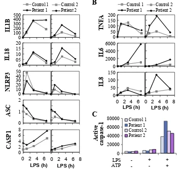
Figure 5 Expression and activation of the NLRP3 inflammasome is altered in peripheral blood mononuclear cells (PBMCs) of
TNFAIP3 p.(Lys91*) carriers. Samples from TNFAIP3 p.(Lys91*) mutation carriers, patients 1 (II-1, woman, 30 years) and 2 (III-1, female, 8 years), were compared with sex-matched and age-matched controls 1 and 2, respectively. Relative expression of (A) NLRP3 inflammasome components and target cytokines and (B) inflammasome-independent proinflammatory cytokines was analysed in PBMCs by quantitative PCR and normalised against housekeeping gene expression (arbitrary units). (C) Whole blood was stimulated with LPS and ATP, followed by the detection of NLRP3 inflammasome-triggered caspase-1 activity in monocytes using a fluorescent FLICA probe; the data are presented as median fluorescence intensity (MFI).
canonical NF-κB signalling. In turn, BRCC3, F175B/ ABRX2, WRIP1 and HUWE1 have specific functions in DNA damage responses,38–41 which could be a signifi-cant finding regarding the association of reduced A20 levels with systemic lupus characterised by autoanti-bodies against nuclear antigens.14 Moreover, BRCC3 and F175B/ABRX2 are also components of the BRISC deubiquitinase complex that cleaves K63-linked ubiq-uitin and regulates mitotic spindle assembly,42 type I interferon signalling43 and NLRP3 inflammasome activation.44 Also, functional close-proximity protein– protein interactions of the A20 p.(Lys91*) mutant, particularly those related to ubiquitinylation and NF-κB signalling, were severely impaired, whereas interac-tions with proteasome, 53BP1-containing DNA repair complex and 14-3-3 regulatory proteins remained partially functional.
The roles of A20 in regulation of the NLRP3 inflam-masome, both NF-κB-dependent and NF-κ B-inde-pendent, are just beginning to be uncovered.15 32 Mice deficient in myeloid A20 develop erosive poly-arthritis specifically due to poor control of NLRP3
inflammasome activation by A20.15 Moreover, increased NLRP3-dependent secretion of IL-1β and IL-18 was reported in HA20 patients,11 17 yet the upstream mech-anism(s) linking this effect to A20 in patient immune cells remain unknown. We found increased IL-1β and IL-18 secretion in PBMCs of TNFAIP3 p.(Lys91*)-posi-tive patients in response to both canonical and alterna-tive NLRP3-activating stimuli. In mouse macrophages, A20 suppresses the NF-κB-induced mRNA expression of NLRP3 receptor and pro-IL-1β, but also blocks RIPK1/3-dependent pro-IL-1β ubiquitination that supports the inflammasome-mediated processing into mature IL-1β.15 32 Our patients’ PBMCs showed elevated LPS-induced transcription of pro-IL-1β, but NLRP3 receptor expression was not consistently increased. The RIPK1 inhibitor necrostatin-1 that reduced aber-rant IL-1β secretion in Tnfaip3-deficient mouse macro-phages32 had no effect on IL-1β nor IL-18 secretion in the patients’ PBMCs, which was, instead, strongly suppressed by caspase-8 inhibition. This suggests a novel caspase-8-dependent mechanism linking reduced A20 function to enhanced NLRP3 inflammasome
on May 7, 2020 by guest. Protected by copyright.
activation. The finding is in line with the increased caspase-8 activity reported in Tnfaip3-deficient mouse cells32 45 and with the caspase-8-suppressing functions of A20,37 45 and further supported by the loss of phys-ical interaction between A20 p.(Lys91*) mutant and BIRC2, which is required for caspase-8 inhibition by A20.37 Previous studies suggest that alongside its apop-totic functions caspase-8 facilitates the priming and activation of NLRP3 inflammasome via multiple mech-anisms.46 In addition, the loss of interaction between A20 p.(Lys91*) mutant and the BRCC3-containing BRISC complex suggests a further novel NLRP3-regu-lating function of A20 that is lost in haploinsufficient cells, as BRCC3 deubiquitinates the NLRP3 receptor directly promoting its activation.44
In summary, we report here a TNFAIP3 p.(Lys91*) loss-of-function mutation leading to HA20 and autoim-mune/inflammatory symptoms. We use this mutation as a model to uncover novel molecular mechanisms related to the role of A20 in disease development. We defined, for the first time, the global physical and func-tional interactome of wt A20 and found severely dimin-ished interactions in the p.(Lys91*) mutant. Moreover, we discovered a novel caspase-8-dependent mechanism linking HA20 to hyperactivation of the NLRP3 inflam-masome in patient immune cells. Collectively, these data greatly deepen our understanding of the molecular mechanisms underlying the clinical disease phenotype of HA20 and other diseases associated with reduced A20 expression or function. We believe that defining the physical and functional interaction changes in (m) any diseases is highly usable for understanding these diseases better on the molecular level and believe such studies will be seen in immense quantity in the near future.
author affiliations
1clinicum, Faculty of Medicine, University of Helsinki, Helsinki, Finland
2institute of Biotechnology, Helsinki institute of life Science (HiliFe), University of
Helsinki, Helsinki, Finland
3immunodeficiency Unit, inflammation center and rare Diseases center, children's
Hospital, Helsinki University and Helsinki University Hospital, Helsinki, Finland
4Department of clinical genetics, PeDegO research Unit, Medical research center,
Oulu University Hospital and University of Oulu, Oulu, Finland
5Department of Pediatrics, PeDegO research Unit, Medical research center, Oulu
University Hospital and University of Oulu, Oulu, Finland
6institute for Molecular Medicine Finland, Helsinki institute of life Science (HiliFe),
University of Helsinki, Helsinki, Finland
7Department of infection control, lapland central Hospital, rovaniemi, Finland 8research Unit of Biomedicine, University of Oulu, Oulu, Finland
9respiratory Diseases, research Unit of internal Medicine, University of Oulu, Oulu,
Finland
10Medical research center Oulu, Oulu University Hospital, Oulu, Finland 11Department of radiology, Oulu University Hospital, Oulu, Finland
12Department of Ophthalmology, PeDegO research Unit, Medical research center,
Oulu University Hospital and University of Oulu, Oulu, Finland
13Department of Medicine and rehabilitation, Helsinki University Hospital and
Helsinki University, Helsinki, Finland
14research Unit of internal Medicine, University of Oulu and Oulu University
Hospital, Oulu, Finland
15Department of rheumatology, inflammation center, Helsinki University and
Helsinki University Hospital, Helsinki, Finland
16research institute, invalid Foundation , Helsinki, Finland 17Orton Orthopaedic Hospital, Helsinki, Finland
18Proteomics Unit, institute of Biotechnology, University of Helsinki, Helsinki, Finland
acknowledgements Sini Miettinen is thanked for expert mass spectrometry skills, and Xiaonan liu for help with chimera.
Contributors Kr and SK designed and performed the experiments, analysed data, prepared the figures and wrote the manuscript. tH identified the index patient. lt and JS performed variant identification. Vg organised clinical samples. MS, OK, PV, aV, PK, rK, aJ, nH, PJ, Dcn and tH provided medical care and sampling. KKe and MV participated in designing experiments, data analysis, figure preparation, and manuscript writing and submission.
Funding the study was supported by Finska läkaresällskapet (KKe, Dn), the canadian institutes of Health research (tHc 135230; KKe), the Stockmann foundation (KKe) and the Paulo foundation (Kr). Molecular graphics and analyses were performed with the UcSF chimera package. chimera is developed by the resource for Biocomputing, Visualization, and informatics at the University of california, San Francisco (supported by nigMS P41-gM103311). exome sequencing of the index patient was conducted in the institute for Molecular Medicine Finland (FiMM) technology centre.
Competing interests none declared.
Patient consent not required.
ethics approval ethical committee for clinical Science of Oulu University Hospital, and coordinating ethics committee of the Hospital District of Helsinki and Uusimaa.
Provenance and peer review not commissioned; externally peer reviewed.
Open access this is an open access article distributed in accordance with the creative commons attribution non commercial (cc BY-nc 4.0) license, which permits others to distribute, remix, adapt, build upon this work non-commercially, and license their derivative works on different terms, provided the original work is properly cited, appropriate credit is given, any changes made indicated, and the use is non-commercial. See: http:// creativecommons. org/ licenses/ by- nc/ 4. 0/
RefeRences
1. Song HY, Rothe M, Goeddel DV. The tumor necrosis factor-inducible zinc finger protein A20 interacts with TRAF1/TRAF2 and inhibits NF-kappaB activation. Proc Natl Acad Sci U S A 1996;93:6721–5. 2. Heyninck K, Beyaert R. The cytokine-inducible zinc finger protein
A20 inhibits IL-1-induced NF-kappaB activation at the level of TRAF6. FEBS Lett 1999;442–147–50.
3. Wertz IE, O'Rourke KM, Zhou H, et al. De-ubiquitination and ubiquitin ligase domains of A20 downregulate NF-kappaB signalling. Nature 2004;430:694–9.
4. Boone DL, Turer EE, Lee EG, et al. The ubiquitin-modifying enzyme A20 is required for termination of toll-like receptor responses. Nat Immunol 2004;5:1052–60.
5. Düwel M, Welteke V, Oeckinghaus A, et al. A20 negatively regulates T cell receptor signaling to NF-kappaB by cleaving malt1 ubiquitin chains. J Immunol 2009;182:7718–28.
6. Shembade N, Ma A, Harhaj EW. Inhibition of NF-kappaB signaling by A20 through disruption of ubiquitin enzyme complexes. Science 2010;327:1135–9.
7. Wertz IE, Newton K, Seshasayee D, et al. Phosphorylation and linear ubiquitin direct A20 inhibition of inflammation. Nature 2015;528:370–5.
8. Skaug B, Chen J, Du F, et al. Direct, noncatalytic mechanism of IKK inhibition by A20. Mol Cell 2011;44:559–71.
9. Verhelst K, Carpentier I, Kreike M, et al. A20 inhibits LUBAC-mediated NF-κB activation by binding linear polyubiquitin chains via its zinc finger 7. Embo J 2012;31:3845–55.
10. Krikos A, Laherty CD, Dixit VM. Transcriptional activation of the tumor necrosis factor alpha-inducible zinc finger protein, A20, is mediated by kappa B elements. J Biol Chem 1992;267:17971–6. 11. Zhou Q, Wang H, Schwartz DM, et al. Loss-of-function mutations
in TNFAIP3 leading to A20 haploinsufficiency cause an early-onset autoinflammatory disease. Nat Genet 2016;48:67–73.
12. Lee EG, Boone DL, Chai S, et al. Failure to regulate TNF-induced NF-kappaB and cell death responses in A20-deficient mice. Science 2000;289:2350–4.
13. Ma A, Malynn BA. A20: linking a complex regulator of ubiquitylation to immunity and human disease. Nat Rev Immunol 2012;12:774–85. 14. Das T, Chen Z, Hendriks RW, et al. A20/Tumor necrosis factor
α-induced protein 3 in immune cells controls development of
on May 7, 2020 by guest. Protected by copyright.
http://rmdopen.bmj.com/
Front Immunol 2018;9:104.
15. Vande Walle L, Van Opdenbosch N, Jacques P, et al. Negative regulation of the NLRP3 inflammasome by A20 protects against arthritis. Nature 2014;512:69–73.
16. Aeschlimann FA, Batu ED, Canna SW, et al. A20 haploinsufficiency (HA20): clinical phenotypes and disease course of patients with a newly recognised NF-kB-mediated autoinflammatory disease. Ann Rheum Dis 2018;77:728–35.
17. Kadowaki T, Ohnishi H, Kawamoto N, et al. Haploinsufficiency of A20 causes autoinflammatory and autoimmune disorders. J Allergy Clin Immunol 2018;141:1485–8.
18. Takagi M, Ogata S, Ueno H, et al. Haploinsufficiency of TNFAIP3 (A20) by germline mutation is involved in autoimmune lymphoproliferative syndrome. J Allergy Clin Immunol 2017;139:1914–22.
19. Duncan CJA, Dinnigan E, Theobald R, et al. Early-onset autoimmune disease due to a heterozygous loss-of-function mutation in TNFAIP3 (A20). Ann Rheum Dis 2018;77:783–6.
20. Ohnishi H, Kawamoto N, Seishima M, et al. A Japanese family case with juvenile onset Behçet's disease caused by TNFAIP3 mutation. Allergol Int 2017;66:146–8.
21. Shigemura T, Kaneko N, Kobayashi N, et al. Novel heterozygous C243Y A20/TNFAIP3 gene mutation is responsible for chronic inflammation in autosomal-dominant Behçet's disease. RMD Open 2016;2:e000223.
22. Trotta L, Hautala T, Hämäläinen S, et al. Enrichment of rare variants in population isolates: single AICDA mutation responsible for hyper-IgM syndrome type 2 in Finland. Eur J Hum Genet 2016;24:1473–8. 23. Trotta L, Martelius T, Siitonen T, et al. ADA2 deficiency: Clonal
lymphoproliferation in a subset of patients. J Allergy Clin Immunol 2018;141:1534–7.
24. Liu X, Salokas K, Tamene F, et al. An AP-MS- and BioID-compatible MAC-tag enables comprehensive mapping of protein interactions and subcellular localizations. Nat Commun 2018;9:1188.
25. Turunen M, Spaeth JM, Keskitalo S, et al. Uterine leiomyoma-linked MED12 mutations disrupt mediator-associated CDK activity. Cell Rep 2014;7:654–60.
26. Heikkinen T, Kämpjärvi K, Keskitalo S, et al. Somatic MED12 nonsense mutation escapes mRNA decay and reveals a motif required for nuclear entry. Hum Mutat 2017;38:269–74. 27. Richards S, Aziz N, Bale S, et al. Standards and guidelines
for the interpretation of sequence variants: a joint consensus recommendation of the american college of medical genetics and genomics and the association for molecular pathology. Genet Med 2015;17:405–23.
28. Rose PW, Prlić A, Altunkaya A, et al. The RCSB protein data bank: integrative view of protein, gene and 3D structural information. Nucleic Acids Res 2017;45–D271–81.
29. Mevissen TET, Kulathu Y, Mulder MPC, et al. Molecular basis of Lys11-polyubiquitin specificity in the deubiquitinase cezanne. Nature 2016;538:402–5.
visualization system for exploratory research and analysis. J Comput Chem 2004;25:1605–12.
31. He Y, Hara H, Núñez G. Mechanism and regulation of NLRP3 inflammasome activation. Trends Biochem Sci 2016;41:1012–21. 32. Duong BH, Onizawa M, Oses-Prieto JA, et al. A20 restricts
ubiquitination of pro-interleukin-1β protein complexes and suppresses NLRP3 inflammasome activity. Immunity 2015;42:55–67.
33. Wang CY, Mayo MW, Korneluk RG, et al. NF-kappaB antiapoptosis: induction of TRAF1 and TRAF2 and c-IAP1 and c-IAP2 to suppress caspase-8 activation. Science 1998;281:1680–3.
34. Zarnegar BJ, Wang Y, Mahoney DJ, et al. Noncanonical NF-kappaB activation requires coordinated assembly of a regulatory complex of the adaptors cIAP1, cIAP2, TRAF2 and TRAF3 and the kinase NIK. Nat Immunol 2008;9:1371–8.
35. Hymowitz SG, Wertz IE. A20: from ubiquitin editing to tumour suppression. Nat Rev Cancer 2010;10:332–41.
36. Yamaguchi N, Oyama M, Kozuka-Hata H, et al. Involvement of A20 in the molecular switch that activates the non-canonical NF-ćB pathway. Sci Rep 2013;3:2568.
37. Yamaguchi N, Yamaguchi N. The seventh zinc finger motif of A20 is required for the suppression of TNF-α-induced apoptosis. FEBS Lett 2015;589:1369–75.
38. Chen X, Arciero CA, Wang C, et al. BRCC36 is essential for ionizing radiation-induced BRCA1 phosphorylation and nuclear foci formation. Cancer Res 2006;66:5039–46.
39. Zhang J, Cao M, Dong J, et al. ABRO1 suppresses tumourigenesis and regulates the DNA damage response by stabilizing p53. Nat Commun 2014;5:5059.
40. Bish RA, Myers MP. Werner helicase-interacting protein 1 binds polyubiquitin via its zinc finger domain. J Biol Chem 2007;282:23184–93.
41. Markkanen E, van Loon B, Ferrari E, et al. Regulation of oxidative DNA damage repair by DNA polymerase ć and MutYH by cross-talk of phosphorylation and ubiquitination. Proc Natl Acad Sci U S A 2012;109:437–42.
42. Yan K, Li L, Wang X, et al. The deubiquitinating enzyme complex BRISC is required for proper mitotic spindle assembly in mammalian cells. J Cell Biol 2015;210:209–24.
43. Zheng H, Gupta V, Patterson-Fortin J, et al. A BRISC-SHMT complex deubiquitinates IFNAR1 and regulates interferon responses. Cell Rep 2013;5:180–93.
44. Py BF, Kim MS, Vakifahmetoglu-Norberg H, et al. Deubiquitination of NLRP3 by BRCC3 critically regulates inflammasome activity. Mol Cell 2013;49:331–8.
45. Jin Z, Li Y, Pitti R, et al. Cullin3-based polyubiquitination and p62-dependent aggregation of caspase-8 mediate extrinsic apoptosis signaling. Cell 2009;137:721–35.
46. Man SM, Kanneganti TD. Converging roles of caspases in inflammasome activation, cell death and innate immunity. Nat Rev Immunol 2016;16:7–21.
on May 7, 2020 by guest. Protected by copyright.