Original Article
Evaluation of non-invasive methods in hepatitis B virus
(HBV)-infected patients with normal liver function
Xu Li1, Shuang Zheng2, Hongqin Xu1,3, Pujun Gao1
Departments of 1Hepatology and 2Radiology, The First Hospital of Jilin University, Changchun, Jilin, China; 3Jilin
Province Key Laboratory of Infectious Disease, Laboratory of Molecular Virology, Changchun, Jilin, China
Received April 16, 2017; Accepted December 14, 2017; Epub February 15, 2018; Published February 28, 2018
Abstract: Background: We aimed to compare the diagnostic performance of four non-invasive methods in detecting liver injury in chronic hepatitis B (CHB) patients with persistently normal liver function, and to develop a combined algorithm to improve current assays for liver injury evaluation. Materials and Methods: We obtained the results of
aspirate aminotransferase (AST)-to-platelet ratio index (APRI), fibrosis index based on the 4 factors (FIB-4), red blood
cell distribution width (RDW) to platelets ratio (RPR) and Fibroscan in a cohort of 58 CHB patients who underwent liver biopsy (LB). Then we combined serum markers with Fibroscan and evaluated their performance in detecting
significant fibrosis. Results: The areas under the receiver operating characteristic curve (AUROC) for APRI, FIB-4, RPR and Fibroscan were 0.696, 0.708, 0.736 and 0.756 respectively for detecting significant liver fibrosis. An improved performance was obtained by combining Fibroscan and RPR, AUROC of whom was 0.836, reducing liver biopsy required for detection of significant fibrosis in 37.9% of patients with an accuracy of 95%. Conclusion: APRI, FIB-4, RPR and Fibroscan show moderate clinical value for detecting significant fibrosis in chronic HBV patients with
normal liver function. The combination of Fibroscan and RPR improved diagnostic performance and reduced the number of patients who need liver biopsy.
Keywords: Aspirate aminotransferase-to-platelet ratio index, fibrosis index based on the 4 factors, red blood cell
distribution width to platelets ratio, Fibroscan, Hepatitis B virus, normal liver function
Introduction
Hepatitis B virus (HBV) infection is a serious global health problem. Approximately 240 mil-lion people are chronically infected with HBV, and are at high risk for developing cirrhosis and hepatocellular carcinoma (HCC) [1]. And chron-ic HBV infection causes a wide spectrum of clinical manifestations. The guidelines for man-aging HBV infection do not recommend antivi-ral treatment for patients with normal alanine aminotransferase (ALT) levels. There is an incr- easing concern about accuracy to reflect liver injury by ALT. Recent studies have found sig- nificant necroinflammation and/or fibrosis in 28-37% of HBV patients with persistent normal ALT [2, 3]. High risk for advanced fibrosis and cirrhosis in patients can be delayed or prevent-ed by antiviral therapy if significant fibrosis is detected earlier [4, 5].
Liver biopsy (LB) based evaluation of liver his-tology has been the most reliable detection of
liver inflammation, stage of liver fibrosis and treatment efficiency. However LB is invasive, expensive and inconvenient for patients. Fur- thermore, sampling error occurs if histological alterations are non-homogenous, and varia-tions between intra and inter-observers pose another challenge [6-9].
HBV-infected patients with elevated ALT [15, 16]. Red blood cell distribution width (RDW) to platelets ratio (RPR) is an index of platelet mor-phology, which can predict significant fibrosis and cirrhosis in CHB patients with relatively high accuracy [17, 18].
However, few studies evaluated detection of significant fibrosis in HBV patients with PNALT using Fibroscan, APRI, FIB-4 and RPR. The aim of this study was to evaluate these four non-invasive assays for detecting significant liver fibrosis using liver histology as a reference in CHB patients with persistent normal liver func-tion. We also tested the models that combin- ed different non-invasive methods to evaluate whether the combination can decrease the rate of LB and have a high accuracy for detecting significant fibrosis in these patients.
Materials and methods
Patients
We retrospectively enrolled 58 patients who were previously diagnosed as CHB and under-went LB from January 2010 to December 2015 in the First Hospital of Jilin University. The pur-pose of LB was to assess severity of liver fibro-sis and inflammation and to determine whether antiviral treatment is warranted. Inclusion crite-ria were as follows: 1) Patients were hepatitis B surface antigen (HBsAg) positive for at least 6 months and chronic hepatitis was confirmed by histology, HBV DNA positive (HBV DNA > 50 IU/ ml by PCR); 2) Liver function was normal, as determined by ALT, AST, and total bilirubin (TBIL), for at least two consecutive measure-ments over a period of 6 months [19], the upper limit of normal (ULN) of ALT, AST and TBIL are 50 U/L for male/40 U/L for female, 40 U/L for male/35 U/L for female and 30 µ mol/L, respectively; 3) Age of patients ≥ 18 years old. Exclusion criteria were: 1) Patients who were co-infected with hepatitis C virus or human immunodeficiency virus (HIV), auto-immune he- patitis, Wilson disease, hepatocelluar carcino-ma, chronic ethanol consumption (> 20 g/day for female, > 40 g/day for male). 2) A history of previous antiviral treatment or a history of malignant disorder. The study was conducted in accordance with the Declaration of Helsinki and was approved by the Ethics Committee in the First Hospital of Jilin University.
Liver biopsy examination
Percutaneous LB was using an 18 G biopsy needle. LB was repeated for patients whose liver tissues were shorter than 13 mm to mini-mize the influence of the length of liver speci-men on the accuracy of diagnosis. The liver tissues were fixed in formalin, embedded in paraffin and stained with hematoxylin and eosin. Every specimen was assessed by one pathologist who was blinded to clinical data. The Scheuer scoring system was used to evalu-ate the samples. Fibrosis was therefore scored on a scale from 0 to 4 (S0 = no fibrosis, S1 = enlarged portal tracts, S2 = periportal or porto-portal septa, S3 = fibrosis with architectural distortion, S4 = cirrhosis). Significant fibrosis was defined as Scheuer score ≥ 2 [20].
Liver stiffness measurement by Fibroscan
Fibroscan (Fibroscan, Echosens SA, Paris, Fra- nce) was performed by an experienced opera-tor (more than 100 determinations in patients with chronic liver diseases) within 1 week be- fore LB. Per instruction, 10 validated measure-ments were taken for each patient. The median value (in kilopascal, kPa) was considered repre-sentative of the liver elastic modulus. The mea-surement was considered reliable unless the interquartile range/median was < 30% and success rate was > 60% as suggested by the manufacturing company [21].
Non-invasive biomarkers
Fasting blood serum samples were used for laboratory tests within one week prior to LB. Platelet (PLT), RDW, AST, ALT, TBIL and gamma-glutamyl transpeptidase (GGT) were analyzed. HBV DNA level was determined with real time PCR with low detection limit of 50 IU/ml. HBsAg, HBsAb, hepatitis B e-antigen (HBeAg), HBe, hepatitis core B antibody (HBcAb), anti-HCV and anti-HIV were also assessed. APRI, FIB-4 and RPR were calculated using the prin-ciple reported formulas, as APRI = AST (U/L)/ [PLT (109/L)], FIB-4 = Age (years) × AST (U/L)/
[PLT (109/L) × ALT1/2 (U/L)], RPR = RDW/PLT
(109/L).
Cut-off values
the cut-off value of Fibroscan was derived from the Manual [12-14]. Patients with APRI > 0.5, FIB-4 > 1.45, RPR > 0.1 or Fibroscan ≥ 7.3 kPa were considered to have no significant fibrosis.
Evaluation of the combined non-invasive methods
Evaluation of non-invasive methods was desi- gned for better detection of significant fibrosis by combining serum markers with Fibroscan, and the results were compared with histology diagnosis. In algorithm APRI+Fibroscan, if the results of APRI and Fibroscan were consist (APRI < 0.5 and Fibroscan < 7.3 kPa or APRI >
0.5 and Fibroscan ≥ 7.3 kPa), the patients were classified as significant fibrosis free(< S2) or to be positive for significant fibrosis (≥ S2). If the results were inconsistent, the patients were considered to need LB. The other algorithms were similar with APRI+Fibroscan.
Statistical analysis
All statistical analyses were processed using SPSS, version 22.0 (SPSS Inc, Chicago, IL). Co- ntinuous variables are expressed as mean ± standard deviations (SD) and the difference was determined by student t test. Categorical variables are expressed as percentages and the difference was determined by the chi-square test. A P value < 0.05 was considered significant. The diagnostic performance of AP- RI, FIB-4, RPR and Fibroscan and their combi-nations were assessed by sensitivity, specifici-ty, positive likelihood ratio (LR+), negative likeli-hood ratio (LR-), diagnostic accuracy, receiver operating characteristic curves (ROC) and AU- ROC. McNemar’s test was performed to evalu-ate the agreement between the combination with best accuracy and liver histology.
Results
Characteristics of enrolled patients
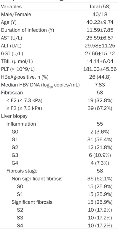
A total of 58 patients were enrolled in this study. The demographic, laboratory, Fibroscan results and histological features are summa-rized in Table 1. Median years of HBV infection were 11.6, 44.8% of the patients were HBeAg positive. There was no significant difference in the years of HBV infection (P = 0.74) between HBeAg positive and negative patients. Signi- ficant fibrosis was detected in 37.3% patients with normal liver function, 8.6% of whom had liver cirrhosis.
Performance of individual non-invasive meth-ods
[image:3.612.91.290.93.482.2]The diagnostic performance of the four non-invasive methods were evaluated for their abil-ity to detect significant fibrosis (Scheuer fibro-sis stage ≥ S2). Cut-off values for APRI, FIB-4, RPR and Fibroscan were 0.5, 1.45, 0.1 and 7.3 kPa respectively. RPR had the highest specific-ity, LR+, diagnostic accuracy and the lowest LR- (Table 2). ROC curves of these methods for detecting significant fibrosis were presented in Figure 1, AUROC of APRI, FIB-4, RPR and Table 1. Characteristics of demography,
labo-ratory index and histology
Variables Total (58)
Male/Female 40/18
Age (Y) 40.22±9.74
Duration of infection (Y) 11.59±7.85
AST (U/L) 25.59±6.87
ALT (U/L) 29.58±11.25
GGT (U/L) 27.66±15.72
TBIL (µ mol/L) 14.14±6.04
PLT (× 10^9/L) 181.03±45.56
HBeAg-positive, n (%) 26 (44.8)
Median HBV DNA (log10 copies/mL) 7.83
Fibroscan 58
< F2 (< 7.3 kPa) 19 (32.8%) ≥ F2 (≥ 7.3 kPa) 39 (67.2%)
Liver biopsy
Inflammation 55
G0 2 (3.6%)
G1 31 (56.4%)
G2 12 (21.8%)
G3 6 (10.9%)
G4 4 (7.3%)
Fibrosis stage 58
Non-significant fibrosis 36 (62.1%)
S0 15 (25.9%)
S1 15 (25.9%)
Significant fibrosis 15 (25.9%)
S2 10 (17.2%)
S3 10 (17.2%)
S4 10 (17.2%)
Fibroscan were 0.696, 0.708, 0.736 and 0.756,
[image:4.612.90.403.99.173.2]respectively. Fibroscan agreed with LB results in 14 cases and RPR agreed with LB in 22 cases. The best Table 2. Performance of APRI, FIB-4, RPR and Fibroscan in excluding
significant fibrosis in livers
Method Cut-off Sensitivity (%) Specificity (%) LR+ LR- (%)DA AUROC (95% CI)
APRI 0.5 31.8 86.1 2.29 0.79 65.5 0.696 (0.55-0.84)
FIB-4 1.45 59.1 80.6 3.05 0.51 72.4 0.708 (0.56-0.86)
RPR 0.1 45.5 91.7 5.48 0.02 74.1 0.736 (0.62-0.89)
Fibroscan 7.3 44.4 86.4 3.26 0.64 60.3 0.756 (0.60-0.87)
APRI: aspirate aminotransferase to platelet ratio index; FIB-4: fibrosis index based on the 4 factors; RPR: red blood cell distribution width to platelets ratio; LR+: positive likelihood ratio; LR-: negative likelihood ratio; DA: diagnostic accuracy; AUROC: area under receiver operating characteristic curve.
Optimal combination algorithms
[image:4.612.92.397.238.486.2]After individual evalua-tions of the four non-invasive methods, we further explored pair- ed combination of th- ese methods. AUROC for predicting significa- nt fibrosis were 0.779, 0.804, 0.836, respec-tively (Table 3). RPR+ Fibroscan had the high-est AUROC (Figure 2), meanwhile the combi-nation improved the pe- rformance to a better level than that achieved by RPR or Fibroscan al- one (ROCs: 0.836 vs 0.736 or 0.756). RPR+ Fibroscan was the be- st performing algorithm with sensitivity, speci-ficity, LR+, LR-, accuracy of 100%, 93.3%, 14.94, 0, 95.5%, respectively. However the consisten-cy of RPR and Fibroscan was mediocre, with only 37.9% of the patients. Next, we performed Mc- Nemar’s test to compa- re the agreement be- tween RPR+Fibroscan and LB. There was an almost perfect agree-ment (Kappa = 0.899, P < 0.001) between RPR+ Fibroscan and LB. RPR and Fibroscan ag- reed on the diagnosis of the stage of fibrosis in 22 patients (37.9%). Th- ere was only 1 discor-dant case who were classified as ≥ S2 by RPR-4+Fibroscan but < S2 by LB. Among the remaining 36 patients, Figure 1. AUROC curves for fibrosis scores (APRI, FIB-4, RPR, Fibroscan) at different
stages of fibrosis: S0-1 vs S ≥ 2, with an area under the receiver operating charac
-teristic curve (AUROC) of 0.696, 0.708, 0.736 and 0.756 respectively.
Table 3. Performance of combinations of APRI, FIB-4, RPR and Fibroscan in detecting significant fibrosis in livers
Method Sensitivity (%) Specificity (%) LR+ LR- DA (%) AUROC (95% CI)
APRI+Fibroscan 70.0 82.4 3.98 0.25 77.8 0.779 (0.65-0.91) FIB-4+Fiberscan 91.7 80.0 4.59 0.10 85.2 0.804 (0.68-0.93) RPR+Fibroscan 100.0 93.3 14.93 0.00 95.5 0.836 (0.73-0.94)
[image:4.612.89.402.576.638.2]diagnostic algorithm for these six combinations is reported in Figure 3.
ods tested in this study was compared based on AUROC and diagnosis accuracy. RPR showed a better diagnostic accuracy among the three serum biomarkers. The best AUROC of these serum biomarkers was 0.736, diagnostic accu-racy was 74.1%, which was lower than the val-ues reported by previous studies [15, 16, 22]. The differences can be explained by varying degrees of fibrosis since AUROC is highly influ-enced by the prevalence of fibrosis stage. The percentage of S0-1 detected in our study was higher compared to other studies. Furthermore, the diagnostic performance of serum biomark-ers in PNALT patients was not as good as in individuals with elevated ALT levels [23-26]. To our knowledge, this study represents the first validation of RPR diagnostic value in HBV pa- tients with normal liver function, and RPR sh- owed the best diagnostic performance among other serum biomarkers.
[image:5.612.92.373.70.308.2]We explored different non-invasive detection combinations of serum markers and Fibroscan to reduce liver biopsy in CHB patients. Among the four non-invasive methods and paired com-binations, the combination of Fibroscan and RPR revealed the best diagnostic performance for significant fibrosis. This combination scan Figure 2. AUROC curves for fibrosis scores (APRI+Fibroscan,
FIB-4+Fibros-can, RPR+Fibroscan) at different stages of fibrosis: S0-1 vs S ≥ 2, with an area under the receiver operating characteristic curve (AUROC) of 0.779,
0.804, 0.836, respectively. Af, APRI+Fibroscan; Ff, FIB-4+Fibroscan; Rf, RPR+Fibroscan.
Figure 3. Proposed algorithms for patient manage-ment using the combination of RPR and Fibroscan
for diagnosing liver fibrosis stages.
Discussion
In this study, we evaluated clinical values of four non-invasive methods, APRI, FIB-4, RPR and Fibroscan as well as paired combinations in a co- hort of 58 CHB patients with persistent normal liver func-tion. We found that a combi- nation of RPR+Fibroscan can detect significant fibrosis with a good accuracy of 95.5%, as confirmed by liver biopsy.
[image:5.612.91.288.390.638.2]meth-covered 37.9% of the cohort patients, 95.5% of patients with S ≥ 2 were detected by Fibroscan+RPR, as well as histology. RPR con-sists of two common hematological parameters and is easy to calculate. Fibroscan presents no risk compared to liver biopsy. Thus, this non-invasive method has potential for wide clinical applications.
To increase the diagnostic accuracy, many com-binations of non-invasive methods have been proposed. The resultant diagnostic accuracies ranged from 84 to 94%, and reduced LB by 48 to 77% in patients with significant fibrosis [27-31]. Since elevated ALT influences the accuracy of serum biomarkers and Fibroscan [25, 26, 32, 33], only patients with normal liver function were selected in this study. We expect that our results could be validated in large cohorts. Furthermore, this study is the first to report that RPR evaluated in patients with normal liver function and used in a combination with anoth-er non-invasive method, resulting in impressive accuracy.
There are a few limitations in this study. The enrolled patients in a retrospective study may have been subjected to a biased selection. And the number of patients was small because only HBV-infected patients with normal liver func-tion were included. The main findings need to be verified in large cohorts.
In conclusion, we evaluated the diagnostic per-formance of four non-invasive methods in HBV patients with normal liver function, and found similar diagnostic performances among these four. A combination of RPR and Fibroscan can improve the performance of each individual non-invasive method, and help predict signifi-cant fibrosis in 40% of patients without LB.
Disclosure of conflict of interest
None.
Address correspondence to: Pujun Gao, Depart-
ment of Hepatology, The First Hospital of Jilin Uni-versity, Jilin UniUni-versity, No. 71, Xinmin Street,
Chang-chun 130021, Jilin, China. Tel :+86 187 4301 2103; E-mail: [email protected]
References
[1] Sarin SK, Kumar M, Lau GK, Abbas Z, Chan HL, Chen CJ, Chen DS, Chen HL, Chen PJ, Chien RN, Dokmeci AK, Gane E, Hou JL, Jafri W, Jia J,
Kim JH, Lai CL, Lee HC, Lim SG, Liu CJ,
Locar-nini S, Al Mahtab M, Mohamed R, Omata M,
Park J, Piratvisuth T, Sharma BC, Sollano J, Wang FS, Wei L, Yuen MF, Zheng SS and Kao
JH. Asian-Pacific clinical practice guidelines on
the management of hepatitis B: a 2015 up-date. Hepatol Int 2016; 10: 1-98.
[2] Lai M, Hyatt BJ, Nasser I, Curry M and Afdhal
NH. The clinical significance of persistently
normal ALT in chronic hepatitis B infection. J Hepatol 2007; 47: 760-767.
[3] Gui HL, Wang H, Yang YH, Wu YW, Zhou HJ, Guo SM, Lin LY, Wang L, Cai W, Chen R, Guo Q, Zhou
XQ, Bao SS and Xie Q. Significant histopathol -ogy in Chinese chronic hepatitis B patients with persistently high-normal alanine
amino-transferase. J Viral Hepat 2010; 17 Suppl 1:
44-50.
[4] Marcellin P, Gane E, Buti M, Afdhal N, Sievert W, Jacobson IM, Washington MK, Germanidis G, Flaherty JF, Aguilar Schall R, Bornstein JD, Kitrinos KM, Subramanian GM, McHutchison JG, Heathcote EJ. Regression of cirrhosis dur-ing treatment with tenofovir disoproxil fuma-rate for chronic hepatitis B: a 5-year open-label follow-up study. Lancet 2013; 381: 468-475. [5] Liaw YF. Impact of therapy on the long-term
outcome of chronic hepatitis B. Clin Liver Dis 2013; 17: 412-423.
[6] Bedossa P, Dargere D and Paradis V. Sampling variability of liver fibrosis in chronic hepatitis C.
Hepatology 2003; 38: 1449-1457.
[7] Regev A, Berho M, Jeffers LJ, Milikowski C, Mo-lina EG, Pyrsopoulos NT, Feng ZZ, Reddy KR, Schiff ER. Sampling error and intraobserver variation in liver biopsy in patients with chronic
HCV infection. Am J Gastroenterol 2002; 97:
2614-2618.
[8] Maharaj B, Maharaj RJ, Leary WP, Cooppan
RM, Naran AD, Pirie D, Pudifin DJ. Sampling variability and its influence on the diagnostic
yield of percutaneous needle biopsy of the liv-er. Lancet 1986; 1: 523-525.
[9] Afdhal NH. Diagnosing fibrosis in hepatitis C: is
the pendulum swinging from biopsy to blood tests? Hepatology 2003; 37: 972-974.
[10] Patel K, Bedossa P and Castera L. Diagnosis of
liver fibrosis: present and future. Semin Liver
Dis 2015; 35: 166-183.
[11] Sharma S, Khalili K and Nguyen GC.
Non-inva-sive diagnosis of advanced fibrosis and cirrho -sis. World J Gastroenterol 2014; 20: 16820-16830.
[12] Forns X, Ampurdanes S, Llovet JM, Aponte J, Quinto L, Martinez-Bauer E, Bruguera M,
[13] Wai CT, Greenson JK, Fontana RJ, Kalbfleisch
JD, Marrero JA, Conjeevaram HS and Lok AS. A simple noninvasive index can predict both
sig-nificant fibrosis and cirrhosis in patients with
chronic hepatitis C. Hepatology 2003; 38: 518-526.
[14] Sterling RK, Lissen E, Clumeck N, Sola R, Cor-rea MC, Montaner J, S Sulkowski M, Torriani FJ, Dieterich DT, Thomas DL, Messinger D, Nelson
M; APRICOT Clinical Investigators. Develop -ment of a simple noninvasive index to predict
significant fibrosis in patients with HIV/HCV
coinfection. Hepatology 2006; 43: 1317-1325. [15] Teshale E, Lu M, Rupp LB, Holmberg SD,
Moor-man AC, Spradling P, Vijayadeva V, Boscarino
JA, Schmidt MA, Gordon SC; CHeCS Investiga-tors. APRI and FIB-4 are good predictors of the
stage of liver fibrosis in chronic hepatitis B: the Chronic Hepatitis Cohort Study (CHeCS). J Viral
Hepat 2014; 21: 917-920.
[16] Mallet V, Dhalluin-Venier V, Roussin C, Bour
-liere M, Pettinelli ME, Giry C, Vallet-Pichard A,
Fontaine H and Pol S. The accuracy of the
FIB-4 index for the diagnosis of mild fibrosis in
chronic hepatitis B. Aliment Pharmacol Ther 2009; 29: 409-415.
[17] Taefi A, Huang CC, Kolli K, Ebrahimi S and Pa -tel M. Red cell distribution width to pla-telet
ra-tio, a useful indicator of liver fibrosis in chronic
hepatitis patients. Hepatol Int 2015; 9: 454-460.
[18] Chen B, Ye B, Zhang J, Ying L and Chen Y. RDW to platelet ratio: a novel noninvasive index
for predicting hepatic fibrosis and cirrhosis in chronic hepatitis B. PLoS One 2013; 8:
e68780.
[19] Marcellin P, Levy S and Erlinger S. Therapy of hepatitis C: patients with normal aminotra- nsferase levels. Hepatology 1997; 26 Suppl 1: 133S-136S.
[20] Scheuer PJ. Classification of chronic viral hepa -titis: a need for reassessment. J Hepatol 1991; 13: 372-374.
[21] Jia J, Hou J, Ding H, Chen G, Xie Q, Wang Y, Zeng M, Zhao J, Wang T, Hu X and Schuppan D. Transient elastography compared to serum
markers to predict liver fibrosis in a cohort of
Chinese patients with chronic hepatitis B. J Gastroenterol Hepatol 2015; 30: 756-762. [22] Sebastiani G, Vario A, Guido M and Alberti A.
Sequential algorithms combining non-invasive markers and biopsy for the assessment of liver
fibrosis in chronic hepatitis B. World J Gastro -enterol 2007; 13: 525-531.
[23] Amorim TG, Staub GJ, Lazzarotto C, Silva AP, Manes J, Ferronato Mda G, Shiozawa MB, Nar-ciso-Schiavon JL, Dantas-Correa EB, Schiavon
Lde L. Validation and comparison of simple
noninvasive models for the prediction of liver
fibrosis in chronic hepatitis C. J Viral Hepat
[24] Colletta C, Smirne C, Fabris C, Toniutto P,
Ra-petti R, Minisini R and Pirisi M. Value of two
noninvasive methods to detect progression of
fibrosis among HCV carriers with normal ami -notransferases. Hepatology 2005; 42: 838-845.
[25] Liu CH, Lin JW, Tsai FC, Yang PM, Lai MY, Chen JH, Kao JH and Chen DS. Noninvasive tests
for the prediction of significant hepatic fibrosis
in hepatitis C virus carriers with persistently normal alanine aminotransferases. Liver Int 2006; 26: 1087-1094.
[26] Sebastiani G, Vario A, Guido M and Alberti A.
Performance of noninvasive markers for liver
fibrosis is reduced in chronic hepatitis C with normal transaminases. J Viral Hepat 2008; 15:
212-218.
[27] Wong GL, Wong VW, Choi PC, Chan AW and
Chan HL. Development of a non-invasive algo-rithm with transient elastography (Fibroscan)
and serum test formula for advanced liver fi -brosis in chronic hepatitis B. Aliment Pharma-col Ther 2010; 31: 1095-1103.
[28] Sebastiani G, Vario A, Guido M, Noventa F, Ple -bani M, Pistis R, Ferrari A and Alberti A. Step-wise combination algorithms of non-invasive
markers to diagnose significant fibrosis in
ch-ronic hepatitis C. J Hepatol 2006; 44: 686-693.
[29] Leroy V, Hilleret MN, Sturm N, Trocme C, Ren -versez JC, Faure P, Morel F and Zarski JP. Pro-spective comparison of six non-invasive scores
for the diagnosis of liver fibrosis in chronic
hepatitis C. J Hepatol 2007; 46: 775-782. [30] Castéra L, Vergniol J, Foucher J, Le Bail B, Ch
-anteloup E, Haaser M, Darriet M, Couzigou P
and de Lédinghen V. Prospective comparison
of transient elastography, Fibrotest, APRI, and
liver biopsy for the assessment of fibrosis in
chronic hepatitis C. Gastroenterology 2005; 128: 343-350.
[31] Castéra L, Sebastiani G, Bail BL, de Lédinghen
V, Couzigou P and Alberti A. Prospective com -parison of two algorithms combining
non-inva-sive methods for staging liver fibrosis in chron -ic hepatitis C. J Hepatol 2010; 52: 191-198. [32] Salkic NN, Cickusic E, Jovanovic P, Denjagic
MB, Iljazovic-Topcic S, Bevanda M and
Ah-metagic S. Online combination algorithm for
non-invasive assessment of chronic hepatitis
B related liver fibrosis and cirrhosis in
resource-limited settings. Eur J Intern Med 2015; 26: 628-634.
[33] Chan HL, Wong GL, Choi PC, Chan AW, Chim
AM, Yiu KK, Chan FK, Sung JJ and Wong VW.
Alanine aminotransferase-based algorithms of liver stiffness measurement by transient