Original Article
α-Asarone ameliorated learning and memory ability in
fragile x syndrome model mice via down-regulating
p-ERK1/2 expression
Weifeng Liao1,2*, Shengqiang Chen3*, Jiequn Huang1, Hui Xiang1, Jiana Wei1, Zelin Pan1, Shaotian Zhang1, Zeming Ye1, Huixia Cai1, Ying Pan1
1The Neurology Department, The Second Affiliated Hospital of Guangzhou Medical University, Guangzhou 510260, PR, China; 2The Third Affiliated Hospital of Sun Yat-Sen University Yuedong Hospital, Meizhou 514000, PR, China; 3Key Laboratory of Neurogenetics and Channelopathies of Guangdong Province, The Ministry of
Educa-tion, Institute of Neuroscience, Second Affiliated Hospital of Guangzhou Medical University, Guangzhou 510260,
PR, China. *Equal contributors.
Received March 21, 2017; Accepted October 20, 2017; Epub March 15, 2018; Published March 30, 2018
Abstract: Objective: To investigate whether α-asarone can modify learning and memory ability in Fmr1 knockout mice. Methods: Seventy FVB mice, including sixty knockout mice (KO) and ten wildtype (WT) mice, with an age of 30 days were used. The KO mice were divided into six groups: blank control group, vehicle group, 0.1 mg/kg α-asarone group, 1 mg/kg α-asarone group, 3 mg/kg α-asarone group, 4.5 mg/kg α-asarone group.The WT mice blank control group was the last group. A passive-avoidance assay was utilized to assess learning and memory performance, while western blot was used to detect the expression level of ERK1/2 and p-ERK1/2 in different brain region of FVB mice. The results were analyzed by independent sample t-test and one-way ANOVA. Results: The results of step-through test demonstrated that the KO mice given 3 mg/kg and 4.5 mg/kg had longer latency and less error count compared to vehicle (P<0.05). The expression of p-ERK1/2 in hippocampal CA1 and CA3 region in KO mice blank control group was profoundly higher than the WT (P<0.05). α-Asarone significantly reduced p-ERK1/2 expression in hippocampus in KO mice at the dose of 1.0 mg/kg, 3.0 mg/kg and 4.5 mg/kg comparing with vehicle (P<0.05). However, α-asarone did not change the ERK1/2 expression level in tissue of KO mice. Conclusion: α-Asarone can ameliorate learning and memory ability of Fmr1 KO mice may be via down-regulating p-ERK1/2 expression. These findings suggest that α-asarone might have a therapeutic effect on FXS and support p-ERK1/2 as a potential thera-peutic target.
Keywords: Fmr1 knockout mice, α-Asarone, ERK1/2, learning and memory
Introduction
Fragile X syndrome, one of the most common X-linked mental retardation monogenic heredi-tary disease, is most often caused by an abnor-mal expansion of a trinucleotide repeat (CGG) in the 5’ terminal untranslated region of fragile X mental retardation 1 gene (FMR1) leading to little or no expression of FMR-1 protein (FMRP) [1]. FMRP is argued to play a crucial role in refin-ing synapses and dendrites durrefin-ing early brain development [2]. Therefore, deficits in cogni-tion and impairment in memory were frequently observed in FXS patients. The Fmr1 KO mouse, an effective model for FXS, was initially
gener-ated via inserting a Neo cassette into exon 5 of the Fmr1 coding region of the mouse ortholog in 1994 [3].
that blocking mGluRs exaggerated spine imma-turity in Fmr1 KO mice. Thus, it is necessary to develop another effective therapy with fewer or no adverse effects for FXS.
α-Asarone (trans-1-propenyl-2,4,5-trimethoxy-benzene) is a principal active oil compound iso-lated from Acorus Gramineus which were reported to be effective in alleviating dementia in Ayurveda, an Indian medicine system [7]. Recently, one research found that α-asarone effectively mitigated cognitive impairment incurred by scopolamine in mice [8]. The other investigators showed that α-asarone could ameliorate spatial memory impairment in β-amyloid (Aβ)-treated rats [9] and meliorate memory deficits in LPS-treated mice [10]. Studies have suggested that lovastatin, a 3-hydroxy-3-methylglutarylcoenzyme A (HMG-CoA) reductase inhibitor, could correct cogni-tive disorder caused by excessive Ras in ne- urofibromatosis type 1 model mice [11] and prevent epilepsy induced by reducing the sig-naling of Ras-ERK1/2 [12]. Lately, an open-label study showed lovastatin dramatically improved the abnormal behaviors in children and adults with fragile X syndrome, especially social skills and learning ability [13]. α-Asarone is also a HMG-CoA reductase inhibitor [14], which has the similar structure to lovastatin. Therefore, in the current study, we attempted to verify whether α-asarone could ameliorate learning and memory deficits in mutant mice and uncover the mechanisms in this process. Importantly, α-asarone may represent the novel drug for FXS therapy in the future.
Materials and methods
Animal
The FVB Fmr1 KO mice and their WT FVB inbred strain mice were obtained from Professor Oostra. Both WT and KO mice were housed separately with a standard environment (23 ± 1°C, 50% ± 5% humidity) and allowed food and water ad libitum with a 12-hour light/dark cycle in the Laboratory Animal Research Center of Guangzhou Medical University.
Experiment procedure
A total of 70 FVB mice (aged 30 d) of both gen-der, including 60 KO and 10 WT, were used in this research. Before experiments, all mice
to a detection method used in previous studies [15]. The KO mice were randomly divided into 4 treatment groups (0.1 mg/kg, 1 mg/kg, 3 mg/ kg, 4.5 mg/kg), one vehicle group and one blank control group. The WT mice were the other blank control group. There were 10 mice in each group. α-Asarone (List Pharmaceutical Co, Chengdu, China) was dissolved in 0.9% saline and injected intraperitoneally (i.p.) once daily between 9 and 10 am for 9 consecutive days. Vehicle group was injected with an equal volume of the 0.9% saline. Behavioral testing for learning and memory ability were done right-ly 30 minutes after injection on day 7 and day 8. And the brain tissues were preparing on day 9. The animal experiments were approved by the Guangzhou Medical University Institution- al Animal Care and Use Committee. Also, all efforts were made to minimize the number of experimental animals and ease their suffering.
Step-through passive avoidance test
Learning and memory performance was evalu-ated in step-through test, which conducted with an experimental box measuring 36×12×12 cm. The experimental box (Chengdu Techno- logy & market Co, Chengdu, China) consisted of six modular shuttle boxes that could start six tests individually at the same time. Each modu-lar shuttle box contained two chambers which were separated from each other by a guillotine door, one chamber was illuminated with bright light and the other was covered with black felt. There was a floor of stainless steel bars in both chambers connecting to an internal shock source.
foot shock in dark chamber. When the mouse entered into the dark chamber with its all four paws, the latency was measured. Then, the times that the mouse crossed to the dark side in 300 seconds were the error counts. The behavioral assay was carried out from 10 to 12 am.
Western blotting
On day 9, the mice were decapitated under deep anesthesia (chloral hydrate, i.p., Kemiou Chemical Reagent Co, Tianjin, China) and their brains were removed rapidly at 30 minutes after finishing α-asarone injection.
The hippocampal CA1 and CA3 region were dis-sected on ice with optical microscope. To gain total tissue lysate, every 100 mg of tissue was mixed with 1000 ul of RIPA lysis buffer, 10 μl phenylmethanesulfonyl fluoride, and 10 μl phosphatase inhibitor (Paragon Biotech Co, Guangzhou, China). Then, the cocktail was homogenized fully on ice and centrifuged at 12000 rpm at 4°C for 5 minutes. After centri-fuging, the supernatant was transferred into other centrifuge tubes and stored at -80°C. Protein concentration of the supernatant was determined in triplicate using the bicinchoninic acid protein assay kit (Paragon Biotech Co, Guangzhou, China).
Equal amounts of protein (30 ug) per lane were fractionated electrophoretically by 10% SDS-polyacrylamide gels, and proteins were trans-ferred to PVDF membranes (Millipore, USA). After blocking in 5% slim milk dissolving in TBST for 1.5 hour, the membranes were incubated with rabbit anti-ERK1/2 (1:6000 dilution, Cell Signaling Technology, Beverly, MA, USA) or rab-bit anti-P-ERK1/2 (1:2000 dilution, Cell Sig-
naling Technology, Beverly, MA, USA) and mouse anti-GAPDH (1:10000 dilution, Protein- tech Group Inc, Chicago, IL, USA) overnight in 4°C. Then, the membranes were developed using horseradish peroxidase-conjugated goat anti-rabbit (1:2000 dilution, Beyotime Institute of Biotechnology, Jiangsu, China) and goat anti-mouse secondary antibodie (1:10000 dilution, Beyotime Institute of Biotechnology, Jiangsu, China) for 2 hour at 25°C, followed by detection with enhanced chemiluminescence by a ECL kit (Bio-Rad, Hercules, CA, USA). The results of western bolt were quantified by Quantity One (Bio-Rad, Hercules, CA, USA). The results of the treatment and control groups were compared on the same gel.
Statistics
Statistical analyses were performed, using the Statistical Package for Social Sciences version 13.0 (SPSS Inc., Chicago, IL USA).
In step-through test, data was presented as mean ± SD. Results between homologous gr- oup of KO and WT mice were evaluated using the Independent-Sample T Test while the res- ults of α-asarone treatment groups versus the vehicle group and blank control group of KO mice were analyzed by one-way ANOVA. A prob-ability value of P<0.05 was considered statisti-cally significant.
In western blot, one-way ANOVA was performed on the comparisons of optical densities among different groups of KO mice. And the Inde- pendent-Sample T Test was used to analyze the data between KO and WT blank control group. A probability value of P<0.05 was considered sta-tistically significant.
Results
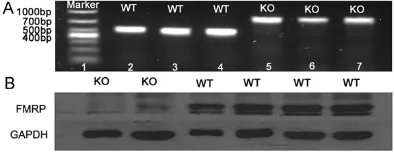
Confirming each experimental mouse is pure
breed
Before each experiment, we used polymerase chain reaction (PCR) and western blot to iden-tify the breed of experimental animals. In PCR, we observed the 468 bp band in WT mice and the 800 bp band in Fmr1 KO mice (Figure 1A). In western blot, WT mice expressed FMRP fully while there was no expression in KO mice (Figure 1B). The genotyping results of PCR were completely consistent with those using western Figure 1. The identification of genotype and Fmrp
[image:3.612.90.287.73.149.2]blot, which confirmed the experimental animals pure breed absolutely.
The Fmr1 KO mice have learning and memory deficit
The step-through test was carried out over two days. Day 1 was the phase of memory acquisi-tion, and then learning ability was tested on day 2. The data that displayed above was from day 2 while data of the 1st day not shown. In the step-through test, the latency for the blank con-trol group of KO mice were significantly sh- orter than the WT mice (P<0.05; Figure 2A). Likewise, the error count were significantly increased in the KO mice compared with the homologous WT group (P<0.05; Figure 2B). α-Asarone prolongs the latency and reduces
the error count in KO mice
Neither the latency nor error count between the blank control group and vehicle group of KO mice was statistically significant (P>0.05; Figure 2A, 2B). And compared with the vehicle group, the latency of α-asarone treatment groups, especially at dose of 0.1 mg/kg, 3 mg/ kg, 4.5 mg/kg, were clearly prolonged, but only at dose of 3 mg/kg and 4.5 mg/kg showed statistical significance (P<0.05; Figure 2A). In- terestingly, α-asarone treatment groups with 3 mg/kg and 4.5 mg/kg significantly reduced the error count (P<0.05; Figure 2B) compared to
vehicle group while the treatment groups with 0.1 mg/kg and 1 mg/kg had no significant dif-ference (P>0.05; Figure 2B).
The p-ERK1/2 is highly expressed in Fmr1 KO
mice
After the behavioral tests, we detected the ERK1/2 and p-ERK1/2 expression in hippo-campal CA1 and CA3 region of FVB KO mice and WT mice. Compared to WT mice, the ERK1/2 level in both CA1 and CA3 region of KO blank control group was slightly decreased but neither of them had the significant differ-ence (P>0.05; Figure 3A). However, the level of p-ERK1/2 expression in both CA1 and CA3 region was markedly increased and both of them had the statistical significance (P<0.05; Figure 3B).
α-Asarone down-regulates the expression of p-ERK1/2 in Fmr1 KO mice
After 9-days consecutive treatment with differ-ent doses of α-asarone, no statistical signifi-cance were found in both ERK1/2 and p-ERK1/2 expression in hippocampal CA1 and CA3 region between vehicle and blank control group of KO mice (P>0.05; Figure 4A, 4B). Interestingly, compared to the vehicle, the expression of p-ERK1/2 in CA1 and CA3 region was decreased profoundly and had statistical significance in treatment groups at the doses Figure 2. Effect of α-asarone on latency and error count of KO mice and WT mice in the step-through test. A. The latency of the blank control group of KO mice were significantly shorter than the WT mice (▲P<0.05); The latency of
α-asarone treatment groups with 3 mg/kg and 4.5 mg/kg were clearly prolonged compared with the vehicle group (*P<0.05). B. The error count of the blank control group of KO mice were significantly increased than the WT mice (▲P<0.05); α-asarone treatment groups with 3 mg/kg and 4.5 mg/kg significantly reduced the error count compared
of 1 mg/kg, 3 mg/kg and 4.5 mg/kg (P<0.05; Figure 4A, 4B), but there was no significant dif-ference in 0.1 mg/kg treatment group (P>0.05; Figure 4A, 4B). However, we did not find any significant changes of ERK1/2 expression in either CA1 or CA3 region in different doses of α-asarone treatment groups when compared to the vehicle (P>0.05; Figure 4A, 4B).
Discussion
Fragile X syndrome (FXS) is the most common inherited cause of intellectual disability that affects all major ethnic groups and races [1]. Thus, it is crucial to find the safe and effective treatment for FXS due to the rapidly growing patient population and the consequent huge burden on affected individuals, their families and care givers, and society as a whole. Fmr1
KO mice share many similar symptoms with FXS patients, which make them be the perfect animal model for studying the features of FXS and drug therapeutics of this disease. Evidence from our early study showed that Fmr1 KO mice exhibited defects in passive avoidance tests [16]. Irwin et al [17] and Galvez et al [18] found that Fmr1 KO mice existed dendritic spine
immaturity and learning and memory deficit. Results in our present research displayed that the latency for the blank control group of KO mice was significantly shorter and the error count was more than WT mice, showing that KO mice had learning and memory impairment which was in accordant with previous studies [19].
[image:5.612.96.516.74.325.2]ligands such as neurotransmitters, growth fac- that can be used to remedy learning and mem-Figure 4. The effect of different doses of α-asarone on ERK1/2 and
p-ERK1/2 levels in hippocampal of Fmr1 knockout mice. A, B. Compared to the vehicle, the expression of p-ERK1/2 in CA1 and CA3 region were decreased profoundly and had statistical significance in treatment groups at the dos-es of 1 mg/kg, 3 mg/kg and 4.5 mg/kg (P<0.05); There was no significant changes of ERK1/2 expression in either CA1 or CA3 region in different doses of α-asarone treatment groups when compared to the vehicle (P>0.05).
quently phosphorylate cyto-plasmic targets or nuclear’s [22]. ERK is abundantly expressed in central nervous system (CNS) and distributed in brain areas that related to learning and memory behav-ior in human, such as neocor-tex, hippocampus and stria-tum. Thomas et al [23] reported that ERK regulated synaptic activity and structur-al and functionstructur-al plastici- ty, and was implicated in both long-term potentiation (LTP) and long-term depres- sion (LTD), which is respec-tively considered to underlie memory and maintain neural plasticity. Mazzucchelli, et al [24] revealed that the latency for the ERK1 mutant mice was significantly prolonged in long-term retention test in avoidance tasks compared to WT mice. These findings ab- ove suggest that ERK1/2 may contribute to cognitive deficit and dendritic spine immatu-rity in FXS.
Experimental studies have reported that the basal level of p-ERK in hippocampal synaptoneurosomes of Fmr1
KO mice increased [25, 26]. Wang, et al [22] likewise found that the p-ERK and MEK1/2 highly expressed in brain tissue both in FXS pa- tients and Fmr1 KO mice. In our work, as expected, com-pared with WT blank control group, the expression of p-ERK1/2 of KO mice profound-ly increased in both hippo-campal CA1 and CA3 region while the expression of ERK1/2 had no significant difference.
[image:6.612.95.368.74.613.2]peritoneal injection of α-asarone in scopol-amine amnesic mice for 15 days, and these mice were tested with step-through passive avoidance test and the Y-maze test. Results showed that α-asarone ameliorated memory and cognitive function as indicated by prolong-ing the transfer latency time and spontaneous alternation time in behavioral testing above. Shin et al [10] did administer α-asarone orally in C57BL/6 mice before the LPS injection once a day for 3 days, and learning and memory defi-cit was evaluated by the Morris water maze test. They showed that α-asarone not only sig-nificantly prolonged the swimming time spent in the target and peri-target zones but also increased the number of target heading and memory score in the Morris water maze, which indicated α-asarone may be beneficial to cogni-tive impairment. Based on previous studies, in present research we used the step-through passive avoidance test to evaluate learning and memory performance of FVB KO mice. After 4 different doses of α-asarone (0.1 mg/kg, 1 mg/kg, 3 mg/kg, 4 mg/kg) i.p. injection for 9 continuous days, the Fmr1 KO mice at dose of 3 mg/kg and 4.5 mg/kg exhibited an increased latency and a decreased error count comparing to vehicle group. The results from the step-through test implied α-asarone’s ameliorating effect on learning and memory deficit in Fmr1
KO mice.
For better understanding the related mecha-nisms of α-asarone’s ameliorating effect on cognitive impairment in Fmr1 KO mice, we used western blotting to detect the expression level of ERK1/2 and p-ERK1/2 in hippocampal tis-sue of mice. In the present study, α-asa- rone down-regulated the expression level of p-ERK1/2 in s both hippocampal CA1 and CA3 region in Fmr1 KO mice, especially at dose of 1 mg/kg, 3 mg/kg and 4.5 mg/kg. From the results of our current study, α-asarone improved the learning and memory dysfunction in Fmr1
knockout mice which may be related to the reduction of p-ERK1/2. However, 1 mg/kg of α-asarone did not rescue the passive avoid-ance deficit in KO mice. According to one of our early research, 9~24 mg/kg of α-asarone made the FVB KO mice sedation, while concen-tration of 3~6 mg/kg was beneficial to improve the abnormal behavior of KO mice [28]. Combining with the results of behavioral test and western blot, we inferred that the dose of
0.1 mg/kg and 1 mg/kg might be too low to change the behavior in KO mice or the nu- mber of the experimental mice in low dose group were not enough to cause a statistically change.
Conclusion
In summary, this study supports the hypothesis that α-asarone effectively ameliorates learning and memory ability of Fmr1 KO mice. And the effect of α-asarone was found to be mediated via by down-regulation of p-ERK1/2 in hippo-campus in KO mice, which indicated that the involvement of p-ERK1/2 contributed to the learning and memory deficit in FXS model mice. More importantly, our findings would provides a new thought, new targets and new theory basis for the treatment of fragile X syndrome. Further studies would be required to establish effective and safe dosage regimen of α-asarone for FXS model mice treatment and for FXS patients in the future.
Acknowledgements
We thank Professor B. A. Oostra (Cellular Bio- logy and Genetics Research Center, Erasmus University, Rotterdam, Netherlands) very much for providing the FVB Fmr1 knockout mice. This study was supported by Science and Technology Planning Project of Guangdong Province, China (2014A020212317).
Disclosure of conflict of interest
None.
Address correspondence to: Ying Pan, The Depart- ment of Neurology, The Second Affiliated Hospital of Guangzhou Medical University, 250 East Changgang Road, Guangzhou 510260, PR, China. Tel: +86138- 02974704; E-mail: xpany@163.com
References
[1] Peprah E. Fragile X syndrome: the FMR1 CGG repeat distribution among world populations. Ann Hum Genet 2012; 76: 178-191.
[2] Christie SB, Akins MR, Schwob JE and Fallon JR. The FXG: a presynaptic fragile X granule ex-pressed in a subset of developing brain cir-cuits. J Neurosci 2009; 29: 1514-1524. [3] BA Oostra, CE Bakker, E Reyniers. Fmr1
[4] Vinueza Veloz MF, Buijsen RA, Willemsen R, Cupido A, Bosman LW, Koekkoek SK, Potters JW, Oostra BA and De Zeeuw CI. The effect of an mGluR5 inhibitor on procedural memory and avoidance discrimination impairments in Fmr1 KO mice. Genes Brain Behav 2012; 11: 325-331.
[5] Westmark CJ, Westmark PR, O’Riordan KJ, Ray BC, Hervey CM, Salamat MS, Abozeid SH, Stein KM, Stodola LA, Tranfaglia M, Burger C, Berry-Kravis EM and Malter JS. Reversal of fragile X phenotypes by manipulation of AbetaPP/Abeta levels in Fmr1KO mice. PLoS One 2011; 6: e26549.
[6] Cruz-Martin A, Crespo M and Portera-Cailliau C. Delayed stabilization of dendritic spines in fragile X mice. J Neurosci 2010; 30: 7793-7803.
[7] Manyam BV. Dementia in Ayurveda. J Altern Complement Med 1999; 5: 81-88.
[8] Kumar H, Kim BW, Song SY, Kim JS, Kim IS, Kwon YS, Koppula S and Choi DK. Cognitive enhancing effects of alpha asarone in amnesic mice by influencing cholinergic and antioxidant defense mechanisms. Biosci Biotechnol Bio-chem 2012; 76: 1518-1522.
[9] Limon ID, Mendieta L, Diaz A, Chamorro G, Es-pinosa B, Zenteno E and Guevara J. Neuropro-tective effect of alpha-asarone on spatial memory and nitric oxide levels in rats injected with amyloid-beta((25-35)). Neurosci Lett 2009; 453: 98-103.
[10] Shin JW, Cheong YJ, Koo YM, Kim S, Noh CK, Son YH, Kang C and Sohn NW. Alpha-asarone ameliorates memory deficit in lipopolysaccha-ride-treated mice via suppression of pro-In-flammatory cytokines and microglial activa-tion. Biomol Ther (Seoul) 2014; 22: 17-26. [11] Li W, Cui Y, Kushner SA, Brown RA, Jentsch JD,
Frankland PW, Cannon TD and Silva AJ. The HMG-CoA reductase inhibitor lovastatin revers-es the learning and attention deficits in a mouse model of neurofibromatosis type 1. Curr Biol 2005; 15: 1961-1967.
[12] Osterweil EK, Chuang SC, Chubykin AA, Sidorov M, Bianchi R, Wong RK and Bear MF. Lovas-tatin corrects excess protein synthesis and prevents epileptogenesis in a mouse model of fragile X syndrome. Neuron 2013; 77: 243-250.
[13] Caku A, Pellerin D, Bouvier P, Riou E and Corbin F. Effect of lovastatin on behavior in children and adults with fragile X syndrome: an open-label study. Am J Med Genet A 2014; 164A: 2834-2842.
[14] Mendieta A, Jimenez F, Garduno-Siciliano L, Mojica-Villegas A, Rosales-Acosta B, Villa-Tana-ca L, Chamorro-Cevallos G, Medina-Franco JL, Meurice N, Gutierrez RU, Montiel LE, Cruz Mdel
C and Tamariz J. Synthesis and highly potent hypolipidemic activity of alpha-asarone- and fi-brate-based 2-acyl and 2-alkyl phenols as HMG-CoA reductase inhibitors. Bioorg Med Chem 2014; 22: 5871-5882.
[15] Xing Z, Sun W, Huang Y, Yi Y, Dai L, Chen S and Li M. Genotype Analysis of Fmr1 Gene Knock-out Mice with Polymerase Chain Reaction. Modern Hospital 2009; 9: 12-14.
[16] Chen S, Luo X, Yang Q, Sun W, Cao K, Chen X, Huang Y, Dai L and Yi Y. Lithium chloride ame-liorates learning and memory ability and inhib-its glycogen synthase kinase-3 beta activity in a mouse model of fragile X syndrome. Neu-ral Regeneration Research 2011; 6: 2452-2459.
[17] Irwin SA, Patel B, Idupulapati M, Harris JB, Crisostomo RA, Larsen BP, Kooy F, Willems PJ, Cras P, Kozlowski PB, Swain RA, Weiler IJ and Greenough WT. Abnormal dendritic spine char-acteristics in the temporal and visual cortices of patients with fragile-X syndrome: a quantita-tive examination. Am J Med Genet 2001; 98: 161-167.
[18] Galvez R and Greenough WT. Sequence of ab-normal dendritic spine development in primary somatosensory cortex of a mouse model of the fragile X mental retardation syndrome. Am J Med Genet A 2005; 135: 155-160.
[19] Qin M, Kang J and Smith CB. Increased rates of cerebral glucose metabolism in a mouse mod-el of fragile X mental retardation. Proc Natl Acad Sci U S A 2002; 99: 15758-15763. [20] Bear MF, Huber KM and Warren ST. The mGluR
theory of fragile X mental retardation. Trends Neurosci 2004; 27: 370-377.
[21] Subramaniam S, Zirrgiebel U, von Bohlen Und Halbach O, Strelau J, Laliberte C, Kaplan DR and Unsicker K. ERK activation promotes neu-ronal degeneration predominantly through plasma membrane damage and independent-ly of caspase-3. J Cell Biol 2004; 165: 357-369.
[22] Wang X, Snape M, Klann E, Stone JG, Singh A, Petersen RB, Castellani RJ, Casadesus G, Smith MA and Zhu X. Activation of the extracel-lular signal-regulated kinase pathway contrib-utes to the behavioral deficit of fragile x-syn-drome. J Neurochem 2012; 121: 672-679. [23] Thomas GM and Huganir RL. MAPK cascade
signalling and synaptic plasticity. Nat Rev Neu-rosci 2004; 5: 173-183.
stria-tum and facilitates striatal-mediated learning and memory. Neuron 2002; 34: 807-820. [25] Hou L, Antion MD, Hu D, Spencer CM, Paylor R
and Klann E. Dynamic translational and prote-asomal regulation of fragile X mental retarda-tion protein controls mGluR-dependent long-term depression. Neuron 2006; 51: 441-454. [26] Price TJ, Rashid MH, Millecamps M, Sanoja R,
Entrena JM and Cervero F. Decreased nocicep-tive sensitization in mice lacking the fragile X mental retardation protein: role of mGluR1/5 and mTOR. J Neurosci 2007; 27: 13958-13967.
[27] Kim JH, Hahm DH, Lee HJ, Pyun KH and Shim I. Acori graminei rhizoma ameliorated ibotenic acid-induced amnesia in rats. Evid Based Com-plement Alternat Med 2009; 6: 457-464. [28] Xing Z, Sun W, Huang Y, Li M, Yi Y, Dai L and