Original Article
TRIM29 promotes gastric cancer cell proliferation,
migration, invasion and predicts the
prognosis of patients
Jinxi Huang1, Yi Zhou2, Chenghu Wang1, Weiwei Yuan1, Zhandong Zhang1, Beibei Chen3, Xiefu Zhang4
1Department of General Surgery, The Affiliated Cancer Hospital of Zhengzhou University, Zhengzhou 450003,
Henan, China; 2Department of Anesthesiology, The Affiliated Cancer Hospital of Zhengzhou University, Zhengzhou
450003, Henan, China; 3Depatment of Oncology, The Affiliated Cancer Hospital of Zhengzhou University,
Zheng-zhou 450003, Henan, China; 4Department of General Surgery, First Affiliated Hospital of Zhengzhou University,
Zhengzhou 450052, Henan, China
Received October 2, 2017; Accepted May 3, 2018; Epub August 15, 2018; Published August 30, 2018
Abstract: Objectives Gastric cancer (GC) is the fourth most common cancer and second leading cause of cancer deaths worldwide. Tripartite motif-containing 29 (TRIM29), a member of the TRIM family of transcription factors, exhibits a contextual function in cancer. The present study aimed to study the expression, function and prognosis significance in GC. Methods Real-time PCR and Western blot were performed to analyze the expression profiles of TRIM29 in GC samples and cell lines. Kaplan-Meier analysis was done to reveal the relationship of TRIM29 expres-sion with overall survival of the patients. Cell-based assays including MTT and flow cytometry and subcutaneous tumorigenicity were performed to investigate the function of TRIM29 in gastric cancer cell proliferation. Transwell assays were performed to investigate the function of TRIM29 in cell migration and invasion. Real-time PCR and Western blot were finally used to explore the mechanism. Results TRIM29 was upregulated in GC samples and cell lines and higher levels of TRIM29 were associated with poor overall survival of patients. RNAi guided TRIM29 knock-down significantly inhibited GC cell proliferation, induced cell cycle arrest, inhibited GC cell migration and invasion, and repressed the tumor formation in nude mice. The expression levels of p53 and β-catenin proteins were impaired by TRIM29 knockdown, indicating a possible linkage underlying the oncogenic role of TRIM29 in GC. Conclusion Our findings provide important evidence that gain of TRIM29 expression contributes to GC cell proliferation and it may serve as a potential prognostic biomarker for GC.
Keywords: Gastric cancer, TRIM29, prognosis, cell proliferation, migration and invasion, p53 and β-catenin signal-ing
Introduction
Gastric cancer (GC) remains as the fourth most frequent type of cancer and the second leading cause of cancer mortality worldwide, although its incidence is declining during the last decades [1, 2]. The treatment option includes surgery, radiotherapy, chemotherapy and mo- lecular targeted therapy [3]. Its progression involves multi-factors and multi-steps, accom-panied by alterations of gene expression, in- cluding activation of oncogenes and suppres-sion of tumor suppressors. Nowadays, more and more novel coding or non-coding genes have been identified to process oncogenic or tumor suppressing potential, we do not make
a breakthrough on the mechanisms of GC occurrence. The 5-year survival rate for GC patients is merely about 20% due to metasta-sis, recurrence and multiple drug resistance (MDR) [4, 5].
immune response [7-9]. Examination of TRIM29 expression in many human cancers has revealed that it is up-regulated in cancers including lung cancer [10], colorectal cancer [11], pancreatic cancer [12], bladder cancer [13], thyroid carcinoma [14] and nasopharyn-geal carcinoma [15]. But it is down-regulated in several other cancers including breast cancer [16] and hepatocellular carcinoma [17]. In gastric cancer, there are two reports ex- pounding the expression, survival correlation and biological functions of TRIM29 [18, 19]. Kosaka et al addressed that TRIM29 expres-sion was far higher in GC tissues and its increased mRNA expression was markedly associated with histological grade, lymph node metastasis and poorer survival rate [18]. Recently, Qiu et al reported that TRIM29 pro-moted cell proliferation, cell cycle and apopto-sis reapopto-sistance via RNA interference (RNAi) tech-noledges. They further demonstrated that TRIM29 functioned by regulating Wnt/β-catenin signaling pathway and was targeted by microR-NA-185 [19]. These findings all support the oncogenic roles of TRIM29 in GC. However, the in vivo data was still absent from the two reports, and the possible role or mechanism of TRIM29 in pathogenesis of GC remains to be further explored.
In the present study, we mainly analyzed the expression profile of TRIM29 in GC samples and cell lines. Then RNAi technology was applied to study the in vitro and in vivo func-tions of TRIM29 in GC cell proliferation, migra-tion and invasion, aiming to provide the basis for new GC therapy methods.
Materials and methods
Patients and tissues
A total of 22 confirmed GC patients were con-secutively recruited between January 2014 and December 2016 for the General Surgery at The Affiliated Cancer Hospital of Zhengzhou University. The normal gastric mucosa tissues, negative lymph node tissues, primary lesion tis-sues, positive lymph node tissues and metas-tasis lesion tissues were obtained by surgery, and examined by a pathologist. Final surgical pathology reports were obtained and recorded. All the patients had provided the written informed consent use of their samples.
Cell lines and cell culture
Five GC cells (SGC-7901, AGS, HGC-27, MGC80-3, NCI-N87) were grown in 1640 or DMEM supplemented with 10% fetal bovine serum (Hyclone) according to ATCC protocols. Cells were maintained in a humidified incubator at 37°C and 5% CO2.
Immunohistochemical staining
The 5 μm tissue sections were dewaxed, rehy-drated, and retrieved in 10 mM citrate buffer pH6.0 (10 mM citrate buffer and 0.05% Tween-20). The sections were immersed in 3% hydro-gen peroxide for 20 min and stained with anti-TRIM29 antibody (1:100) with the non-im- munized goat serum at 4°C overnight. After incubating with secondary antibodies (1:3000, Beyotime) for 1 h, the sections were stained with DAB (Beyotime). The sections were photo-graphed under a microscope.
Reverse transcription (RT) and quantitative real-time PCR
Total RNA was isolated from tissues using TRIzol reagent (Life Technologies). First-strand cDNA was synthesized from 2 mg of total RNA using oligo (dT)n (Takara) and reverse transcrip-tase (Takara). Quantitative real-time PCR analy-sis of TRIM29 expression was carried out using 2 ml of cDNA and the SYBR Green Master Mix (TOYOBO), as recommended by the manufac-turer. GAPDH was used as internal controls.
SDS-PAGE and western blot
ed by a chemiluminescence detection system (Millipore).
Short interfering RNAs and transfections
TRIM29 siRNA and control siRNA were pur-chased from Santa Cruz. Transfections were performed by Lipofectamin-2000 reagent (Life Technology) followed by the manufacturer’s instruction.
MTT assay
SGC-7901 or MGC80-3 cells were transfect- ed with control siRNA or TRIM29 siRNA at vary-ing time: 12 h, 24 h, 48 h and 72 h. Then 20 μl of sterile MTT (5 mg/ml, sigma) was added and incubation for another 4 h at 37°C. After that, 150 μl of DMSO was added to each well
and the plates were thoroughly mixed for 20 min. Spectrometric absorbance at a wave-length of 490 nm was measured on a micro-plate reader.
Cell cycle analysis
SGC-7901 or MGC80-3 cells were fixed by 70% ethanol and stained with propidium iodide for 30 min. After that, the cells were immediately analyzed by cell flow cytometry. Histograms represent the percentage of cells in each phase of the cell cycle (G0/G1 phase, S phase, and G2/M phase).
Transwell assay
Gastric cells were harvested after transfect- ed with control siRNAs or TRIM29 siRNAs for 24 h. Then cells were suspended with serum-free medium, then seeded in the upper cham-ber of 8 μm pore size Transwell (Corning Inc., Coring, NY, USA) at the density of 1 × 10⁵ cells
per chamber for migration and invasion assay, respectively. For invasion assay, the upper chambers were precoated with BD Matrigel. The lower chambers were filled with medium containing 10% FBS and incubated for 24 h. Cells remaining on the upper chamber were removed carefully by swabs, the migrating cells in the lower chamber were fixed with 4% formal-dehyde, and stained with 0.5% crystal violet solution. The stained cells were imaged with an inverted microscope, and counted at five ran-domly selected visual fields for each experi-mental condition. Each experiment was per-formed in triplicate.
In vivo xenograft tumor growth
4-week-old female nude mice [BALB/cA-nu (nu/ nu)] were obtained from Shanghai Experimental Animal Center and maintained in pathogen-free conditions. The mice were separated into two groups and were subcutaneously injected with control or TRIM29 knockdown MGC80-3 cells (2 × 10⁶) at each flank. Tumor volumes were
measured with a caliper every 6 days. The mice were killed and the tumor weight was measured with a scale at the sixtieth day.
Statistical analysis
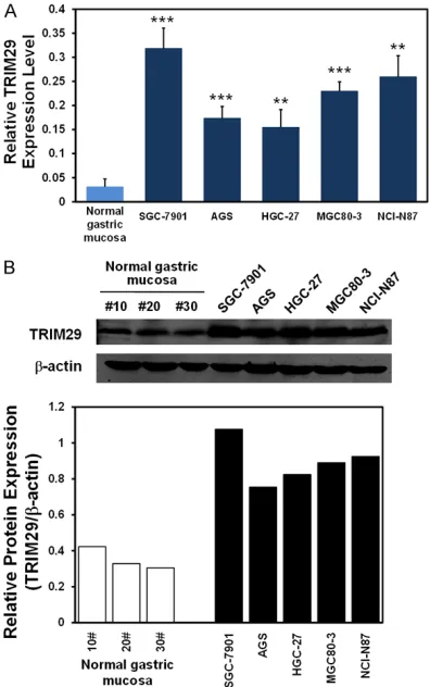
[image:4.612.90.288.72.388.2]All data were analyzed by SAS v9.2 software and presented as mean ± SD. Kaplan-Meier analysis was performed to analysis the survival time between TRIM29 negative and positive Figure 2. The expression of TRIM29 protein is highly
groups. Differences between any two groups were assessed by the Student’s t-test. P<0.05 was considered as a statistically significant difference.
Results
TRIM29 protein expression is increased in GC samples
We firstly examined the mRNA and protein expression profile of TRIM29 in 22 sets of clini-cal GC samples, including normal gastric mu- cosa, negative lymph node, primary lesion, pos-itive lymph node and metastasis lesion of 22 individuals. The results of real-time PCR and
3, NCI-N87) and the human normal gastric mucosa cells. As shown in Figure 2A and 2B, in comparison with normal gastric mucosa cells, TRIM29 mRNA and protein levels were signifi-cantly increased in the 5 different GC cell lines (P<0.01~0.001). Together with the above results of TRIM29 expression in GC samples, these data all strongly suggested that highly expressed TRIM29 might be important for GC progression.
TRIM29 knockdown attenuated cell malignant progression in vitro
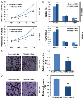
[image:5.612.88.376.70.409.2]To investigate the function of TRIM29 in GC cells, we applied RNAi to knockdown intra- Figure 3. Knockdown of TRIM29 inhibited cell proliferation and cell cycle
in vitro. A. Inhibition of cell proliferation in SGC-7901 and MGC80-3 cells after TRIM29 knockdown. Cell proliferation was measured using MTT assay at 490nm. B. Inhibition of cell cycle (G1 arrest) in SGC-7901 and MGC80-3 cells after TRIM29 knockdown. Cell proliferation was measured using cell flow cytometry. Data had three repeats. *: P<0.05, **: P<0.01, compared to control group. C. Inhibition of cell migration in SGC-7901 cells after TRIM29 knockdown. Cell migration was measured using transwell Assay. D. Inhibition of cell invasion in MGC80-3 cells after TRIM29 knockdown. Cell invasion was measured using transwell Assay.
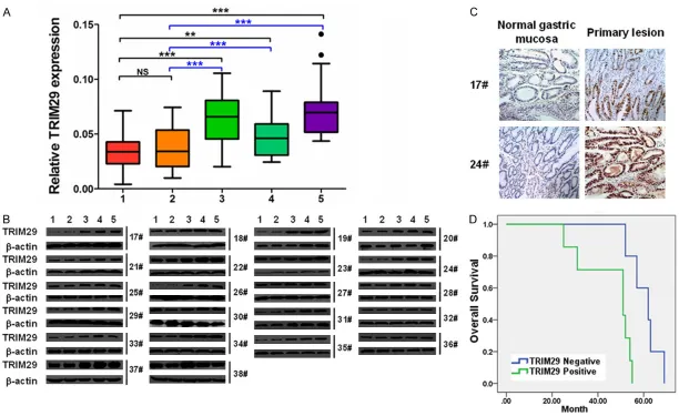
Western blot revealed that TRIM29 mRNA and protein were both significantly incr- eased in primary lesions, pos-itive lymph nodes and me- tastasis lesions of GC sam-ples, when compared with that of normal gastric mucosa and negative lymph nodes (P<0.01~0.001, Figure 1A
and 1B). The results of im- munohistochemistry staining revealed that, TRIM29 was mainly localized in the nuclei of GC cells, and its expres- sion was much more domi-nant in the primary lesion than that in the neighbor- ing normal gastric mucosa (Figure 1C). By Kaplan-Meier analysis, we found that GC patients with higher TRIM29 expression had a significantly shorter overall survival time than those with lower TRIM29 levels (P<0.01, Figure 1D). These results indicated a tu- mor promoting function of TRIM29 in GC, finely in line with the former studies [18, 19].
TRIM29 protein is highly expressed in GC cell lines
MGC80-cellular TRIM29 expression in two different GC cell lines SGC-7901 and MGC80-3. Then MTT assay was performed to evaluate the cell proliferation potential of these cells. The plotted growth curves indicated that knock-down of TRIM29 (TRIM29 siRNA-transfect- ed) resulted in dereased proliferation in both SGC-7901 and MGC80-3 cells when compar- ed with Control siRNA groups (P<0.05~0.001,
Figure 3A). We further analyzed the effect of TRIM29 knockdown on cell cycle using flow cytometry. TRIM29 silencing cells dis-played significant G0/G1 arrest and G2/M phase reduction (P<0.05~0.01, Figure 3B), which indicated that TRIM29 knockdown in- hibited GC proliferation as the result of G0/ G1 arrest. Furthermore, transwell assay with-out or with BD Matrigel was performed to validate the effect of TRIM29 knockdown on cell migration and invasion. The results show- ed significant inhibitions of cell migration ab- ility in SGC-7901 cells and cell invasion abi- lity in MGC80-3 cells (P<0.01, Figure 3C and
3D).
TRIM29 knockdown inhibited xenograft growth of GC cells in vivo
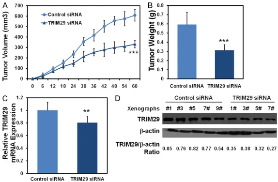
To further confirm the role of TRIM29 in the tumorigenesis of GC, a subcutaneous
tumori-(Figure 4D and 4E). These results indicated that TRIM29 knockdown repressed the tumor formation of GC cells in vivo.
Molecular mechanisms of TRIM29 in the cell growth of GC
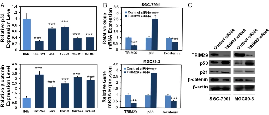
[image:6.612.89.376.71.259.2]We finally investigated the molecular mecha-nisms of TRIM29 in controlling GC cell prolifera-tion. According to the previous literature, TRIM29 might negatively regulate p53 [20] and positively regulate β-catenin expression [19], two important molecules in cancer de- velopment. The expression of p53 and β- catenin was determined in the above men-tioned GC cell lines and normal gastric mu- cosa cells. As shown in Figure 5A and 5B, in comparison with normal gastric mucosa cells, p53 mRNA was significantly decreas- ed, while β-catenin mRNA was obviously in- creased in the 5 different GC cell lines (P< 0.001). In SGC-7901 and MGC80-3 cells, TRIM29 siRNA transfection led to elevated p53, p21 and reduced β-catenin mRNAs/pro-teins (Figure 5C and 5D). Consistent with the former reports, our data suggested that inhibi-tion of p53 and activainhibi-tion of β-catenin signal-ing might be required for TRIM29’s oncogenic effects.
Figure 4. Knockdown of TRIM29 represses xenograft growth of GC cells in vivo. 2 × 10⁶ MGC80-3 cells which were either infected with silenced TRIM29
were injected subcutaneously into the each flank of nude mice. After 60 days, the mice were killed and tumors were harvested and weighted. A. Tu-mor growth was monitored over two months. B. TuTu-mors were dissected for analysis of the differences mass. C, D. Tumor samples were subjected to quantitative real-time PCR and western blot analysis for TRIM29 expression. Data are means ± SD, N=5 or 4, **: P<0.01, ***: P<0.001.
Discussion
TRIM29 has been found to be associated with many forms of aggressive disease including human cancers. Abnormal expression of TRIM29 was observed in a several cancers [10-17], among which, an association was found between TRIM29 expression and reduced patient survival [18, 21]. Finely in accordance with these, we also found that TRIM29 was upregulated in GC samples and cell lines and higher levels of TRIM29 were associated with poor overall survival of patients. Actually, in GC, Kosaka’s finding was almost the same with ours, except that they found increased TRIM29 expression markedly associated with lymph node metastasis [18]. In the present study, we compared the expression of TRIM29 in 22 sets of GC samples, including normal gastric muco-sa, negative lymph node, primary lesion, posi-tive lymph node and metastasis lesion. However, we did not find significant expression difference of TRIM29 mRNA or protein between paired primary lesions and metastasis lesions. Therefore, we did not concentrate on the role of TRIM29 in GC cell migration and invasion. This might attribute to the sample size we used here, or it might be specific to human species. In support of the oncogenic role of TRIM29, we found that TRIM29 knockdown inhibited GC cell proliferation, induced cell cycle arrest, inhibited GC cell migration and invasion and repressed
the tumor formation in nude mice. Although it was clear in the previous report that TRIM29 knockdown resulted in significantly reduced cell proliferation, colony formation, as well as G1-S cell cycle arrest [18]. Our present study added the first evidence of the in vivo function of TRIM29 in GC.
Regarding to the molecular mechanisms of TRIM29’s role in GC, we learned from the litera-tures that overexpression of TRIM29 in pan- creatic cancer lines promoted cell growth in vitro and metastatic activity in vivo stemming from stimulation of Wnt/β-catenin signaling [22]. On the other hand, TRIM29 could bind to p53, the famous tumor suppressor by in- hibiting cancer cell proliferation and cell cycle, and inhibit its transcriptional activity [23]. We validated the expression of p53, p21 and β-catenin in a panel of GC cell lines, and TRIM29-silenced cells. The results revealed that p53 and p21 expression was negatively while β-catenin expression was positively cor-related with TRIM29 expression, indicating a possible linkage underlying the oncogenic role of TRIM29 in GC.
In summary, we documented that TRIM29 expression is increased in GC samples and cell lines, and that TRIM29 knockdown elevated p53, p21 expression while reduced β-catenin expression, thus to drive enhanced cell prolif-eration and cell cycle. Our study strengthened the oncogenic role of TRIM29 in GC and pro-Figure 5. Molecular mechanisms regulated by TRIM29 in GC cells. A. The mRNA expression of p53 and β-catenin was increased in five GC cell lines compared with normal gastric mucosa by quantitative real-time PCR assay. GAPDH was used as internal controls. B. The mRNA expression of p53 and β-catenin was analyzed by quantitative real-time PCR when knockdown of TRIM29 in SGC-7901 and MGC80-3 cells. C. The protein expression of p53, p21 and β-catenin was analyzed by western blot when knockdown of TRIM29 in SGC-7901 and MGC80-3 cells. ***:
[image:7.612.91.520.69.253.2]vided the molecular basis by inhibiting tumor suppressive p53 signalingmeanwhile activating β-catenin signaling.
Acknowledgements
The present work was supported by the Basic and Advanced Technology Research Project of Science and Technology Department of Henan Province (NO.142300410276).
Disclosure of conflict of interest
None.
Address correspondence to: Yi Zhou, Department of Anesthesiology, The Affiliated Cancer Hospital of Zhengzhou University, Zhengzhou 450003, Henan, China. Tel: +86-037165587230; E-mail: yizhouhn- [email protected]
References
[1] Jemal A, Bray F, Center MM, Ferlay J, Ward E, Forman D. Global cancer statistics. CA Cancer J Clin 2011; 61: 69-90.
[2] Siegel R, Ma J, Zou Z, Jemal A. Cancer statis-tics, 2014. CA Cancer J Clin 2014; 64: 9-29. [3] Kato M, Asaka M. Recent development of
gas-tric cancer prevention. Jpn J Clin Oncol 2012; 42: 987-994.
[4] Lin M, Lin JX, Huang CM1, Zheng CH, Li P, Xie JW, Wang JB, Lu J. Prognostic analysis of gas-tric gastrointestinal stromal tumor with syn-chronous gastric cancer. World J Surg Oncol 2014; 12: 25.
[5] Yu P, Du Y, Yang L, Fan S, Wu J, Zheng S. Sig-nificance of multidrug resistance gene-related proteins in the postoperative chemotherapy of gastric cancer. Int J Clin Exp Pathol 2014; 7: 7945-7950.
[6] Hatakeyama S. TRIM proteins and cancer. Nat Rev Cancer 2011; 11: 792-804.
[7] Reymond A, Meroni G, Fantozzi A, Merla G, Cairo S, Luzi L, Riganelli D, Zanaria E, Messali S, Cainarca S, Guffanti A, Minucci S, Pelicci PG, Ballabio A. The tripartite motif family identifies cell compartments. EMBO J 2001; 20: 2140-2151.
[8] Napolitano LM, Meroni G. TRIM family: pleiot-ropy and diversification through homomultimer and heteromultimer formation. IUBMB Life 2012; 64: 64-71.
[9] Zeng SX, Cai QC, Guo CH, Zhi LQ, Dai X, Zhang DF, Ma W. High expression of TRIM29 (ATDC) contributes to poor prognosis and tumor me-tastasis by inducing epithelial mesenchymal transition in osteosarcoma. Oncol Rep 2017; 38: 1645-1654.
[10] Song X, Fu C, Yang X, Sun D, Zhang X, Zhang J. Tripartite motif-containing 29 as a novel bio-marker in non-small cell lung cancer. Oncol Lett 2015; 10: 2283-2288.
[11] Jiang T, Tang HM, Lu S, Yan DW, Yang YX, Peng ZH. Up-regulation of tripartite motif-containing 29 promotes cancer cell proliferation and pre-dicts poor survival in colorectal cancer. Med Oncol 2013; 30: 715.
[12] Sun H, Dai X, Han B. TRIM29 as a novel bio-marker in pancreatic adenocarcinoma. Dis Markers 2014; 2014: 317817.
[13] Tan ST, Liu SY, Wu B. TRIM29 overexpression promotes proliferation and survival of bladder cancer cells through NF-κB signaling. Cancer Res Treat 2016; 48: 1302-1312.
[14] Xu J, Li Z, Su Q, Zhao J, Ma J. TRIM29 promotes progression of thyroid carcinoma via activating P13K/AKT signaling pathway. Oncol Rep 2017; 37: 1555-1564.
[15] Zhou XM, Sun R, Luo DH, Sun J, Zhang MY, Wang MH, Yang Y, Wang HY, Mai SJ. Upregu-lated TRIM29 promotes proliferation and me-tastasis of nasopharyngeal carcinoma via PTEN/AKT/mTOR signal pathway. Oncotarget 2016; 7: 13634-50.
[16] Liu J, Welm B, Boucher KM, Ebbert MT, Ber-nard PS. TRIM29 functions as a tumor sup-pressor in nontumorigenic breast cells and in-vasive ER+ breast cancer. Am J Pathol 2012; 180: 839-847.
[17] Jiang N, Chen WJ, Zhang JW, Xu C, Zeng XC, Zhang T, Li Y, Wang GY. Downregulation of miR-432 activates Wnt/β-catenin signaling and promotes human hepatocellular carcinoma proliferation. Oncotarget 2015; 6: 7866-7879. [18] Kosaka Y, Inoue H, Ohmachi T, Yokoe T, Matsu-moto T, Mimori K, Tanaka F, Watanabe M, Mori M. Tripartite motif-containing 29 (TRIM29) is a novel marker for lymph node metastasis in gastric cancer. Ann Surg Oncol 2007; 14: 2543-2549.
[19] Qiu F, Xiong JP, Deng J, Xiang XJ. TRIM29 func-tions as an oncogene in gastric cancer and is regulated by miR-185. Int J Clin Exp Pathol 2015; 8: 5053-5061.
[20] Sho T, Tsukiyama T, Sato T, Kondo T, Cheng J, Saku T, Asaka M, Hatakeyama S. TRIM29 neg-atively regulates p53 via inhibition of Tip60. Biochim Biophys Acta 2011; 1813: 1245-1253.
[23] Yuan Z, Villagra A, Peng L, Coppola D, Glozak M, Sotomayor EM, Chen J, Lane WS, Seto E. The ATDC (TRIM29) protein binds p53 and an-tagonizes p53-mediated functions. Mol Cell Biol 2010; 30: 3004-3015.