Original Article
L-3-n-butylphthalide may attenuate Aβ
1-42-induced
neuronal apoptosis and synaptotoxicity in primary
cultured cortical neurons possibly via modulating
both PI3K/AKT and ERK signaling pathways
Yunxia Zhao1, Yong Zhang1, Xiuying Chen1, Wei Dong2, Yifeng Du1
Departments of 1Neurology, 2Thoracic Surgery, Shandong Provincial Hospital Affiliated to Shandong University,
Jinan 250021, P.R. China
Received November 10, 2017; Accepted July 21, 2018; Epub September 15, 2018; Published September 30, 2018
Abstract: Background: Alzheimer’s disease (AD), which is characterized by progressively cognitive decline and neu-ronal loss, is one of the most common neurodegenerative diseases. The β-amyloid (Aβ)-mediated mitochondrial dysfunction and eventually neuronal apoptosis contribute to the pathogenesis of AD. L-3-n-butylphthalide (L-NBP) has been proven to have significant protective effects in many stoke and AD models. Our study is to investigate the protective effects and mechanisms of L-NBP against Aβ induced cell injury in cortical neurons. Material and method: The primary cortical neurons treated with Aβ1-42 were used as the AD cell model. Then the cells were treated with or without L-NBP. MTT assay were used to assess the cell viability. Apoptosis of cortical neurons was analyzed by Hoechst 33342 staining. The flow cytometry was used to assess the mitochondrial membrane potential (Δψm). The apoptotic and signaling associated protein expression levels were measured using Western blotting. Results: Pretreatment with L-NBP effectively inhibited Aβ1-42-induced cytotoxicity, apoptosis, mitochondrial dysfunction, and synaptotoxicity in primary cultured cortical neurons in a dose-dependent way. The protective effects of L-NBP were accompanied by increased phosphorylation of AKT and decreased phosphorylation of the extracellular protein ki-nase (ERK). The neuroprotective effects could be abolished by LY294002, an inhibitor of phosphoinositide 3-kiki-nase (PI3K)-dependent protein kinase B (AKT). Furthermore, ERK pathway inhibitor U0126 enhanced the beneficial ef-fects of L-NBP in inhibiting Aβ-induced apoptosis. Conclusions: L-NBP may exert protective efef-fects on Aβ induced cell injury in cortical neurons through regulating both PI3K/AKT and ERK signaling pathways.
Keywords: Alzheimer’s disease (AD), β-amyloid (Aβ), apoptosis, PI3K/AKT pathway, ERK pathway
Introduction
Alzheimer’s disease (AD) is an age-related neu-rodegenerative disease featured by extracellu-lar β-amyloid (Aβ) peptide deposition and intra-cellular neurofibrillar tangles, which is followed by loss of cortex and hippocampus neurons and cognitive dysfunction [1]. The mechanisms underlying AD pathogenesis largely remain elu-sive. However, increasing evidence suggests that Aβ deposition can initiate a cascade and lead progressively to inflammation, tau hyper-phosphorylation, synaptic dysfunction, neuro-nal apoptosis, and fineuro-nally dementia [2]. The hip-pocampi of AD patients and cultured neurons under Aβ exposure show characteristics of apoptosis [3]. However, the mechanism
under-lying Aβ induced neural apoptosis remains to be elucidated.
dys-function and neuron apoptosis by inhibiting the activation of caspase-3 and caspase-9 [10]. The beneficial effect of neuroglobin was acco- mpanied by an increased expression of p-AKT and an inhibitor of PI3K (LY294002) could block the protective effect [10]. Previous study has shown that gastrodin protected against neurotoxicity induced by Aβ1-42 in primary neu-ral progenitor cells and improved hippocampal neurogenesis of C57BL/6 mice injected with Aβ1-42 through reverse phosphorylation of MEK-1/2, ERK, and c-Jun terminal kinase (JNK). Fur- thermore, the MEK inhibitor U0126 and JNK inhibitor SP600125 showed a synergistic effect with gastrodin [11].
L-3-n-butylphthalide (L-NBP) was originally ex- tracted from the seeds of Apium graveolens
Linn (Chinese celery). Studies have shown that L-NBP protects against ischemic stroke through multiple mechanisms, such as inhibiting plate-let aggregation, improving microcirculation in arterioles [12, 13], decreasing oxidative dam-age and neuronal apoptosis [14], improving mitochondrial function and inhibiting inflam- matory response [15]. Furthermore, L-NBP can improve Aβ-induced learning and memory im- pairments, oxidative damage, and mitochon-drial dysfunction [16, 17]. Lei et al. found that L-NBP attenuated Aβ-induced toxicity through modulating mitochondrial apoptosis and MAPK signaling pathway in SH-SY5Y cells [18]. How- ever, it is not yet known whether L-NBP can pre-vent Aβ1-42 induced apoptosis and synaptotoxic-ity through modulating the PI3K/AKT and/or ERK pathway.
The present study was designed to analyze the protective effects of L-NBP against neuronal apoptosis and synaptotoxicity induced by Aβ
in vitro. In addition, the potential mechanisms including mitochondrial function and possible signaling pathway underlying the neuroprotec-tive effects of L-NBP were further analyzed and discussed.
Materials and methods
Animals
Pregnant Sprague-Dawley rats on gestation day 18 were provided by the Laboratory Animal Re- search Center of Shandong University, China. All experimental procedures involving animals were carried out in accordance with the Guid- ance Suggestion for the Care and Use of
Laboratory Animals. All efforts were made to minimize animal suffering.
Primary cortical neuron isolation and culture
Primary cortical neurons were cultured as pre-viously described [19]. Briefly, the cerebral cor-tices were separated from embryonic Sprague-Dawley rat fetuses (E18D), cut into pieces, and then digested with 0.25% trypsin for 20 min in 37°C. After adding DMEN medium (with 10% fetal bovine serum) for 10 min, tissues were dissociated mechanically with a fire-polished pipette. Then after centrifugation and re-sus-pension, about 1×10⁵ cells in 1 ml were plated on 6-well plates coated with poly-D-lysine. The medium was replaced by Neurobasal medium (Gibco, CA, USA) supplemented with 2% B-27 supplement (Gibco) and 0.5 mM L-glutamine 4 h later. For maintenance, the medium was changed by half every 4 days. Cells were cul-tured in a humidified incubator containing 5% CO2 at 37°C for 8 days before experiments.
Cell treatment
Aβ1-42 (Sigma-Aldrich Inc, St. Louis, MO, USA) was dissolved in distilled water and aged for 6 days at 37°C. L-NBP (purity >98%), which was synthesized by the Department of Synthe- tic Medicinal Chemistry, Institute of Materia Medica (Shijiazhuang, China), was dissolved in dimethyl sulfoxide (DMSO) as 200 mM stock solution. The LY294002 and U0126 were from Cell Signaling Technology, Inc. (Beverly, MA, USA) and were dissolved by DMSO. Cells were divided into control group (without treatment), Aβ group (10 μM Aβ1-42 for 24 h), Aβ+L-NBP group (first treated with 0.1, 1.0, or 10 μM L-NBP for 4 h and then treated with 10 μM Aβ1-42 for 24 h), Aβ+L-NBP+LY294002 group (first treated with 60 uM LY294002 for 1 h, then with 10 μM L-NBP for 4h and finally with 10 μM Aβ1-42 for 24 h), and Aβ+L-NBP+U0126 group (first treated with 10 uM U0126 for 1 h, then with 10 μM L-NBP for 4h and finally with 10 μM Aβ1-42 for 24 h).
MTT assay
incu-bated for 4 h. DMSO (200 μl) was added to the plates and vibrated for 10 minutes at 37°C to solubilize MTT formazan crystals. Then the treatment wells were quantified spectrophoto-metrically at 570 nm using a LAS-4000 micro-plate reader (FUJIFILM, Japan). The cell viability was presented as the ratio of the experimental group to the control group.
Immunofluorescence assay
For immunofluorescence analysis, cortical neu-rons were plated on coverslips coated with poly-L-lysine. Then the cells were fixed with freshly prepared 4% paraformaldehyde in PBS for 30 min, permeabilized with Triton X-100 (0.1%) for 15 min, and rinsed with PBS for 3 times. After blocking with 5% goat serum for 30 min, cells were incubated with the primary antibody of rabbit anti-synaptophysin antibody (1:100, Abcam, UK) overnight at 4°C. After washing with PBS, secondary antibodies of Alexa Fluor 568 goat anti-rabbit IgG (1:500, Abcam, UK) were added and incubated for 1 h under darkness. Then, cells were incubated in 10 μg/ml DAPI for 10 min. After rinsed with PBS for 3 times, the cortical neurons were analyzed on an inverted fluorescence microscopy (Zeiss Jenalumar, Jena, Germany). Cell incubated without primary antibodies was as a negative control. The fluorescence intensity of synapto-physin was quantified using Image J 1.48u soft-ware (National Institutes of Health).
Hoechst 33342 staining
The cortical neurons were seeded on cover-slips. After treatment described above, the cells were fixed with freshly prepared 4% pa- raformaldehyde for 30 min. Then, Hoechst 33342 (1 μM) was added to the cells and in- cubated for 30 min in the dark. After washed with PBS for 3 times, the cortical neurons were analyzed on an inverted fluorescence micros-copy (UV excitation and emission at 360 and 450 nm, respectively). The cells with conden- sed nuclear chromatin and fragmentation were confirmed as apoptotic cells. The apoptotic ratio was calculated as the apoptotic cells to the total cells counted.
Flow cytometry to assess mitochondrial
mem-brane potential (Δψm)
After treatments, 5 μM of JC-1 was added into the neurons and incubated for 15 min at 37°C under darkness. Then a flow cytometer was
applied to analyze the ratio of red (590 nm)/ green (529 nm) fluorescence and determine the percentage of cells with Δψm collapse.
Western blotting
Cells were harvested and incubated with lysis buffer containing phosphatase and protease inhibitors on the ice. The lysed cells were cen-trifuged (12,000× g) for 10 min at 4°C. The supernatants were collected and the total pro-tein was qualified by a BCA propro-tein assay kit (Beyotime, Nanjing, China). Then, proteins were electrophoresed on SDS-PAGE and then trans-ferred to PVDF membrane. Membranes were blocked with blocking buffer (Thermo Scientific, Waltham, USA) for 1 h at room temperature and then incubated with primary antibodies against pro-caspase-3, cleaved caspase-3 (c- caspase-3), synaptophysin, phospho-Tau (Ser 199/202), Akt (Ser 473), phospho-Erk1/2 (Thr202/thy204), Akt, phospho-Erk1/2, total Tau and β-actin (Abcam, Cambridge, UK) overnight at 4°C, respectively. After rinsed with PBS, membranes were incubated with the corre-sponding secondary antibodies for 1 h at room temperature. Finally, the antibody-bound pro-teins were detected by an ECL Western blot detection kit (Santa Cruz, CA, USA). The densi-ties of protein bands were measured by Image J software.
Statistical analysis
All the results are expressed as mean ± SD and statistical analysis was performed using SPSS statistical software (version 17.0). The differ-ences were analyzed by one-way variance anal-ysis combined with Bonferroni-Dunn’s test or Student’s t test. A P value less than 0.05 was considered as statistically significant.
Results
L-NBP can attenuate the cytotoxicity, apopto-sis, mitochondrial dysfunction, and
synapto-toxicity of primary cultured neurons under Aβ
exposure
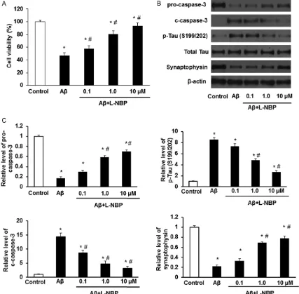
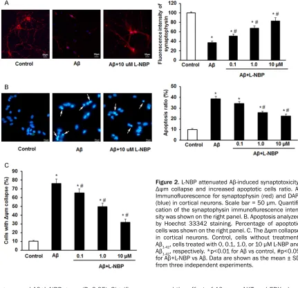
in cortical neurons. Next, the expression of pro-caspase-3, c-pro-caspase-3, p-Tau and synapto-physin was detected by Western Blot. Results showed that 10 μM of Aβ1-42 markedly increa- sed the expression of c-caspase-3 and p-Tau, and decreased the expression of pro-caspase- 3, synaptophysin in cortical neurons (Figure 1B and 1C). Immunofluorescence assay also detected the Aβ1-42 induced decreased synap- tophysin expression (Figure 2A). The Hoechst 33342 staining showed Aβ1-42 increased the ratio of apoptotic cells significantly (Figure 2B).
[image:4.612.92.519.73.492.2]group and Aβ+L-NBP group (P<0.05). Significa- nt differences were also found between con- trol group and Aβ group, and, between control group and Aβ+L-NBP group (P<0.05). The basal levels of cortical cell viability, pro-caspase-3, c-caspase-3, p-Tau, Δψm collapse and synap-tophysin were affected by L-NBP insignificantly (p>0.05; data are not shown here). These resu- lts demonstrate that L-NBP can protect cortical neurons against injuries induced by Aβ1-42.
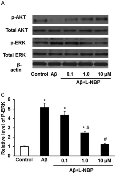
The protective effects of L-NBP are accompa-nied by increased phosphorylation of AKT and decreased phosphorylation of ERK
To further explore the neuroprotective mecha-nism of L-NBP, the phosphorylation of AKT and ERK was detected by Western blot. We found that Aβ1-42 inhibited the level of AKT phosphory-lation whereas increased the level of ERK pho- sphorylation. However, L-NBP treatment rever-
sed the effect of Aβ1-42 on AKT and ERK phos-phorylation in a dose-dependent way (Figure 3A-C). There were significant differences am- ong control group, Aβ group and Aβ+L-NBP group (P<0.05). Additionally, the basal levels of AKT or ERK were insignificantly affected by L-NBP (p>0.05; data are not shown here). The- se results indicate that L-NBP pretreatment could reverse the decreased phosphorylation of AKT and increased phosphorylation of ERK induced by Aβ1-42.
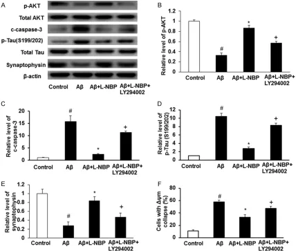
LY294002 can block the protective effects of L-NBP
[image:5.612.88.519.74.491.2]function induced by Aβ (Figure 4A-F). Pretreat- ment with L-NBP could significantly attenuate 10 μM Aβ1-42 induced increased expression of c-caspase-3 and p-Tau, and decreased the ex- pression of synaptophysin and phosphorylation of AKT in cortical neurons. However, Aβ+L-NBP+LY294002 group showed increased ex- pression of c-caspase-3 and p-Tau and decre- ased phosphorylation of AKT than Aβ+L-NBP group. And the Aβ+L-NBP+LY294002 group sh- owed increased Δψm collapse in contrast with Aβ+L-NBP group. There were significant differ-ences among control group, Aβ group, Aβ+L-NBP group and Aβ+L-Aβ+L-NBP+LY294002 group (P<0.05). Additionally, Aβ-induced decreased phosphorylation of AKT in primary cultured neu-rons could be reversed by LY294002. These results show that L-NBP may protect against Aβ induced cytotoxicity, apoptosis, synaptotoxicity, and mitochondrial dysfunction by increasing the phosphorylation of AKT.
U0126 showed a significant synergistic effect with L-NBP against Aβ1-42-induced apoptosis
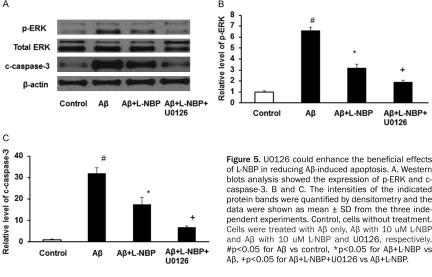
To determine the change of ERK signaling path-way underlying the neuroprotective of L-NBP, Western blot was performed. The ERK pathway inhibitor U0126 showed a significant
synergis-tic effect with L-NBP against Aβ1-42-induced in- crease expression of p-ERK and c-caspase-3 (Figure 5A-C). Pretreat with L-NBP could signifi-cantly decrease the increased expression of p-ERK and c-caspase-3 induced by Aβ, and the Aβ+L-NBP+U0126 group showed lower expres-sion of p-ERK and c-caspase-3 than Aβ+L-NBP group. There were significant differences am- ong control group, Aβ group, Aβ+L-NBP group and Aβ+L-NBP+U0126 group (P<0.05). These results show that L-NBP may protect against Aβ induced apoptosis by decreasing the phos-phorylation of ERK in cortical neurons.
Discussion
[image:6.612.92.293.73.375.2]ished the beneficial role of L-NBP in reducing the cytotoxicity, apoptosis, synaptotoxicity, and mitochondrial dysfunction induced by Aβ. And, the ERK inhibitor, U0126, which can inhibit the ERK pathway, enhanced the beneficial effects of L-NBP in reducing Aβ-induced apoptosis. As a result, we conclude that L-NBP may protect against the neuronal apoptosis, mitochondrial dysfunction and synaptotoxicity induced by Aβ through modulating PI3K/AKT and ERK signal-ing pathways.
Accumulated studies demonstrate that both extracellular and intracellular Aβ1-42 can acti-vate caspases, which lead to synaptic and neu-ronal degeneration [21]. The activation of cas-pases by Aβ1-42 is mediated by both extrinsic
[image:7.612.92.523.71.434.2]c-caspase-3 were associated with Aβ1-42 indu- ced neurotoxicity in cortical neurons. The L-NBP pre-incubation can significantly increase the p-AKT level and decrease c-caspase-3 and p- Tau expression. And, these beneficial effects were blocked by LY294002. Our results are partly consistent with previous researches [28, 29]. For instance, curcumin can improve synap-tic plassynap-ticity and cognitive function in rats via the PI3K/AKT signaling pathway [28]. Further- more, melatonin protects against Aβ-induced neurotoxicity through decreasing memory dete-rioration, synaptic disorder, tau hyperphosphor-ylation, and neurodegeneration through PI3K/ AKT/GSK3β signaling in the Aβ1-42-treated mou- se model of AD [29]. These results suggest that PI3K/AKT/GSK-3β pathway may be used as an effective therapeutic target in treating AD. MAPK pathways, the critical mediators that propagate extracellular signals from the mem-brane to the nucleus, include ERK, JNK/stress-activated protein kinase, and p38. It is gener-ally accepted that ERK activity is typicgener-ally in- volved in cell survival, proliferation, differentia-tion, and memory formation in nervous system [30]. Accumulating evidence [31, 32] have sh- own that the phosphorylation of ERK is actively involved in AD pathology. Nevertheless, views about the function of ERK under Aβ1-42
expo-sure are not conclusive. Some studies demon-strate that decreased ERK activity induced by Aβ1-42 can protect against cytotoxicity and apop-tosis [33, 34]. However, others studies indicate that Aβ-induced ERK activation leads to neural apoptosis [35, 36]. The current study showed that Aβ1-42-induced increased expression of both p-ERK and c-caspase-3 in cortical neu-rons and that these increases were attenuat- ed by L-NBP significantly. Meanwhile, the ERK pathway inhibitor U0126 enhanced beneficial role of L-NBP in inhibiting Aβ1-42-induced apop-tosis. These results suggest that L-NBP exhibit protective effects through inhibition of ERK ph- osphorylation in Aβ1-42-induced neuronal apop-tosis. Our results are partly consistent with pre-vious investigations, which reported that in iso- lated hippocampal cell culture, increased phos-phorylated ERK by Aβ exposure was in parallel with that in cell death and apoptosis and insulin could inhibit ERK phosphorylation induced by Aβ1-42 [37].
As the source of energy, mitochondria play a crucial role in regulating cell apoptosis and redox balance, and the “mitochondrial cascade hypothesis” has been proposed to be involved in the pathogenesis of AD [38]. Increasing evi-dence has reported that Aβ1-42 exposure impairs mitochondrial permeability and that mitochon-Figure 5. U0126 could enhance the beneficial effects of L-NBP in reducing Aβ-induced apoptosis. A. Western blots analysis showed the expression of p-ERK and c-caspase-3. B and C. The intensities of the indicated protein bands were quantified by densitometry and the data were shown as mean ± SD from the three inde-pendent experiments. Control, cells without treatment.
Cells were treated with Aβ only, Aβ with 10 uM L-NBP and Aβ with 10 uM L-NBP and U0126, respectively.
[image:8.612.90.522.74.351.2]drial permeability transition pore openings lead to Δψm collapse and pro-apoptotic factors re- lease from mitochondrial to cytosol [39, 40]. Accumulating evidence suggests that mito-chondrial perturbation plays a critical role in synaptic failure and degeneration in AD [41, 42]. The present study further showed that L-NBP significantly attenuated Aβ1-42-induced Δψm collapse and synaptotoxicity, and that LY294002 blocked this effect of L-NBP. The present results are consistent to the study of Zhang Yu et al. [43], which has found that L-NBP can reconstruct synaptic and spine function in aged APP/PS1 AD transgenic mice by suppress-ing Aβ1-42 plaques deposition and neuroinflam-matory response and that Wnt/β-catenin sig-naling pathway may be involved [43].
In conclusion, our findings indicate that L-NBP has protective effects on Aβ1-42 induced cell injury possibly by regulating both PI3K/AKT and ERK signaling pathways. The PI3K/AKT and ERK pathways may become potential therapeu-tic targets in AD treatment.
Disclosure of conflict of interest
None.
Address correspondence to: Yifeng Du, Department of Neurology, Shandong Provincial Hospital Affili- ated to Shandong University, No. 324 Jingwu Road, Jinan 250021, P.R. China. Tel: +86-0531-68776451; Fax: +86-0531-68776451; E-mail: duyifengzr@163. com; Wei Dong, Department of Thoracic Surgery, Shandong Provincial Hospital Affiliated to Shandong University, No. 324 Jingwu Road, Jinan 250021, P.R. China. Tel: 68776387; Fax: +86-0531-68777100; E-mail: dongwei318@163.com
References
[1] Cordonnier C, van der Flier WM. Brain micro-bleeds and Alzheimer’s disease: innocent ob-servation or key player? Brain 2011; 134: 335-44.
[2] De Strooper B, Karran E. The cellular phase of Alzheimer’s disease. Cell 2016; 164: 603-15. [3] Majd S, Zarifkar A, Rastegar K and Takhshid
MA. Different fibrillar Abeta 1-42 concentra-tions induce adult hippocampal neurons to re-enter various phases of the cell cycle. Brain Res 2008; 1218: 224-229.
[4] Rohn TT. The role of caspases in Alzheimer’s disease; potential novel therapeutic opportuni-ties. Apoptosis 2010; 15: 1403-1409.
[5] Li J, Ding X, Zhang R, Jiang W, Sun X, Xia Z, Wang X, Wu E, Zhang Y and Hu Y. Harpagoside ameliorates the amyloid-beta-induced cogni-tive impairment in rats via up-regulating BDNF expression and MAPK/PI3K pathways. Neuro-science 2015; 303: 103-114.
[6] Morishima Y, Gotoh Y, Zieg J, Barrett T, Takano H, Flavell R, Davis RJ, Shirasaki Y and Green-berg ME. Beta-amyloid induces neuronal apop-tosis via a mechanism that involves the c-Jun N-terminal kinase pathway and the induction of Fas ligand. J Neurosci 2001; 21: 7551-7560.
[7] Modi PK, Komaravelli N, Singh N and Sharma P. Interplay between MEK-ERK signaling, cyclin D1, and cyclin-dependent kinase 5 regulates cell cycle reentry and apoptosis of neurons. Mol Biol Cell 2012; 23: 3722-3730.
[8] Srinivasan M, Bayon B, Chopra N and Lahiri DK. Novel nuclear factor-kappaB targeting peptide suppresses beta-amyloid induced in-flammatory and apoptotic responses in neuro-nal cells. PLoS One 2016; 11: e0160314. [9] Zheng Y, Wang J, Li D, Guo M, Zhen M and
Chang Q. Wnt/ss-catenin signaling pathway against Abeta toxicity in PC12 cells. Neurosig-nals 2016; 24: 40-47.
[10] Ashraghi MR, Pagano G, Polychronis S, Nicco-lini F and Politis M. Parkinson’s disease, diabe-tes and cognitive impairment. Recent Pat En-docr Metab Immune Drug Discov 2016; 10: 11-21.
[11] Li M and Qian S. Gastrodin protects neural pro-genitor cells against amyloid beta (1-42)-in-duced neurotoxicity and improves hippocam-pal neurogenesis in amyloid beta (1-42)-in- jected mice. J Mol Neurosci 2016; 60: 21-32. [12] Peng Y, Zeng X, Feng Y and Wang X.
Antiplate-let and antithrombotic activity of L-3-n-bu-tylphthalide in rats. J Cardiovasc Pharmacol 2004; 43: 876-881.
[13] Ye J, Zhai L, Zhang S, Zhang Y, Chen L, Hu L and Ding Z. DL-3-n-butylphthalide inhibits pla- telet activation via inhibition of cPLA2-mediat-ed TXA2 synthesis and phosphodiesterase. Platelets 2015; 26: 736-744.
[14] Chang Q and Wang XL. Effects of chiral 3-n-butylphthalide on apoptosis induced by tran-sient focal cerebral ischemia in rats. Acta Phar-macol Sin 2003; 24: 796-804.
[15] Xu HL and Feng YP. Inhibitory effects of chiral 3-n-butylphthalide on inflammation following focal ischemic brain injury in rats. Acta Phar-macol Sin 2000; 21: 433-438.
[17] Peng Y, Hu Y, Xu S, Li P, Li J, Lu L, Yang H, Feng N, Wang L and Wang X. L-3-n-butylphthalide re-duces tau phosphorylation and improves cog-nitive deficits in Abeta PP/PS1-Alzheimer’s transgenic mice. J Alzheimers Dis 2012; 29: 379-391.
[18] Lei H, Zhao CY, Liu DM, Zhang Y, Li L, Wang XL and Peng Y. l-3-n-Butylphthalide attenuates beta-amyloid-induced toxicity in neuroblasto-ma SH-SY5Y cells through regulating mito-chondrion-mediated apoptosis and MAPK sig-naling. J Asian Nat Prod Res 2014; 16: 854-864.
[19] Troy CM, Rabacchi SA, Friedman WJ, Frappier TF, Brown K and Shelanski ML. Caspase-2 me-diates neuronal cell death induced by beta-amyloid. J Neurosci 2000; 20: 1386-1392. [20] Lesne S, Gabriel C, Nelson DA, White E,
Mack-enzie ET, Vivien D and Buisson A. Akt-depen-dent expression of NAIP-1 protects neurons against amyloid-{beta} toxicity. J Biol Chem 2005; 280: 24941-24947.
[21] Gotz J, Eckert A, Matamales M, Ittner LM and Liu X. Modes of Abeta toxicity in Alzheimer’s disease. Cell Mol Life Sci 2011; 68: 3359-3375.
[22] Dickson DW. Apoptotic mechanisms in Alzhei- mer neurofibrillary degeneration: cause or ef-fect? J Clin Invest 2004; 114: 23-27.
[23] Salminen A, Ojala J, Suuronen T, Kaarniranta K and Kauppinen A. Amyloid-beta oligomers set fire to inflammasomes and induce Alzheimer’s pathology. J Cell Mol Med 2008; 12: 2255-2262.
[24] Lustbader JW, Cirilli M, Lin C, Xu HW, Takuma K, Wang N, Caspersen C, Chen X, Pollak S, Chaney M, Trinchese F, Liu S, Gunn-Moore F, Lue LF, Walker DG, Kuppusamy P, Zewier ZL, Arancio O, Stern D, Yan SS and Wu H. ABAD directly links Abeta to mitochondrial toxicity in Alzheimer’s disease. Science 2004; 304: 448-452.
[25] Kitagishi Y, Nakanishi A, Ogura Y and Matsuda S. Dietary regulation of PI3K/AKT/GSK-3beta pathway in Alzheimer’s disease. Alzheimers Res Ther 2014; 6: 35.
[26] Manning BD and Cantley LC. AKT/PKB signal-ing: navigating downstream. Cell 2007; 129: 1261-1274.
[27] Lee KY, Koh SH, Noh MY, Kim SH and Lee YJ. Phosphatidylinositol-3-kinase activa-tion blocks amyloid beta-induced neurotoxi- city. Toxicology 2008; 243: 43-50.
[28] Wang R, Li YH, Xu Y, Li YB, Wu HL, Guo H, Zhang JZ, Zhang JJ, Pan XY and Li XJ. Curcumin produces neuroprotective effects via activating brain-derived neurotrophic factor/TrkB-depen-dent MAPK and PI-3K cascades in rofactor/TrkB-depen-dent corti-cal neurons. Prog Neuropsychopharmacol Biol Psychiatry 2010; 34: 147-153.
[29] Ali T and Kim MO. Melatonin ameliorates amy-loid beta-induced memory deficits, tau hyper-phosphorylation and neurodegeneration via PI3/Akt/GSk3beta pathway in the mouse hip-pocampus. J Pineal Res 2015; 59: 47-59. [30] Roskoski R Jr. ERK1/2 MAP kinases: structure,
function, and regulation. Pharmacol Res 2012; 66: 105-143.
[31] Kirouac L, Rajic AJ, Cribbs DH and Padmanab-han J. Activation of Ras-ERK signaling and GSK-3 by amyloid precursor protein and amy-loid beta facilitates neurodegeneration in Al-zheimer’s disease. eNeuro 2017; 4.
[32] Deng LJ, Cheng C, Wu J, Wang CH, Zhou HB and Huang J. Oxabicycloheptene sulfonate pro-tects against beta-amyloid-induced toxicity by activation of PI3K/Akt and ERK signaling path-ways via GPER1 in C6 cells. Neurochem Res 2017; 42: 2246-2256.
[33] Fan CD, Li Y, Fu XT, Wu QJ, Hou YJ, Yang MF, Sun JY, Fu XY, Zheng ZC and Sun BL. Reversal of beta-amyloid-induced neurotoxicity in PC12 cells by curcumin, the important role of ROS-mediated signaling and ERK pathway. Cell Mol Neurobiol 2017; 37: 211-222.
[34] Savage MJ, Lin YG, Ciallella JR, Flood DG and Scott RW. Activation of c-Jun N-terminal kinase and p38 in an Alzheimer’s disease model is as-sociated with amyloid deposition. J Neurosci 2002; 22: 3376-3385.
[35] Frasca G, Carbonaro V, Merlo S, Copani A and Sortino MA. Integrins mediate beta-amyloid-in-duced cell-cycle activation and neuronal de- ath. J Neurosci Res 2008; 86: 350-355. [36] Yang H, Wang S, Yu L, Zhu X and Xu Y.
Esculen-toside A suppresses Abeta(1-42)-induced neu-roinflammation by down-regulating MAPKs pathways in vivo. Neurol Res 2015; 37: 859-866.
[37] Ghasemi R, Moosavi M, Zarifkar A, Rastegar K and Maghsoudi N. The interplay of Akt and ERK in Abeta toxicity and insulin-mediated pro-tection in primary hippocampal cell culture. J Mol Neurosci 2015; 57: 325-334.
[38] Swerdlow RH, Burns JM and Khan SM. The Al-zheimer’s disease mitochondrial cascade hy-pothesis: progress and perspectives. Biochim Biophys Acta 2014; 1842: 1219-1231. [39] Santos RX, Correia SC, Wang X, Perry G, Smith
MA, Moreira PI and Zhu X. Alzheimer’s disease: diverse aspects of mitochondrial malfunction-ing. Int J Clin Exp Pathol 2010; 3: 570-581. [40] Lu Y, Wang R, Dong Y, Tucker D, Zhao N, Ahmed
ME, Zhu L, Liu TC, Cohen RM and Zhang Q. Low-level laser therapy for beta amyloid toxicity in rat hippocampus. Neurobiol Aging 2017; 49: 165-182.
[42] Du H, Guo L, Fang F, Chen D, Sosunov AA, McK-hann GM, Yan Y, Wang C, Zhang H, Molkentin JD, Gunn-Moore FJ, Vonsattel JP, Arancio O, Chen JX and Yan SD. Cyclophilin D deficiency attenuates mitochondrial and neuronal pertur-bation and ameliorates learning and memory in Alzheimer’s disease. Nat Med 2008; 14: 1097-1105.