Original Article
Gene expression profiling and identification of key
genes involved in neonatal hypoxic-ischemic brain injury
Jing Shi1,2*, Yi Zhou3*, Xiaoyan Yang1,2, Dapeng Chen1,2
1Department of Pediatrics, West China Second University Hospital, Sichuan University, Chengdu 610041, Sichuan
Province, P.R. China; 2Key Laboratory of Birth Defects and Related Disease of Women and Children (Sichuan
University), Ministry of Education, Chengdu 610041, Sichuan Province, P.R. China; 3No. 4 West China Teaching
Hospital of Sichuan University, Chengdu, Sichuan Province, P.R. China. *Equal contributors.
Received August 23, 2016; Accepted October 14, 2016; Epub January 15, 2017; Published January 30, 2017
Abstract: This study aimed to get a better understanding on the molecular circuitry and identify potential critical genes as therapeutic targets for neonatal hypoxic-ischemic (HI) encephalopathy. The microarray data of GSE37777, including 4 HI samples and 4 controls, was downloaded from the GEO database. Differentially expressed genes (DEGs) were screened in the contralateral cerebral cortices of mature rats (8 weeks old) after neonatal HI brain insult. Pathway clustering analysis was performed and a functionally grouped pathway network of DEGs was con-structed. Besides, a protein-protein interaction (PPI) network was concon-structed. Total 973 DEGs (599 up- and 374 down-regulated) were identified in HI group compared with the controls. Furthermore, a functionally grouped path -way network of DEGs was constructed. Hedgehog signaling path-way was identified and path-way-related genes
SHH, DHH, WNT1, WNT2B, and WNT4 were up-regulated in HI group. Furthermore, CCND1, SHH and RET were hub proteins in the PPI network. Wnt signaling pathway may be activated by the hedgehog signaling pathway in the
contralateral cerebral cortices of mature rats after neonatal HI injury. CCND1 may be involved in apoptosis and cell cycle regulation in neonatal HI encephalopathy. Besides, RET may play a role in neonatal HI encephalopathy.
Keywords: Neonatal hypoxic-ischemic brain injury, differentially expressed genes, pathway clustering analysis, protein-protein interaction network, hedgehog signaling pathway
Introduction
Neonatal hypoxia-ischemia (HI) brain injury still remains an major issue as it is a frequent cause of acute mortality and chronic disability in new-borns [1]. Neonatal HI encephalopathy can ca- use long-lasting morbidity, including seizure, cerebral palsy, and cognitive retardation in newborns and children [2, 3]. Currently, there is no definitive therapeutic intervention that can minimize brain disorder induced by HI except that few studies indicated the possible benefits of hypothermia in some moderate cases [4, 5]. The treatment strategies are restricted due to the incomplete understanding of the underlying pathogenesis in neonatal HI brain insult. Th- erefore, further investigations on the molecular mechanisms of neonatal HI brain injury remain a high priority.
Many studies have been performed to investi-gate the mechanisms of HI brain damage in the
both neuroprotective and neurotoxic properties in HI brain injury by its participation in distinct events such as neovascularization, neuropro-tection and the apoptotic process [10]. Besides, Tian et al. showed that cytoglobin (CYGB) was up-regulated by HI in the neonatal brain and CYGB might exhibit neuroprotective effects possibly by antioxidant and anti-apoptotic func-tions as well as through stimulating angiogene-sis [11]. In addition, the work of Li et al. demon-strated that the activation of the phosphati- dylinositol 3-kinase/protein kinase B (PI3K/ Akt) signaling pathway could protect the brain in the neonatal rat model with HI brain damage [12]. Moreover, Wang et al. suggested that the increase in neural stem cells might be regulat-ed by the hregulat-edgehog signaling pathway in HI neonatal rats and thus alleviated brain damage [13]. However, the molecular mechanisms un- derlying the development and progression of HI encephalopathy deserve further research. Unravelling the complex functions of more criti-cal genes and pathways involved in the mecha-nisms of HI brain injury may be important for exploring novel and effective therapies.
The Rice model of unilateral HI has been widely used as a useful experimental tool to study neonatal HI brain insult [14], but the resulting brain damage is reported to be mainly restrict-ed to one hemisphere basrestrict-ed on the histological analysis and behavioral tests [15, 16]. However, neonatal unilateral ischemia and bilateral hypoxia was identified to induce a long-term deficit in the reducing ability in the contralateral hemisphere by using the electron paramagnet-ic resonance imaging technique, lasting at least until the age of 8 weeks in the Rice model [17]. Recently, Kojima et al. performed comprehen-sive gene expression and network analysis using a DNA microarray system in contralateral cerebral cortices of mature rats (8 weeks old) after neonatal HI brain insult. They revealed that many up-regulated genes were related to cell death signaling even within the contralat-eral cerebral hemisphere [18]. In this study, we analyzed the differentially expressed genes (DEGs) in the contralateral cerebral cortex of mature rats following HI brain injury using the same gene expression profiling. Comprehensive bioinformatics analysis was used to analyze the significant pathways and to construct the pro -tein-protein interaction (PPI) network for inves-tigating the critical DEGs associated with HI
brain injury. We aimed to get a better under-standing of the molecular circuitry in neonatal HI brain insult and identify potentially critical genes as therapeutic targets for neonatal HI encephalopathy.
Materials and methods
Affymetrix microarray data
The microarray data of GSE37777 [18] depos-ited by Kojima et al. was downloaded from the Gene Expression Omnibus (GEO) database [19] in NCBI (http://www.ncbi.nlm.nih.gov/geo/). The platform information is GPL7294 Agilent- 014879 Whole Rat Genome Microarray 4x44K G4131F (Probe Name version). Eight samples were included in this dataset. As described in the original study [18], pregnant Wistar rats were purchased and the pups were reared with their dams until the start of the experiment. A total of 8 rat pups from different litters were used in the experiment. Thereinto, 4 Wistar rats (7-day-old) were subjected to the Rice model construction procedure to induce HI brain injury. Briefly, these 4 rats in the HI group were anesthetized with ether, and the left carotid artery was isolated and ligated with sur-gical silk. These rats were allowed to recover for 1-2 hours and then exposed to 1 hour of hypox-ia condition by being placed in a plastic cham-ber which was perfused with a mixture of humidified 8% oxygen balanced with nitrogen. While HI brain insult was not induced in the other 4 Wistar rats (7-day-old) that was used as controls. The left carotid artery of the control rats was isolated but not ligated. Animals were killed 7 weeks after the induction of HI brain insult. The cerebral cortices contralateral to the HI brain insult in the HI group and the cerebral cortices on the same side as the Rice models in the control animals were removed. Total RNA was then extracted from the tissues.
Data preprocessing and screening of DEGs
22,503 sequences. Up-regulated and down-regulated genes were identified in the HI group compared with the control group using limma package [21]. Fold change (FC) was used to evaluate whether genes were differentially ex- pressed. The FC was expressed as a log2 value in keeping with the microarray output format [22]. In order to complete the significance of a gene’s differential expression estimation, we attempted to get the most robust or reliable lists of DEGs by simultaneously filtering our results on p-value and a minimum cutoff of absolute log2 FC. In this study, p-value < 0.05 and FC ≥ 1.5 were chosen as the threshold for identifying DEGs.
Pathway enrichment analysis
ClueGO, an easy-to-use Cytoscape plug-in, strongly improves biological interpretation of a large list of genes. ClueGO can integrate Kyoto Encyclopedia of Genes and Genomes (KEGG) pathways and create a functionally organized pathway term network [23]. For biological net -works, CluePedia can calculate the correlation for DEGs based on four tests, Spearman’s rank, Pearson correlation, distance correlation and maximal information coefficient, to investigate linear and non-linear dependencies between the implemented variables [24].
In this study, we used the Cytoscape [25] plug-in, ClueGO+Cluepedia [23] to integrate KEGG pathway terms to create a functionally grouped pathway network of the DEGs. KEGG pathway terms served as the clustering criterion with a two-sided (enrichment/depletion) hypergeo-metric test followed by Bonferroni correction (significance level of 0.05) to identify signifi -cantly affected pathways. The network repre-sented each pathway as an individual node, while the edges between pathways denoted an approximation of biological interaction between the pathways based on the cross-pathway fea-ture overlap. The node size corresponded to the statistical significance for each term enriched. Additionally, the most significant pathways (with bigger node size) identified in this network were selected as our focus and were subse-quently carried out for DEG distribution presen-tation using cytoKEGG [26], a Cytoscape plug-in.
PPI network construction
[image:3.612.94.520.72.296.2]existence of an interaction and the interactions are provided with a confidence score [27]. Different cut-off score was calculated to distin-guish three confidence categories (low-, medi -um- and high-confidence) [28]. A protein with an evidence score greater than 0.4 is consid-ered to have medium confidence of interaction with the other proteins [27]. In our study, the PPIs were identified by using data extracted from the STRING database. The PPIs with a confidence score > 0.4 (medium confidence) were selected to construct the final PPI network [29]. In addition, the Cytoscape software [25] was applied to create the network visualiza-tions, where nodes represented proteins and edges represented physical interactions. The extended degree of each node in the network was calculated. The nodes with higher degree were identified to be hub proteins.
Results
Data preprocessing and DEGs screening
The gene expression profile after normalization was shown in Figure 1A. A t test in limma pack-age with a significance level set at p-value < 0.05 was carried out. Moreover, the DEGs were selected using the other criterion of FC ≥ 1.5 in expression between the HI group and the con-trol group. As a result, a total of 973 DEGs were identified in the contralateral cerebral cortex of mature rats (8 weeks old) in the HI group
com-pared with those in the control group, including 599 up-regulated genes and 374 down-regulat-ed genes. Heat-map of the DEGs was shown in
Figure 1B.
Pathway enrichment analysis
In this study, KEGG pathway terms served as the clustering criterion with a two-sided hyper-geometric test followed by Bonferroni correc-tion (significance level of 0.05) to identify sig -nificantly affected pathways. An annotation network based on the identified KEGG path -ways of the DEGs was constructed as a group of functionally organized pathway terms net-work. The term network information was des- cribed in detail as Figure 2. The network com-prised several significantly overrepresented terms, such as Cytokine-cytokine receptor in- teraction, Endocytosis, Antigen processing and presentation, Complement and coagulation ca- scades, and Hedgehog signaling pathway. Be- sides, since the node size represented the term enrichment significance as described above (Figure 2), we found that the hedgehog signal-ing pathway with bigger node size seemed to be most significantly involved.
Additionally, further sub-network analysis of the pathway network was visualized specifically to understand the significant pathway involved in the contralateral cerebral cortex after HI brain damage. Genes that were involved in the Figure 2. Pathway network analysis of DEGs. The network represents each pathway as an individual node, while the edges between pathways denote an approximation of biological interaction between the pathways based on the cross-pathway feature overlap. DEGs are in red text. The node size represents the term enrichment significance. Hedgehog signaling pathway with bigger node size seemed to be most significant involved in the contralateral cere -bral cortex of mature rats (8 weeks old) after neonatal HI brain injury.
[image:5.612.94.523.147.311.2]Hedgehog signaling pathway were shown in
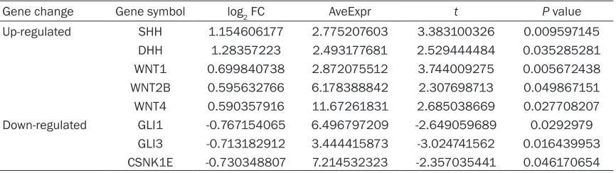
Figure 3. Besides, the changes of DEGs invo- lved in the Hedgehog signaling pathway were represented in Table 1. The results showed that the hedgehog signaling pathway-related genes sonic hedgehog (SHH), desert hedgehog (DHH), wingless-type MMTV integration site family, member 1 (WNT1), WNT2B, and WNT4
were up-regulated in HI samples.
PPI network construction
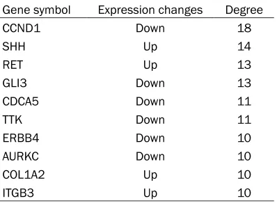
The PPI network (a confidence score > 0.4) based on the DEGs was created, consisting of 395 nodes and 528 interactions (edges) ( Fi-gure 4). Among these 395 nodes, 257 up-regu-lated genes and 138 down-reguup-regu-lated genes were included. After analysis of the node de- grees, we found that the degrees were expo-nentially distributed and the network was scale-free. The top 10 hub proteins with higher node degrees in the PPI network were present-ed in Table 2, such as cyclin D1 (CCND1) (de- gree = 18), SHH (degree = 14), ret
proto-onco-gene (RET) (degree = 13), glioma-associated (GLI) family zinc finger 3 (GLI3) (degree = 13), and cell division cycle associated 5 (CDCA5) (degree = 11).
Discussion
Neonatal HI brain injury can lead to serious brain damage and it is a common cause of neu-rological handicaps in adulthood [30]. Major efforts are needed to understand neonatal HI brain injury at a molecular level. Kojima et al.
[image:6.612.91.524.84.206.2]suggested that progressive neuronal damage may occur in the contralateral cerebral cortex of mature rats [18] though previous studies mostly focus on the investigations of the ipsilat-eral side of the brain at early stages of neonatal HI encephalopathy. In the current study, a total of 973 DEGs were identified in cerebral cortices of mature rats (8 weeks old) after neonatal HI brain insult compared with the corresponding controls. Furthermore, a functionally grouped pathway network of DEGs was constructed and hedgehog signaling pathway was identified. The Table 1. The changes of DEGs that were involved in the Hedgehog signaling pathway
Gene change Gene symbol log2 FC AveExpr t P value
Up-regulated SHH 1.154606177 2.775207603 3.383100326 0.009597145 DHH 1.28357223 2.493177681 2.529444484 0.035285281 WNT1 0.699840738 2.872075512 3.744009275 0.005672438 WNT2B 0.595632766 6.178388842 2.307698713 0.049867151 WNT4 0.590357916 11.67261831 2.685038669 0.027708207 Down-regulated GLI1 -0.767154065 6.496797209 -2.649059689 0.0292979
[image:6.612.93.524.244.411.2]GLI3 -0.713182912 3.444415873 -3.024741562 0.016439953 CSNK1E -0.730348807 7.214532323 -2.357035441 0.046170654 AveExpr, average expression value.
hedgehog signaling pathway-related genes
SHH, DHH, WNT1, WNT2B, and WNT4 were up-regulated in HI group. Furthermore, CCND1,
SHH, RET and GLI3 were hub proteins in thes-cale-free PPI network.
The hedgehog signaling pathway is highly con-served and crucial for the development of the normal embryo [31]. Hedgehog signaling can regulate both the patterning and polarity events in early embryogenesis and the morphogenesis of specific tissues and organs in mammals [32]. The pathway is then silenced in most adult tissues but can be reactivated after injury to accelerate repair and regeneration [32]. Rece- ntly, Wang et al. had demonstrated that umbili-cal cord blood mononuclear cells (UCBMC) could promote neuronal differentiation and reduce glial differentiation in neonatal HI rats via the hedgehog signaling pathway [13]. In the present study, we found that the hedgehog sig-naling pathway with bigger node size in the pathway network of DEGs was significantly identified (Figure 2), suggesting that hedgehog signaling pathway might be involved in the con-tralateral cerebral cortex of mature rats (8 we- eks old) after neonatal HI brain insult to accel-erate repair and regeneration. Besides, the study of Ferent et al. revealed that SHH tran-scripts were not detected in control rats but were up-regulated at a time-dependent manner in the oligodendroglia lineage within the central nervous system (CNS) lesion [33]. In accorda- nce with previous findings, our results showed that SHH was up-regulated in the HI group, indi-cating the activation of hedgehog signaling pathway in the contralateral cerebral cortex of mature rats in neonatal HI brain insult. On the other hand, the downstream molecules WNT1,
WNT2B, and WNT4 were up-regulated in HI
group (Figure 3). WNT1, WNT2B, and WNT4
were members of WTN gene family which had been implicated in some developmental pro-cesses, such as regulation of cell fate and pat-terning during embryogenesis [34]. Wnt signal-ing also activates non-canonical pathways which regulate planar cell polarity via stimulat-ing cytoskeletal reorganization and can also result in calcium mobilization [34]. Furthermore, excessive entry of Ca2+ into cells was recog-nized as an important mechanism of HI brain injury. In this context, we suggested that the Wnt signaling pathway might be activated by the hedgehog signaling pathway and might play an essential role in the contralateral cerebral cortex of mature rats after neonatal HI enceph-alopathy via participating in the compensatory processes.
CCND1 is a member of the highly conserved cyclin family whose members are characterized by a dramatic periodicity in protein abundance throughout the cell cycle [35]. Cai et al. had reported that CCND1 contributed to cell prolif-eration and repression of CCND1 could induce apoptosis and cell cycle arrest in osteosarcoma [36]. Moreover, Northington et al. had demon-strated that apoptosis could significantly con -tribute to delayed cell death in perinatal HI [37]. In line with previous study, our study showed that CCND1 was a hub protein in the PPI net-work and was down-regulated in HI group (Figure 4), suggesting that CCND1 may play a significant role via involving in apoptosis and cell cycle regulation in the contralateral cere-bral cortex of mature rats after neonatal HI encephalopathy.
In addition, RET, a member of the cadherin superfamily, encodes one of the receptor tyro-sine kinases which are involved in many cellular mechanisms including cell proliferation, neuro-nal navigation, cell migration, and cell differen-tiation on binding with glial cell derived neuro-trophic factor (GDNF) family ligands [38]. Duarte et al. had revealed that the neuropro-tective effect of exogenous GDNF could be observed in different experimental models of brain ischemia [39]. Furthermore, Xu et al.
showed that increased expression of GDNF family receptor RET was identified in the isch -emic cortex after electro acupuncture on HI brain injury, which at least in part was
attribut-Table 2. Top 10 hub genes of the PPI network
Gene symbol Expression changes Degree
CCND1 Down 18
SHH Up 14
RET Up 13
GLI3 Down 13
CDCA5 Down 11
TTK Down 11
ERBB4 Down 10
AURKC Down 10
COL1A2 Up 10
[image:7.612.91.290.85.231.2]ed to the activation of PI3-K/Akt signaling path-way [40]. Additionally, our study identified that RET was a hub protein in the PPI network (Figure 4). Thus, we suggested that up-regulat-ed RET might play an essential role in the pro-gression of neonatal HI encephalopathy. In conclusion, the critical genes (CCND1, SHH,
RET, WNT1, WNT2B and WNT4) in the contralat-eral cerebral cortex of mature rats following neonatal HI encephalopathy had been identi-fied based on the gene expression profile. The Wnt signaling pathway may be activated by the hedgehog signaling pathway and play an essen-tial role in the contralesional cerebral cortex of mature rats after neonatal HI encephalopathy via participating in the compensatory process-es. Besides, CCND1 may play a significant role via involving in apoptosis and cell cycle regula-tion in the contralesional cerebral cortex after neonatal HI brain injury. Moreover, the up-regu-lated RET may play an essential role in the pro-gression of neonatal HI encephalopathy. Be- cause of the relatively small number of samples in the current study, further studies with a larg-er sample size to validate and detlarg-ermine the role of the DEGs identified are needed. Further investigations on the molecular mechanism of neonatal HI encephalopathy may facilitate the development of novel diagnostic and therapeu-tic applications.
Acknowledgements
This work was supported by National Natural Science Foundation of China (grant number: 81000263) and National Key Development Program of Clinical Specialties (neonatology) (grant number: 1311200003303), Applied Ba- sic Research Programs of Science and Techno- logy Department of Sichuan Province (grant number: 2016JY0180).
Disclosure of conflict of interest
None.
Address correspondence to: Dapeng Chen, Depart- ment of Pediatrics, West China Second University Hospital, Sichuan University, 20 Section 3 South Renming Road, Chengdu 610041, Sichuan Province, P.R. China. Tel: +85503743; Fax: 86-28-85503784; E-mail: dapengch89@163.com
References
[1] Liu S, Zhu S, Zou Y, Wang T and Fu X. Knock -down of IL-1beta improves hypoxia-ischemia brain associated with IL-6 up-regulation in cell and animal models. Mol Neurobiol 2015; 51: 743-752.
[2] Glass HC, Hong KJ, Rogers EE, Jeremy RJ, Bon-ifacio SL, Sullivan JE, Barkovich AJ and Ferriero DM. Risk factors for epilepsy in children with neonatal encephalopathy. Pediatr Res 2011; 70: 535-540.
[3] Thornton C, Rousset CI, Kichev A, Miyakuni Y, Vontell R, Baburamani AA, Fleiss B, Gressens P and Hagberg H. Molecular mechanisms of neonatal brain injury. Neurol Res Int 2012; 2012: 506320.
[4] Shankaran S, Barnes PD, Hintz SR, Laptook AR, Zaterka-Baxter KM, McDonald SA, Ehren-kranz RA, Walsh MC, Tyson JE and Donovan EF. Brain injury following trial of hypothermia for neonatal hypoxic-ischaemic encephalopathy. Arch Dis Child Fetal Neonatal Ed 2012; 97: F398-F404.
[5] Choi HA, Badjatia N and Mayer SA. Hypother-mia for acute brain injury-mechanisms and practical aspects. Nat Rev Neurol 2012; 8: 214-222.
[6] Perlman JM. Summary proceedings from the neurology group on hypoxic-ischemic encepha-lopathy. Pediatrics 2006; 117: S28-33. [7] Northington FJ, Chavez-Valdez R and Martin LJ.
Neuronal cell death in neonatal hypoxia-isch-emia. Ann Neurol 2011; 69: 743-758.
[8] Lara-Celador I, Goñi-de-Cerio F, Alvarez A and Hilario E. Using the endocannabinoid system as a neuroprotective strategy in perinatal hy-poxic-ischemic brain injury. Neural Regen Res 2013; 8: 731.
[9] Chavez-Valdez R, Martin L, Flock D and Northington F. Necrostatin-1 attenuates mito -chondrial dysfunction in neurons and astro-cytes following neonatal hypoxia-ischemia. Neuroscience 2012; 219: 192-203.
[10] Fan X, Heijnen CJ, van der Kooij MA, Groenendaal F and van Bel F. The role and regulation of hypoxia-inducible factor-1α ex -pression in brain development and neonatal hypoxic-ischemic brain injury. Brain Res Rev 2009; 62: 99-108.
[11] Tian SF, Yang HH, Xiao DP, Huang YJ, He GY, Ma HR, Xia F and Shi XC. Mechanisms of neuropro -tection from hypoxia-ischemia (HI) brain injury by up-regulation of cytoglobin (CYGB) in a neo-natal rat model. J Biol Chem 2013; 288: 15988-16003.
brain damage through the PI3K/Akt pathway. Int J Clin Exp Med 2015; 8: 8197.
[13] Wang X, Zhao Y and Wang X. Umbilical cord blood cells regulate the differentiation of en-dogenous neural stem cells in hypoxic isch-emic neonatal rats via the hedgehog signaling pathway. Brain Res 2014; 1560: 18-26. [14] Rice JE, Vannucci RC and Brierley JB. The influ
-ence of immaturity on hypoxic-ischemic brain damage in the rat. Ann Neurol 1981; 9: 131-141.
[15] Arteni N, Pereira L, Rodrigues A, Lavinsky D, Achaval M and Netto C. Lateralized and sex-dependent behavioral and morphological ef-fects of unilateral neonatal cerebral hypoxia-ischemia in the rat. Behav Brain Res 2010; 210: 92-98.
[16] Sameshima H and Ikenoue T. Hypoxic-isch-emic neonatal encephalopathy: animal experi-ments for neuroprotective therapies. Stroke Res Treat 2013; 2013: 659374.
[17] Yokoyama H, Ueda Y, Itoh O, Ikeda T, Noor JI and Ikenoue T. EPR imaging to estimate the in vivo intracerebral reducing ability of mature rats after neonatal hypoxic-ischemic brain in-jury. Magn Reson Imaging 2004; 22: 1305-1309.
[18] Kojima T, Ueda Y, Sato A, Sameshima H and Ikenoue T. Comprehensive gene expression analysis of cerebral cortices from mature rats after neonatal hypoxic-ischemic brain injury. J Mol Neurosci 2013; 49: 320-327.
[19] Barrett T, Troup DB, Wilhite SE, Ledoux P, Rudnev D, Evangelista C, Kim IF, Soboleva A, Tomashevsky M and Edgar R. NCBI GEO: min-ing tens of millions of expression profiles--data -base and tools update. Nucleic Acids Res 2007; 35: D760-765.
[20] Irizarry RA, Hobbs B, Collin F, Beazer-Barclay YD, Antonellis KJ, Scherf U and Speed TP. Ex-ploration, normalization, and summaries of high density oligonucleotide array probe level data. Biostatistics 2003; 4: 249-264.
[21] Smyth GK. Limma: linear models for microar-ray data. In: editors. Bioinformatics and com-putational biology solutions using R and Bio-conductor. Springer; 2005. pp. 397-420. [22] Allison DB, Cui X, Page GP and Sabripour M.
Microarray data analysis: from disarray to con-solidation and consensus. Nat Rev Genet 2006; 7: 55-65.
[23] Bindea G, Mlecnik B, Hackl H, Charoentong P, Tosolini M, Kirilovsky A, Fridman WH, Pages F, Trajanoski Z and Galon J. ClueGO: a cytoscape plug-in to decipher functionally grouped gene ontology and pathway annotation networks. Bioinformatics 2009; 25: 1091-1093.
[24] Bindea G, Galon J and Mlecnik B. CluePedia cytoscape plugin: pathway insights using
inte-grated experimental and in silico data. Bioin-formatics 2013; 29: 661-663.
[25] Shannon P, Markiel A, Ozier O, Baliga NS, Wang JT, Ramage D, Amin N, Schwikowski B and Ideker T. Cytoscape: a software environ-ment for integrated models of biomolecular interaction networks. Genome Res 2003; 13: 2498-2504.
[26] Goltsov A, Deeni Y, Khalil HS, Soininen T, Kyria-kidis S, Hu H, Langdon SP, Harrison DJ and Bown J. Systems analysis of drug-induced re-ceptor tyrosine kinase reprogramming follow-ing targeted mono-and combination anti-can-cer therapy. Cells 2014; 3: 563-591.
[27] Szklarczyk D, Franceschini A, Kuhn M, Simo -novic M, Roth A, Minguez P, Doerks T, Stark M, Muller J, Bork P, Jensen LJ, von Mering C. The STRING database in 2011: functional interac-tion networks of proteins, globally integrated and scored. Nucleic Acids Res 2011; 39: D561-D568.
[28] Fazekas D, Koltai M, Türei D, Módos D, Pálfy M, Dúl Z, Zsákai L, Szalay-Bekő M, Lenti K and Farkas IJ. SignaLink 2-a signaling pathway re -source with multi-layered regulatory networks. BMC Syst Biol 2013; 7: 7.
[29] Von Mering C, Huynen M, Jaeggi D, Schmidt S, Bork P and Snel B. STRING: a database of pre-dicted functional associations between pro-teins. Nucleic Acids Res 2003; 31: 258-261. [30] Kojima T, Ueda Y, Sato A, Sameshima H and
Ikenoue T. Comprehensive gene expression analysis of cerebral cortices from mature rats after neonatal hypoxic-ischemic brain injury. J Mol Neurosci 2013; 49: 320-327.
[31] Ingham PW and McMahon AP. Hedgehog sig-naling in animal development: paradigms and principles. Genes Dev 2001; 15: 3059-3087. [32] McMillan R and Matsui W. Molecular
path-ways: the hedgehog signaling pathway in can-cer. Clin Cancer Res 2012; 18: 4883-4888. [33] Ferent J, Zimmer C, Durbec P, Ruat M and Traif
-fort E. Sonic hedgehog signaling is a positive oligodendrocyte regulator during demyelin-ation. J Neurosci 2013; 33: 1759-1772. [34] Willert K and Nusse R. Wnt proteins. Cold
Spring Harb Perspect Biol 2012; 4: a007864. [35] Huerta M, Muñoz R, Tapia R, Soto-Reyes E,
Ramírez L, Recillas-Targa F, González-Mariscal L and López-Bayghen E. Cyclin D1 is transcrip -tionally down-regulated by ZO-2 via an E box and the transcription factor c-Myc. Mol Biol Cell 2007; 18: 4826-4836.
[37] Northington FJ, Graham EM and Martin LJ. Apoptosis in perinatal hypoxic-ischemic brain injury: how important is it and should it be in-hibited? Brain Res Rev 2005; 50: 244-257. [38] Schlessinger J. Cell signaling by receptor
tyro-sine kinases. Cell 2000; 103: 211-225. [39] Duarte EP, Curcio M, Canzoniero LM and
Du-arte CB. Neuroprotection by GDNF in the isch -emic brain. Growth Factors 2012; 30: 242-257.