Original Article
Clinical features of 12 cases with peripheral primitive
neuroectodermal tumors of soft tissue
Xuexia Yuan1*, Lihong Zhang2*, Linsheng Wang1, Deguo Liu1
1Medical Imaging Center, Affiliated Hospital of Jining Medical University, Jining City, Shandong Province, P. R.
China; 2CT Department, Jining No. 1 People’s Hospital, Jining City 272011, Shandong Province, P. R. China. *Equal
contributors and co-first authors.
Received April 13, 2018; Accepted May 6, 2018;Epub June 15, 2018; Published June 30, 2018
Abstract: Objective: To clarify the clinical characteristics including basic data, imaging, and pathological features of peripheral primitive neuroectodermal tumors (pPNETs) in patients. Methods: A retrospective analysis was
con-ducted on the clinical data of 12 pathologically-confirmed pPNETs patients treated in the Affiliated Hospital of Jining
Medical University from January 2012 to December 2017. All the patients were examined by CT and MRI imaging,
and the imaging findings were evaluated. The pooled tumor specimens were subject to routine H&E staining and
immunohistochemical staining for observation of pathological features. Nine patients who had received surgical re-section were followed for 1 year and their rates of tumor recurrence and metastasis were assessed. Results: Twelve enrolled patients had a mean age of 22.2 years (range, 8-50 years), with much more males than females (at a ratio of 2 to 1). The tumors were all pPNETs of soft tissue. The manifestations of pPNETs on computed tomography (CT) images showed uneven density nodules with necrosis and cystic changes, and heterogeneous enhancement mass. On MR images, the tumors were isointense or hypo-intense on T1-weighed image (T1WI), and isointense or hyper-intense on T2-weighed image (T2WI). The regions with tumor cystic degeneration or necrosis showed hypo-hyper-intense
on T1W images and hyper-intense on T2W images. Typical Homer-Wright rosette forming was noted by H&E staining. The results of immunohistochemical staining demonstrated that the positive rates of CD99, neuron-specific enolase
(NSE), Vimentin, synaptophysin (Syn), and S-100 proteins were 100%, 50%, 33.3%, 58.3% and 16.7%, respectively. After one-year follow-up, the recurrence rate of pPNETs was 33.3% and the metastasis rate was 25% in patients
with surgical resection of pPNETs. Conclusion: pPNETs of soft tissue were not specific on CT and MRI images, and the definitive diagnosis was still dependent on pathological examination.
Keywords: Primitive neuroectodermal tumor, peripheral type, computed tomography, magnetic resonance imaging
Introduction
Peripheral primitive neuroectodermal tumor (pPNET) is a clinically rare malignant tumor of the nervous system, and a group of highly malignant small round cell tumors arising from neural crest cells beyond the central system and the sympathetic nervous system [1, 2]. The tumor is characteristic of potential multi-direc-tional differentiation, invasive growth, poor prognosis, and recurrent tendency [3]. Clini- cal epidemiological studies demonstrate that pPNET accounts for approximately 4% of soft tissue sarcomas, with a 5-year survival rate of 45%, and a mortality rate of over 70% in patients [4]. Children and adolescents are at high risk for the disease. The majority of pa-
Therefore, in order to further investigate the characteristics of CT and MRI studies regarding pPNETs, in the present study ranging from January 2012 to December 2017, a retrospec-tive analysis was made to elucidate the clinical features of 12 patients with pPNETs confirmed by pathologic examinations in The Affiliated Hospital of Jining Medical University. The goal was to provide more clinical evidence for the clinical diagnosis and treatment of the tumor in patients.
Materials and methods
Study participants
All the enrolled patients in this study provided written informed consent, and this study was approved by the medical ethics committee of The Affiliated Hospital of Jining Medical Uni-versity. From January 2012 to December 2017, 12 patients with pPNETs admitted to The Affiliated Hospital of Jining Medical University were recruited in this retrospective study, and their conditions were confirmed by surgical resection or biopsy. The patients ranged in the course of disease from 1 week to 1 year (mean, 5.2 ± 1.6 months). The major findings showed one case of abdominal pain and distention, 2 cases of pelvic swelling, 4 cases of superficial soft tissue masses, 2 cases of chest tightness and pain, and 3 cases of progressive local pain and tumor compression without evident incen-tives. Nine patients were treated with surgical resection followed by adjuvant chemotherapy or radiotherapy. Three patients who could not undergo surgical resection received chemo-therapy or radiochemo-therapy. Chemochemo-therapy utilized a cyclophosphamide plus doxorubicin-based regimen, while radiotherapy adopted three-di- mensional therapy or intensity-modulated con-formal radiation therapy.
Imaging examination
Plain CT scanning and enhanced scanning were carried out using a 64-row spiral CT scanner (Philips, the Netherlands). The parameters of the 64-row spiral CT scanner were tube voltage 120 kV, current 250 mA, acquisition matrix 256 * 256, slice thickness and gap 4-8 mm, and reconstructed slice thickness 2.5 mm. The scanning range exceeded the superior and infe-rior borders of the lesions, and enhanced CT scanning was performed subsequent to plain
scanning. Prior to enhanced scanning, non-ion-ic iodine contrast agent (80-100 ml) was inject-ed at an injection speinject-ed of 3 mL/s via the cubi-tal vein using a double cylinder high-pressure injector for all the patients. A dual-phase CT scan was conducted, namely arterial phase (30 seconds after contrast injection) and venous phase (70 seconds after contrast injection). MRI examination was performed for scanning the transverse, sagittal, and coronal planes of the lesion sites using a GE 1.5-T MR system (USA). Conventional magnetic resonance exam-inations were performed on T1-weighed images (T1WI) and T2-weighed images (T2WI). TR of T1WI in spin echo sequence was 530 ms and TE was 15 ms. TR of T2WI in spin echo sequence was 4800 ms, and TE was 120 ms; slice thick-ness was 4 mm; slice gap was 1.5 mm. As con-trast agent of enhanced scan, Gd-DTPA was injected via the antecubital vein at a dose of 0.1 mmol/kg and at a rate of 2.5 ml/s.
Pathological examination
Nine patients received surgical resection, and 3 patients underwent biopsy. H&E-stained sec -tions and immunohistochemistry stained sec-tions of tumor tissue were examined for diagno-sis. The neuronal markers used in immuno- histochemistry included CD99, neuron-specific enolase (NSE), vimentin, S-100 protein, and synaptophysin (Syn). The criteria for diagnosing pPNETs were microscopically typical undiffer-entiated small round cells with less cytoplasm, hyperchromatic nuclei, and a high nucleo-cyto-plasmic ratio, visible nuclear division with or without visible rosette formation or expression of two or more positive neuronal markers.
Follow-up
Nine patients who had undergone surgical resection were followed for 1 year by means of clinic appointments and telephone calls. Imaging examinations were performed at 3, 6 and 12 months postoperatively.
Statistical analysis
Results
Basic data of patients
Of the 12 enrolled patients, 8 were males and 4 were females, with an age ranging from 8 to 50 years. The smallest nodule was 4.2 * 3.5 * 5.1 cm3 in diameter, whereas the largest nod-ule was 15 * 20 * 13 cm3 in diameter. The pPNET lesions were located in lower extremi-ties (2 cases), pelvic cavity (5 cases), abdomi-nal cavity (1 case), the lung (2 cases), and the chest walls (2 cases), respectively. All nodules were of soft-tissue type, as shown in Table 1.
CT and MRI findings
CT scan showed heterogeneous density in the tumor with necrosis and cystic changes, but no
and hyper-intense on T2W images. Contrast-enhanced MRI scans demonstrated moderate or significant enhancement in the arterial phase and sustained homogeneous or hetero-geneous enhancement in the parenchymal phase (Figure 2).
Pathological examination
[image:3.612.87.380.75.425.2]The lesions demonstrated diversified lobulated or nodular masses in volume. Between the tumor margins and surrounding tissue, vague boundaries were noted and affected the sur-rounding bone. The cut surface of pPNETs was fish-like, grayish-white, or gray-yellow, and tena -cious, with local visible necrosis. Under a light microscope, the tumor cells were small and uni-form in size, with less cytoplasm, hyperchro-matic nuclei, and apparent nuclear division Table 1. Basic patient data
Case Age (year) Sex Size (cm3) Site
1 17 Male 4.2 * 3.5 * 5.1 Right chest wall
2 21 Female 11 * 8 * 9 Left ovary
3 8 Male 7 * 5 * 4 Left chest wall
4 38 Male 12.3 * 8.6 * 7.5 Left thigh
5 12 Female 5.5 * 6.3 * 3.7 Right ovary
6 15 Male 5.2 * 4.8 * 5.8 Prostate
7 36 Male 8 * 9 * 12 Pelvic cavity
8 50 Male 11 * 12 * 7 Right lung
9 16 Female 15*20*13 Abdominal cavity
10 19 Male 7 * 8 * 13 Right thigh
11 11 Female 8 * 11 * 6 Left lung
12 23 Male 6 * 4 * 10 Pelvic cavity
calcification. The tumor foci were heterogeneous after en- hancement scan. They were moderate or showed signifi -cant heterogeneous enhan- cement in the arterial phase, and maintained incremental homogeneous enhancement in the parenchymal phase (Figure 1). The lesions in the chest wall (2 cases) invaded the surrounding ribs, which gave rise to hyperplasia and sclerosis of ribs. The lesions in the lung (2 cases) led to ipsilateral compression to the lung that was incomplete dila-tion. The lesions in the ab- dominal cavity (1 case) and in the pelvic cavity (5 cases) with clear boundary resulted in displacement and com-pression to the surrounding organs, which included the spleen, pancreas, kidney, rec-tum, and uterus, but no in- vasion.
MRI scans revealed isoin-tense or hypo-inisoin-tense opaci-ties on T1WI, and isointense or hyper-intense opacities on T2WI. The regions with tumor cystic degeneration or necro-sis showed the tumor was hypo-intense on T1W images Figure 1. Plain CT and contrast-enhanced CT images of pPNET of the right
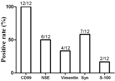
with typical Homer-Wright rosette forming (Figure 3). The results of immunohistochemical staining indicated that the positive rates were 100% (12/12) for CD99, 50% (6/12) for NSE, 33.3% (4/12) for Vimentin, 58.3% (7/12) for Syn protein, and 16.7% (2/12) for S-100 protein (Table 2, and Figure 4).
Follow-up results
At 1-year follow-up, 4 patients had local recur-rent lesions, with a recurrence rate of 33.3%, with 3 patients (including 1 case of tive bone metastasis and 1 case of
postopera-years old. Most of them were adolescents, with more males than females. This might be attrib-utable to the small sample size of the current study. Our results indicated that the lesions were located in lower extremities (2 cases), the pelvic cavity (5 cases), abdominal cavity (1 case), lung (2 cases), and chest wall (2 cases), consistent with the affected sites reported in the previous literature [14]. The vast majority of the soft tissue masses in the present study were larger than 5 cm in diameter, which might be related to the facts that the tumor masses are occult and grow in a big space as they are present in the abdominal cavity, pelvic cavity or the thoracic cavity, which was basically consis-tent with the result reported by Park et al. [15]. pPNETs grow rapidly due to their high degree of malignancy, and the tumors at an early stage may be associated with the symptoms of pain arising from soft tissue compression or destruc-tion of bone tissue. Of the 12 patients in the current study, one had abdominal distention and pain, two had pelvic swellings, four had superficial soft-tissue masses, two had chest tightness and pain, and three had progressive local pain and symptoms of tumor compression without obvious incentives.
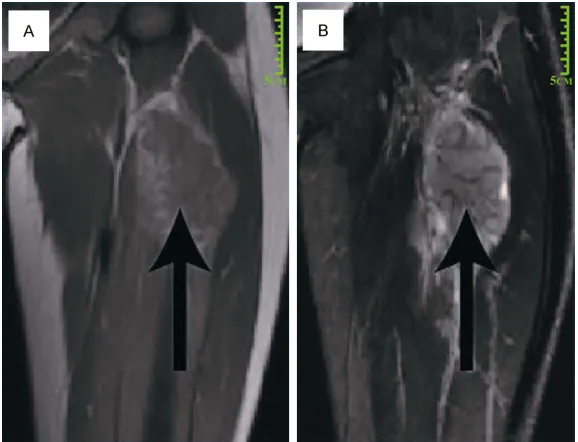
[image:4.612.89.378.71.292.2]Histologically, pPNETs are extremely similar to other types of small round malignancies which include Ewing’s sarcoma, but they are still dif-ferent in nature [16]. Currently, the difdif-ferential Figure 2. MRI images of pPNET of the right thigh. The tumor mass (Arrow)
showed vague boundaries and are lobulated. A: Isointense signals on T1WI; B: Mildly hyper-intense signals on T2WI.
Figure 3. HE staining of pPNET (200×). The arrow in -dicates Homer-Wright type rosette forming.
tive double-lung metastasis at 6 months, and 1 case of brain metastasis at 12 mon), with a metastasis rate of 25%. Discussion
[image:4.612.89.290.352.518.2]diagnosis of pPNETs depends on pathological examination. Additionally, under a light micro-scope, the cells of pPNET were small and uni-form in size, with less cytoplasm, hyperchro-matic nuclei, and apparent nuclear division with typical rosette forming. The immunohisto-chemical staining showed the expression of at least two differentiated neural antigens. The results of immunohistochemical staining in the current study revealed that CD99 was positive in 100% of the pPNET cases (12/12); NSE posi-tive was in 50% (6/12); Vimentin was posiposi-tive in 33.3% (4/12); Syn protein was positive in 58.3% (7/12); S-100 protein was positive in 16.7% (2/12); and at least two differentiat- ed neural antigens were expressed in each tumor, suggesting that the patients enrolled in
the present study met diagnostic criteria for pPNETs.
[image:5.612.90.291.97.266.2]The current study also demonstrated that on CT images, pPNET lesions in soft tissue were characterized uneven density nodules with necrosis and cystic changes, but no calcifica -tion. The tumor foci were evidently heteroge-neous after enhancement scan. They were mo- derate or significantly heterogeneous enhance -ment in the arterial phase, and maintained incrementally homogeneous enhancement in the parenchymal phase. MRI scans revealed isointense or mildly hypo-intense opacities on T1W images, and isointense or hyper-intense opacities on T2W images. The regions with tumor cystic degeneration or necrosis showed hypo-intense on T1W images and hyper-intense on T2W images. Contrast-enhanced MRI scans showed moderate or significant enhancement in the arterial phase and sustained homoge-neous or heterogehomoge-neous enhancement in the parenchymal phase. CT scanning in the current study revealed intra-tumor cystic degeneration and necrosis which might be related to necro-sis in blood-supply areas arising from infiltra -tion of tumors into the mass or the peripheral blood vessels. This is basically in line with the findings reported in previous studies [17, 18]. Surgical resection is the primary modality for treating pPNETs. It has been reported that sur-gical resection in combination with adjuvant radiotherapy and chemotherapy benefit the patients with appropriately prolonged survival [19]. pPNETs are highly malignant tumors. Although postoperative adjuvant radio-chemo-therapy can properly prolong the patients’ sur-vival, the rates of local recurrence and metas-tasis remain high after surgery [20]. In the current study, we paid follow-up visits to the patients with surgically resected pPNETs for 1 year and found that the recurrence rate was 33.3% and the metastasis rate was 25%. In conclusion, as pPNETs do not have specific manifestations on CT and MRI images, the definitive diagnosis is still dependent on patho -logical examinations. CT and MRI imaging clearly show the tumor density and signal char-acteristics, and define the scope and metasta -sis of tumors, which can provide reliable evi-dence for further development of surgical protocols and assessment of the therapeutic effects.
Table 2. Results of immunohistochemical staining in 12 patients
Case CD99 NSE Vimentin Syn S-100
1 + - + +
-2 + - - + +
3 + + - -
-4 + - + +
-5 + + - +
-6 + - + -
-7 + - - +
-8 + + - - +
9 + + - -
-10 + + - +
-11 + - + -
-12 + + - +
-Note: NSE, denotes neuron-specific enolase; Syn, synap
-tophysin.
[image:5.612.91.286.313.450.2]Disclosure of conflict of interest
None.
Address correspondence to: Lihong Zhang, CT
Department, Jining No. 1 People’s Hospital, No. 6,
Jiankang Road, Jining City 272011, Shandong Province, P. R. China. Tel: +86-0537-6056666; Fax: +86-0537-6056666; E-mail: lihongzhang61@163. com
References
[1] Fu J, Song J, Zhao Y, Wang F and Shao G. Triple-phase (99m) Tc-3P-RGD2 imaging of peripher-al primitive neuroectodermperipher-al tumor in the hip muscle group with bone metastasis. Mol Clin Oncol 2017; 6: 197-200.
[2] Chiang S, Snuderl M, Kojiro-Sanada S, Quer
Pi-Sunyer A, Daya D, Hayashi T, Bosincu L, Ogawa F, Rosenberg AE, Horn LC, Wang L, Iafrate AJ
and Oliva E. Primitive neuroectodermal tumors of the female genital tract: a morphologic, im-munohistochemical, and molecular study of 19 cases. Am J Surg Pathol 2017; 41: 761-772.
[3] Jin X, Cao J, Liu Y, Bian F, Zhao Q, Wang Y, Lv X
and Huang Y. Primitive neuroectodermal tumor
originating from the lung: a case report. Oncol Lett 2016; 12: 2692-2695.
[4] Hou W, Xu L, Zhan H, Wang H, Xu M and Yu Y.
Computed tomography and magnetic reso-nance imaging characteristics of peripheral primitive neuroectodermal tumor: a retrospec-tive analysis of 16 cases. J Comput Assist To-mogr 2017; 41: 224-230.
[5] Thoriya PJ, Watal P, Bahri NU and Rathod K. Primary spinal primitive neuroectodermal tu-mor on MR imaging. Indian J Radiol Imaging 2015; 25: 459-463.
[6] Dick EA, McHugh K, Kimber C and Michalski A.
Imaging of non-central nervous system primi-tive neuroectodermal tumours: diagnostic fea-tures and correlation with outcome. Clin Radiol 2001; 56: 206-215.
[7] Ba L, Tan H, Xiao H, Guan Y, Gao J and Gao X. Radiologic and clinicopathologic findings of pe -ripheral primitive neuroectodermal tumors. Acta Radiol 2015; 56: 820-828.
[8] Kumar V, Singh A, Sharma V and Kumar M. Pri-mary intracranial dural-based Ewing sarcoma/ peripheral primitive neuroectodermal tumor mimicking a meningioma: a rare tumor with review of literature. Asian J Neurosurg 2017; 12: 351-357.
[9] Fan C, Kong D, Tan C and Yang J. Isolated car-diac peripheral primitive neuroectodermal tu-mor: a case report. Cancer Biol Ther 2017; 18: 4-7.
[10] Zhang Y, Cai P, Chen M, Yi X, Li L, Xiao D, Liu W,
Li W and Li Y. Imaging findings of adrenal prim -itive neuroectodermal tumors: a series of sev-en cases. Clin Transl Oncol 2017; 19: 641-649.
[11] Khmou M, Malihy A, Lamalmi N, Rouas L and Alhamany Z. Peripheral primitive neuroecto-dermal tumors of the spine: a case report and review of the literature. BMC Res Notes 2016; 9: 438.
[12] Yan Y, Xu T, Chen J, Hu G and Lu Y. Intraspinal
Ewing’s sarcoma/primitive neuroectodermal tumors. J Clin Neurosci 2011; 18: 601-606. [13] Qi W, Deng X, Liu T, Hou Y, Yang C, Wu L, Fang
J, Tong X, Yang J and Xu Y. Comparison of pri-mary spinal central and peripheral primitive neuroectodermal tumors in clinical and imag-ing characteristics and long-term outcome. World Neurosurg 2016; 88: 359-369.
[14] Tan Y, Zhang H, Ma GL, Xiao EH and Wang XC.
Peripheral primitive neuroectodermal tumor: dynamic CT, MRI and clinicopathological char-acteristics--analysis of 36 cases and review of the literature. Oncotarget 2014; 5: 12968-12977.
[15] Park JY, Lee S, Kang HJ, Kim HS and Park SY.
Primary Ewing’s sarcoma-primitive neuroecto-dermal tumor of the uterus: a case report and literature review. Gynecol Oncol 2007; 106: 427-432.
[16] Qian X, Kai X, Shaodong L, Gaohong C, Hong M
and Jingjing L. Radiological and clinicopatho-logical features of pPNET. Eur J Radiol 2013; 82: e888-893.
[17] Akkaya Z, Peker E, Gulpinar B, Karadag H and Erden A. CT and MRI findings in a rare case of
renal primitive neuroectodermal tumor. Pol J Radiol 2016; 81: 401-406.
[18] Xiao H, Bao F, Tan H, Wang B, Liu W, Gao J and Gao X. CT and clinical findings of peripheral
primitive neuroectodermal tumour in children. Br J Radiol 2016; 89: 20140450.
[19] Wang C, Li B, Yu XF, Xuan M, Gu QQ, Qian W, Qiu TT, Shen ZJ and Zhang MM. Radiological
and clinical findings of osseous peripheral
primitive neuroectodermal tumors. Oncol Lett 2015; 10: 553-559.