Original Article
Development and
characterization of experimental models of
oligometastatic and polymetastatic progression
Zhiwei Sun*, Shixia Zhou*, Junling Tang, Ting Ye, Jingyuan Li, Jianyu Wang#, H Rosie Xing#
Laboratory of Translation Cancer Stem Cell Research, Institute of Life Sciences, Chongqing Medical University, Chongqing, P.R. China. *Equal contributors. #Co-senior authors.
Received June 15, 2017; Accepted March 13, 2018; Epub June 15, 2018; Published June 30, 2018
Abstract: Clinical oligometastases are characterized by a limited number of metastases which are a form of stable metastatic dissemination that presents the window for curative treatment. A better understanding of the molecular mechanisms underlining the differences between the stable oligometastases from the widespread polymetastases requires the development of clinically-relevant animal models for mechanistic investigation. In this study, we
cre-ated from a MDA-MB-435 human tumor, the first mouse xenograft model of oligo- and polymetastases. Here, using
this novel xenograft model to study two distinct types of metastatic progression, we report detailed characterization of experimental oligo- and polymetastases models. The oligometastatic model remained stable after serial rounds of in vivo passage, had limited organ involvement and had less than 5 discrete metastatic foci. In contrast, the polymetastatic progression model showed multiple metastatic foci in the lung, or involved multiple anatomic sites. In summary, the new xenograft models of metastasis that we reported here recapitulated phenotypic features of two clinically relevant human metastasis phenotypes: the oligometastatic and polymetastatic progression. These models should prove useful for mechanistic investigation as well as for evaluating therapeutic targeting of sustained oligometastases to achieve the clinical goal of long-term disease free survival.
Keywords: Oligometastases, polymetastases, metastasis, MDA-MB-435
Introduction
Metastases are the leading cause of cancer related mortality. However clinical studies have demonstrated the effectiveness of metastasis-directed therapies such as surgery or radiother-apy in treating patients exhibiting a limited number of metastases, i.e. oligometastases progression [1-11]. Approximately 75% of pa- tients, initially presenting with limited metasta-ses, will progress to a widespread metastatic state. We and our collaborators reported priori-tized features of a potential microRNA classifier associated with a true oligometastatic state (cured or remain <5 metastases after metasta-sis directed treatment) in patients who present-ed with 5 or less metastases before being treated with stereotactic body radiotherapy (SBRT) [12]. These clinical observations sug-gest that molecular differences exist between tumors that remain oligometastatic following
treatment and tumors that progress to poly- metastases.
In order to study oligometastases as well as oligo- to poly-metastatic progression, we devel-oped and characterized an oligometastases model of MDA-MB-435 human tumors in nude mice in which the clinical oligometastatic phe-notype (≤ 5 total body macroscopic metasta -ses) remained stable during consecutive in vivo
characterization. In parallel, we also develop- ed an MDA-MB-435 polymetastatic model in which the distribution of metastases demon-strated multiple foci in the lung, or involved more than five anatomic sites including lung, heart, muscle, peritoneal cavity, and pleura.
Materials and methods
Cell cultures
The ATCC origin of the parental MDA-MB-435-GFP cell line and its authentication was des- cribed in our recent study. MDA-MB-435 cells
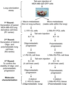
[image:2.612.90.375.73.424.2]435-based models of oligo- and polymetasta-ses. Generation of lung derivative cell lines at each round of in vivo modeling was described in our recent study [12]. Briefly, we first pro -duced paired cell lines derived from lung mac-rometastases (L1-R1-OL, L: lung, R1: Round 1 of in vivo modeling; OL: Oligometastatic) and micrometastases (L1Mic-R1-POL, L: lung; Mic: micrometastases; POL: polymetastatic). We define micro-metastatic foci as those that can only be identified by microscopic histological examination. In contrast, macro-metastatic foci are visible under external fluorescence imag-ing usimag-ing Sellstrom Z87 fluorescence goggles and an LDP 470 nm bright blue flashlight. Sub-sequently, we established individual MDA-MB-435-GFP sub-lines from distinct lungs of oli- go- and poly-metastatic animals injected with L1-R1-OL and L1Mic-R1-POL cells, respectively. All four oligometastatic L1-R2-OL (R2: Round 2 Figure 1. Experimental scheme of generation and in vivo characterization of
metastatic MDA-MB-435-GFP lung derivative cell line series.
stably expressing green fluo -rescent protein (GFP) were generated as previously des- cribed [27, 28]. Cells were maintained in DMEM high glucose supplemented with 10% FBS + 200 µg/ml G418 (Gibco). Cells in the linear phase of growth in culture were harvested and prepar- ed for intravenous tumor in- jection.
Generation of MDA-MB-435 lung oligometastatic (L1-OL) or polymetastatic (L1Mic-POL) sub-lines
All animal work was conduct-ed in accordance with a proto-col approved by the Institu- tional Animal Care and Use Committee (IACUC) at the Ch- ongqing Medical University. Tail-vein injection of tumor cells and identification of polymetastatic animals over the course of 12 weeks. The number of lung metastases was determined by fluores -cence imaging at necropsy.
Figure 1 summarizes the
MDA-MB-of in vivo modeling) cell lines and four polymet-astatic L1Mic-R2-POL cell lines will be used for future molecular characterization. Cell lines ge- nerated in the second round of in vivo modeling were tested for oligo- and polymetastatic pro-gression in an additional round of in vivo test-ing [12].
Non-invasive in vivo whole-body fluorescence imaging of metastasis
The Olympus OV100 Small Animal Imaging System that affords variable magnification and high-resolution visualization capability from whole animal to a single cell was used for non-invasive imaging of tumor growth in live mice as we previously described [29-31]. 8-bit format images were acquired, converted into RGB co-
MB-435-based xenograft models of oligo- and polymetastatic progression. A general descrip-tion of the incidence and time kinetics of me- tastasis of the oligo- and poly-metastatic lung cell lines summarized in Figure 1. Here, we pro-vide a detailed characterization of distinct in vivo metastatic phenotypes of the oligo- and polymetastatic cell lines.
Development of oligometastatic and polymeta-static lung derivative cell lines
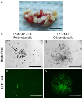
Green fluorescent protein (GFP) labeled MDA-MB-435 (435-P) cancer cells transplanted in the mouse fat pad develop a high incidence of spontaneous lung metastases [28-30]. We uti-lized the high lung colonization efficiency fea -ture of this human tumor model to develop Figure 2. Development and characterization of MDA-MB-435-GFP xenograft
models of oligo- and polymetastases. A. Polymetastatic lung used for estab-lishing paired oligometastatic L1-R1-OL and polymetastatic L1Mic-R1-POL
MDA-MB-435-GFP cell lines (scale bar, 0.5 cm). B. Isolation, purification and
generation of oligometastatic L1-R1-OL (from macrometastses on the lung surface) and polymetastatic L1Mic-R1-POL cells lines (from dormant or
mi-crometastases inside the lung) (scale bars, 200 μm).
lor TIFF images using Image J, and imported into Photo- shop in which contrast and brightness adjustment were applied to the whole image when necessary.
Histological analysis
Metastatic tissues harboring visible macro-metastases (in- cluding lungs, bone, muscle, heart, peritoneum, kidney) we- re excised and fixed in 10% formalin for 12 hours. Paraf- fin embedding, sectioning and H&E staining were perform- ed for histological examina-tion of the presence of both macro- and micro-metasta-ses. Lungs were harvested from all animals. A total of 5 lungs from each treatment group were examined.
Statistical analysis
Statistical significance was assessed by unpaired two-tailed Student’s t-test. P< 0.05 was accepted as sta- tistically significant.
[image:3.612.90.377.69.413.2]Results
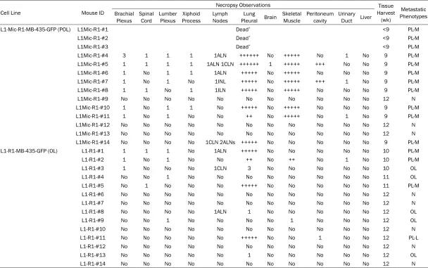
MDA-Table 1. Characterization of metastatic outcome of L1-R1-OL and L1Mic-R1-POL MDA-MB-435-GFP lung derivative cell lines in vivo (the 2nd
round of in vivo passage)
Cell Line Mouse ID
Necropsy Observations Tissue
Harvest (wk)
Metastatic Phenotypes Brachial
Plexus Spinal Cord Lumber Plexus ProcessXiphoid Lymph Nodes Pleural BrainLung Skeletal Muscle Peritoneum cavity Urinary Duct Liver
L1-Mic-R1-MB-435-GFP (POL) L1Mic-R1-#1 Dead* <9 PL-M
L1Mic-R1-#2 Dead* <9 PL-M
L1Mic-R1-#3 Dead* <9 PL-M
L1Mic-R1-#4 3 1 1 1 1ALN ++++++ No +++++ No 1 No 9 PL-M
L1Mic-R1-#5 1 1 1 1 1ALN 1CLN ++++++ 1 +++++ +++ No No 9 PL-M
L1Mic-R1-#6 1 No 1 1 1ALN +++++ No +++++ No No No 9 PL-M
L1Mic-R1-#7 1 No 1 No 1INL +++++ No +++++ +++ 1 No 9 PL-M
L1Mic-R1-#8 1 1 No 1 1ILN +++++ No +++++ No No No 9 PL-M
L1Mic-R1-#9 No No No No No No No No No No No 12 N
L1Mic-R1-#10 1 No 1 1 No +++++ No +++++ No No No 9 PL-M
L1Mic-R1-#11 1 No 1 No No ++ No +++++ No 1 No 9 PL-M
L1Mic-R1-#12 No No No No No No No No No No No 12 N
L1Mic-R1-#13 No No No No No No No No No No No 12 N
L1Mic-R1-#14 No No No No 1CLN 2ALNs +++++ No No No No No 9 PL-M
L1-R1-MB-435-GFP (OL) L1-R1-#1 1 1 1 No 1ALN +++++ No No No No No 10 PL-M
L1-R1-#2 1 No 1 No No ++ No ++ No 1 No 10 PL-M
L1-R1-#3 1 No No No 1CLN 3 No No No No No 10 OL
L1-R1-#4 No No 1 No No No No No No No No 11 OL
L1-R1-#5 No 1 No No No +++++ No No No No No 11 PL-M
L1-R1-#6 No No No No No No No No No No No 12 N
L1-R1-#7 No No No No No No No No No No No 12 N
L1-R1-#8 No No No No 1ALN 1 No No No No No 12 OL
L1-R1-#9 No No 1 No No No No 1 No No No 12 OL
L1-R1-#10 No No No No No No No No No No No 12 N
L1-R1-#11 No No No No No +++++ No No 1 No No 12 PL-L
L1-R1-#12 No No No No No No No No No No No 12 N
L1-R1-#13 No No No No No 1 No No No No No 12 OL
L1-R1-#14 No No No No No No No No No No No 12 N
[image:4.792.92.700.95.476.2]stable MDA-MB-435-GFP xenograft models of oligometastatic and polymetastatic progres-sion. Three consecutive rounds of lung coloni-zation assays were performed in which lung and whole body macroscopic metastases were evaluated during the course of 12 weeks after tail vein injection of 2 × 10⁶ cancer cells (Figure
1 and Methods).
In order to minimize the extrinsic influence of the host tissue, such as mouse lungs, on meta-static progression, we isolated metastasizing MDA-MB-435-GFP cells, in the same animal, either from distinct macro-metastatic nodules on the lung surface (L1-R1-OL cells; L: lung, R1: Round 1 of in vivo modeling; OL: Oligometasta- tic) (Figures 1 and 2A) or from micro-metasta-ses containing viable, slowly proliferating MDA-MB-435 cells within the lung parenchyma (L1- Mic-R1-POL cells; L: lung; Mic:
micrometasta-astatic progression
We then tested whether the L1-R1-OL, and L1Mic-R1-POL MB-435-GFP cells remain meta-static in the lung colonization assay and wheth-er they produce diffwheth-erent pattwheth-erns of metasta-ses. Metastatic outcome and phenotype of each animal injected with either L1-R1-OL or L1Mic-R1-POL MDA-MB-435-GFP lung deriva-tive cell lines in the second round of in vivo test-ing was summarized in details in Table 1. Among 14 animals receiving L1-R1 cells, 10 de- veloped either oligometastases or no metasta-ses (Table 1, Oligometastases: defined as 5 or less, n=5; No metastases: n=5). Polymetastas- es (defined as more than 5 metastases) devel -oped in 4 of 14 mice and occurred at 10-12 weeks post tumor cell injection. In contrast 11 of 14 mice receiving L1Mic-R1-POL cells devel-Figure 3. Widespread polymetastases in animals injected with
L1Mic-R1-POL cells. A. OV-100 fluorescent imaging detected widespread polymetasta -ses in animals injected with L1Mic-R1-POL cells. Shown is a representative animal that developed widespread polymetastases (scale bars, 1 cm). B. H&E characterization of polymetastases in different organs (scale bars, i-iii,
500 μm; iv-ix, 200 μm; M, metastasis).
ses; POL: polymetastatic) (Fi- gures 1 and 2A). L1-R1-OL and L1Mic-R1-POL MDA-435-GFP cells were recovered in culture with G418 selection to increase GFP expression (Figure 2B). The two resultant cell lines had distinct cellular morphology and growth rate. L1-R1-OL cells exhibited rapid cell proliferation and consist-ed of a mixture of small, round cells as well as larger, elon-gated cells (Figure 2B). In contrast, L1Mic-R1-POL cul-tured cells were mainly small, round, and morphologically less differentiated (Figure 2B). Expanded L1Mic-R1-OL cultures contained a mixture of both morphologically dis-tinct cell types, though a sig-nificant portion of cells re-main small and round and were either floating or loose-ly attached (data not shown). L1-R1-OL and L1Mic-R1-POL MB-435-GFP cells lines were used subsequently for devel-oping oligometastases- and polymetastases-like xenogra- ft models.
[image:5.612.91.376.68.401.2]polymet-oped metastases, all of which were all polyme-tastases (Table 1). Polymetastases were either poly-foci in the lung (Table 1, POL-L: defined as >5 foci at lung only) or had multi-organ involve-ment in addition to lung polymetastases (Table 1, POL-M: >5 foci at lung and involved multi-organs). The time kinetics of metastasis in poly-metastatic L1Mic-R1-POL cells was significant
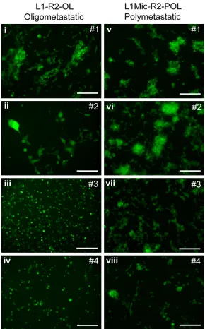
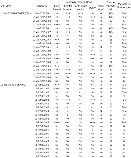
[image:6.612.88.378.71.537.2]-metastases and poly-metastases, respectively. Four L1-R2-OL and four L1Mic-R2-POL lung me- tastases-derived cell lines were used (Table 2). Among 19 mice that received the L1Mic-R2-POL cell lines (Figure 4), three failed to develop macroscopic metastasis. Of 16 animals that developed metastases, all had polymetastatic foci defined as in the lungs as well as in other Figure 4. Generation of primary cell lines in the second round in vivo lung
colonization assay. L1-R1-OL or L1Mic-R1-POL cells were injected via the tail vein. Metastasized L1-R1-OL or L1Mic-R1-POL cells to the lungs of oligomet-astatic and polymetoligomet-astatic animals were isolated, respectively. Cancer cells were then recovered and expanded in culture. Four L1-R2-OL (i-iv) and four L1Mic-R2-POL cell lines (v-viii) were isolated for further in vivo testing (scale
bars, 100 μm).
oligo-Table 2. Characterization of metastatic outcome of L1-R2-OL and L1Mic-R2-POL MDA-MB-435-GFP lung derivative cell lines in vivo (the 3rd round of in vivo passage)
Cell Line Mouse ID
Necropsy Observations Tissue
Harvest (wk)
Metastatic Phenotypes Lung
Pleural Skeletal Muscle Peritoneum cavity Ovary Other sites*
L1Mic-R2-MB-435-GFP (POL) L1Mic-R2-PL1-#1 ++++ ++++ ++++ +++ 4 6.5 PL-M
L1Mic-R2-PL1-#2 ++ ++++ No ++++ No 6.5 PL-M
L1Mic-R2-PL1-#3 No No No No No 12 N
L1Mic-R2-PL1-#4 ++++ No No No No 12 PL-M
L1Mic-R2-PL2-#1 ++++ ++++ No No 4 6.5 PL-M
L1Mic-R2-PL2-#2 ++++ ++++ No +++ 4 6.5 PL-M
L1Mic-R2-PL2-#3 ++++ +++ No No 3 10 PL-M
L1Mic-R2-PL2-#4 +++ No No No 1 11 PL-M
L1Mic-R2-PL2-#5 ++++ No No No 1 12 PL-M
L1Mic-R2-PL3-#1 ++++ ++++ No +++ 3 7 PL-M
L1Mic-R2-PL3-#2 + ++ No ++ 1 8 PL-M
L1Mic-R2-PL3-#3 ++++ +++ No ++++ 1 8 PL-M
L1Mic-R2-PL3-#4 ++++ No No +++ No 11 PL-M
L1Mic-R2-PL3-#5 ++++ No No +++ No 12 PL-M
L1Mic-R2-PL4-#1 +++ ++ ++ No 2 6.5 PL-M
L1Mic-R2-PL4-#2 ++++ ++++ No +++ 3 8 PL-M
L1Mic-R2-PL4-#3 ++++ ++++ ++++ ++++ 3 9 PL-M
L1Mic-R2-PL4-#4 No No No No No 12 N
L1Mic-R2-PL4-#5 No No No No No 12 N
L1-R2-MB-435-GFP (OL) L1-R2-OL1-#1 +++ +++ No ++ 3 6 PL-M
L1-R2-OL1-#2 ++++ No No No No 9 PL-M
L1-R2-OL1-#3 No +++ ++ +++ 3 11 PL-M
L1-R2-OL1-#4 4 No No No No 12 OL
L1-R2-OL2-#1 ++++ ++++ ++ ++++ 4 7 PL-M
L1-R2-OL2-#2 No No No No No 12 N
L1-R2-OL2-#3 ++++ +++ ++ ++ 3 7 PL-M
L1-R2-OL2-#4 ++++ +++ No No 3 7 PL-M
L1-R2-OL2-#5 No 1 No No No 12 OL
L1-R2-OL2-#6 No No No No No 12 N
L1-R2-OL3-#1 ++++ +++ No ++++ No 6 PL-M
L1-R2-OL3-#2 No No No No No 12 N
L1-R2-OL3-#3 1 No No + No 12 OL
L1-R2-OL3-#4 No No No No No 12 N
L1-R2-OL3-#5 No No No No No 12 N
L1-R2-OL4-#1 No No No No No 12 N
L1-R2-OL4-#2 No No No No No 12 N
L1-R2-OL4-#3 No No No No No 12 N
L1-R2-OL4-#4 No No No No No 12 N
L1-R2-OL4-#5 No No No No No 12 N
POL-M: multi-organ polymetastases; OL: oligometastasis(es); N: no metastasis; *: other sites include brachial plexus, spinal cord, lumber plexus and xiphoid process; +: semi-quantitative assessment of polymetastases in an organ when the metastatic foci were diffused.
organs (Table 2, POL-M). The widespread poly-metastases could be detected before 7 weeks after tumor cell injection in 6 animals. In con-trast of 20 mice injected with the L1Mic-R2-POL cell lines (Figure 4), 10 mice failed to de-
[image:7.612.90.519.96.614.2]extent of metastasis, the outcome of oligo- and polymetastases as well as the kinetics of estab-lishing macro-metastases between the L1-R2-OL and L1Mic-R2-PL1-R2-OL lung cell lines. Therefore, the differences between the two metastatic phenotypes were stably maintained in vivo over three successive passages.
Discussion
Mounting clinical observations begin to recog-nize oligometastases as a distinct clinical met-astatic state where metastases are limited in number and destination organ [32-35] and oli- gometastases may represent a potentially cur-able subset of metastatic diseases [5, 6, 36]. However, only a subset of patients diagnosed with oligometastases will remain oligometa-static following treatment. Identification of this subset of patients at their initial presentation could help direct appropriate therapy.
Two models of tumor metastases have been proposed: the “late” and the “early” models of metastatic dissemination [37]. In the “late me- tastasis” model, late disseminating cells are genetically similar to the primary tumor. How- ever, in the “early metastasis” model, the early metastatic tumor cells are genetically distinct from the non-disseminating tumor cells within the primary tumor. It is conceivable that the two models may not be mutually exclusive, and human solid tumors may employ both strate-gies in order to optimally progress and dissemi-nate. Therefore, oligo- and polymetastatic state could be reached via both the early- or late models of metastases.
While various single organ-specific or sponta -neous experimental models of metastases have been developed, they are selected for enhanced metastatic efficiency in contrast to the non-invasive or minimally invasive parental cancer lines or primary tumors [13-19]. Xeno- graft models of multi-organ polymetastases that resembles late-stage clinical metastatic dissemination are lacking. Prior to our study, animal models for human cancer oligometasta-ses were not available. In order to investigate mechanisms underlying differences between oligometastases and polymetastases, we de- veloped oligometastases and polymetastases models of the MDA-MB-435 human tumor in nude mice. These models have some unique features.
The oligometastatic phenotype of the L1-OL MDA-MB-435-GFP model remains stable in vivo after three consecutive rounds of passage (each round of testing lasted 12 weeks) and meets the clinical criteria of ≤ 5 total body mac -roscopic metastases. In contrast, metastatic efficiency of the existing metastasis models is invariably enhanced after in vivo selection [20-23], as also observed in our L1Mic-POL MDA-MB-435-GFP polymetastases model.
Tail-vein injection of polymetastatic L1Mic-R1-POL cells produced a high frequency of multi-organ metastases in the brain, peritoneal me- mbrane and cavity, muscles, bone, kidneys, heart, and lymph nodes, with intramuscular metastases to the back and hind legs as the second most frequent event (Figure 3A, 3B). Therefore, cells metastasized to organs other than lung likely have completed the full cas-cade of metastatic dissemination. The dissemi-nation pattern of our polymetastatic MDA-MB-435-GFP breast cancer model while overlaps in part (lung, brain, bone, visceral) but is not iden-tical to that observed in human breast cancer (Table 1). Most notably are the high incidence of muscle metastasis seen in our model that is uncommon in human cancer, and the lack of liver metastasis that is frequent in human breast cancer. Such discrepancies reflect dif -ferences in tumor cell and host microenviron-ment at the site of metastasis in mouse and in human. Nevertheless, the metastatic dissemi-nation patterns of polymetastatic lung and multi-organ involvement are similar to clinical polymetastases.
In summary, our study demonstrated the devel-opment and characterization of new xenograft models of metastases. They recapitulated phe-notypic features of two clinically relevant hu- man metastases phenotypes we identified: the oligometastatic and polymetastatic progres-sion. These models should prove useful for me- chanistic investigation as well as for evaluating therapeutic targeting of sustained oligometas-tases to achieve the clinical goal of long-term disease free survival.
Acknowledgements
This study was supported by grants from the Programs of National Natural Science of China (81272405, 81672908, 81602596).
Disclosure of conflict of interest
None.
Address correspondence to: Dr. H Rosie Xing, La- boratory of Translational Cancer Stem Cell Rese- arch, Chongqing Medical University, 1 Yi Xue Yuan Road, Yuzhong District, Chongqing, P.R. China. Tel: 023-68485106; Fax: 023-68486646; E-mail: [email protected]
References
[1] Staren ED, Salerno C, Rongione A, Witt TR and Faber LP. Pulmonary resection for metastatic breast cancer. Arch Surg 1992; 127: 1282-1284.
[2] Selzner M, Morse MA, Vredenburgh JJ, Meyers WC and Clavien PA. Liver metastases from breast cancer: long-term survival after cur- ative resection. Surgery 2000; 127: 383-389. [3] Fong Y, Cohen AM, Fortner JG, Enker WE,
Turn-bull AD, Coit DG, Marrero AM, Prasad M, Blumgart LH and Brennan MF. Liver resection for colorectal metastases. J Clin Oncol 1997; 15: 938-946.
[4] Tanvetyanon T, Robinson LA, Schell MJ, Strong VE, Kapoor R, Coit DG and Bepler G. Outcomes of adrenalectomy for isolated synchronous ver-sus metachronous adrenal metastases in non-small-cell lung cancer: a systematic review and pooled analysis. J Clin Oncol 2008; 26: 1142-1147.
[5] Hellman S and Weichselbaum RR. Oligometas-tases. J Clin Oncol 1995; 13: 8-10.
[6] Weichselbaum RR and Hellman S. Oligometas-tases revisited. Nat Rev Clin Oncol 2011; 8: 378-382.
[7] Salama JK, Chmura SJ, Mehta N, Yenice KM, Stadler WM, Vokes EE, Haraf DJ, Hellman S
and Weichselbaum RR. An initial report of a radiation dose-escalation trial in patients with
one to five sites of metastatic disease. Clin
Cancer Res 2008; 14: 5255-5259.
[8] Milano MT, Katz AW, Muhs AG, Philip A, Buch-holz DJ, Schell MC and Okunieff P. A prospec-tive pilot study of curaprospec-tive-intent stereotactic body radiation therapy in patients with 5 or fewer oligometastatic lesions. Cancer 2008; 112: 650-658.
[9] Rusthoven KE, Kavanagh BD, Burri SH, Chen C, Cardenes H, Chidel MA, Pugh TJ, Kane M, Gaspar LE and Schefter TE. Multi-institutional phase I/II trial of stereotactic body radiation therapy for lung metastases. J Clin Oncol 2009; 27: 1579-1584.
[10] Norihisa Y, Nagata Y, Takayama K, Matsuo Y, Sakamoto T, Sakamoto M, Mizowaki T, Yano S and Hiraoka M. Stereotactic body radiotherapy for oligometastatic lung tumors. Int J Radiat Oncol Biol Phys 2008; 72: 398-403.
[11] Hoyer M, Roed H, Traberg Hansen A, Ohlhuis L, Petersen J, Nellemann H, Kiil Berthelsen A, Grau C, Aage Engelholm S and Von der Maase H. Phase II study on stereotactic body radio-therapy of colorectal metastases. Acta Oncol 2006; 45: 823-830.
[12] Lussier YA, Xing HR, Salama JK, Khodarev NN, Huang Y, Hasselle MD, Zhang Q, Yang X, Khan SA, Malik R, Darga TE, Fan H, Perakis S, Filippo M, Lee Y, Posner MC, Chmura SJ, Hellman S and Weichselbaum RR. microRNA expression characterizes oligometastases Clinical Cancer Research 2011; Submitted, under consider-ation.
[13] Fidler IJ. Models for spontaneous metastasis. Cancer Res 2006; 66: 9787.
[14] Fidler IJ, Balasubramanian K, Lin Q, Kim SW and Kim SJ. The brain microenvironment and cancer metastasis. Mol Cells 30: 93-98. [15] Fidler IJ and Kripke ML. Metastasis results
from preexisting variant cells within a malig-nant tumor. Science 1977; 197: 893-895. [16] Morikawa K, Walker SM, Nakajima M, Pathak
S, Jessup JM and Fidler IJ. Influence of organ
environment on the growth, selection, and me-tastasis of human colon carcinoma cells in nude mice. Cancer Res 1988; 48: 6863-6871. [17] Nguyen DX, Bos PD and Massague J.
Metasta-sis: from dissemination to organ-specific colo -nization. Nat Rev Cancer 2009; 9: 274-284. [18] Sung V, Cattell DA, Bueno JM, Murray A,
Zwieb-el JA, Aaron AD and Thompson EW. Human breast cancer cell metastasis to long bone and soft organs of nude mice: a quantitative assay. Clin Exp Metastasis 1997; 15: 173-183. [19] Bos PD, Nguyen DX and Massague J. Modeling
[20] Bruns CJ, Harbison MT, Kuniyasu H, Eue I and Fidler IJ. In vivo selection and characterization of metastatic variants from human pancreatic adenocarcinoma by using orthotopic implanta-tion in nude mice. Neoplasia 1999; 1: 50-62. [21] Morikawa K, Walker SM, Jessup JM and Fidler
IJ. In vivo selection of highly metastatic cells from surgical specimens of different primary human colon carcinomas implanted into nude mice. Cancer Res 1988; 48: 1943-1948. [22] Naito S, Walker SM and Fidler IJ. In vivo
selec-tion of human renal cell carcinoma cells with high metastatic potential in nude mice. Clin Exp Metastasis 1989; 7: 381-389.
[23] Poste G, Doll J, Hart IR and Fidler IJ. In vitro selection of murine B16 melanoma variants with enhanced tissue-invasive properties. Can-cer Res 1980; 40: 1636-1644.
[24] Fidler IJ and Nicolson GL. Organ selectivity for implantation survival and growth of B16 mela-noma variant tumor lines. J Natl Cancer Inst 1976; 57: 1199-1202.
[25] Fidler IJ and Nicolson GL. Tumor cell and host properties affecting the implantation and sur-vival of blood-borne metastatic variants of B16 melanoma. Isr J Med Sci 1978; 14: 38-50. [26] Fidler IJ. Selection of successive tumour lines
for metastasis. Nat New Biol 1973; 242: 148-149.
[27] Li X, Wang J, An Z, Yang M, Baranov E, Jiang P, Sun F, Moossa AR and Hoffman RM. Optically imageable metastatic model of human breast cancer. Clin Exp Metastasis 2002; 19: 347-350.
[28] Zhang Q, Fan H, Shen J, Hoffman RM and Xing HR. Human breast cancer cell lines co-express neuronal, epithelial, and melanocytic differen-tiation markers in vitro and in vivo. PLoS One 5: e9712.
[29] Yamauchi K, Yang M, Jiang P, Xu M, Yamamoto N, Tsuchiya H, Tomita K, Moossa AR, Bouvet M and Hoffman RM. Development of real-time subcellular dynamic multicolor imaging of
can-cer-cell trafficking in live mice with a variable-magnification whole-mouse imaging system.
Cancer Res 2006; 66: 4208-4214.
[30] Zhang Q, Yang M, Shen J, Gerhold LM, Hoff-man RM and Xing HR. The role of the intravas-cular microenvironment in spontaneous me-tastasis development. Int J Cancer 2010; 126: 2534-41.
[31] Zhang Q, Bindokas V, Shen J, Fan H, Hoffman RM and Xing HR. Time-course imaging of ther-apeutic functional tumor vascular normaliza-tion by antiangiogenic agents. Mol Cancer Ther 10: 1173-1184.
[32] Rusthoven KE, Hammerman SF, Kavanagh BD, Birtwhistle MJ, Stares M and Camidge DR. Is there a role for consolidative stereotactic body
radiation therapy following first-line systemic
therapy for metastatic lung cancer? A patterns-of-failure analysis. Acta Oncol 2009; 48: 578-583.
[33] Mehta N, Mauer AM, Hellman S, Haraf DJ, Co-hen EE, Vokes EE and Weichselbaum RR. Anal-ysis of further disease progression in meta-static non-small cell lung cancer: implications for locoregional treatment. Int J Oncol 2004; 25: 1677-1683.
[34] Yoon SS and Tanabe KK. Surgical treatment and other regional treatments for colorectal cancer liver metastases. Oncologist 1999; 4: 197-208.
[35] House MG, Ito H, Gonen M, Fong Y, Allen PJ, DeMatteo RP, Brennan MF, Blumgart LH, Jar-nagin WR and D’Angelica MI. Survival after he-patic resection for metastatic colorectal can-cer: trends in outcomes for 1,600 patients during two decades at a single institution. J Am Coll Surg 210: 744-752, 752-745.
[36] Macdermed DM, Weichselbaum RR and Sala-ma JK. A rationale for the targeted treatment of oligometastases with radiotherapy. J Surg Oncol 2008; 98: 202-206.
[37] Coghlin C and Murray GI. Current and emerg-ing concepts in tumour metastasis. J Pathol 222: 1-15.
[38] Chaffer CL and Weinberg RA. A perspective on cancer cell metastasis. Science 331: 1559-1564.