organic papers
Acta Cryst.(2005). E61, o2299–o2300 doi:10.1107/S1600536805019707 Seik Weng Ng C
6H5ClN2O2
o2299
Acta Crystallographica Section E Structure Reports Online
ISSN 1600-5368
5-Chloro-2-nitroaniline
Seik Weng Ng
Department of Chemistry, University of Malaya, 50603 Kuala Lumpur, Malaysia
Correspondence e-mail: [email protected]
Key indicators
Single-crystal X-ray study T= 295 K
Mean(C–C) = 0.002 A˚ Rfactor = 0.041 wRfactor = 0.122
Data-to-parameter ratio = 13.4
For details of how these key indicators were automatically derived from the article, see http://journals.iucr.org/e.
#2005 International Union of Crystallography
Printed in Great Britain – all rights reserved
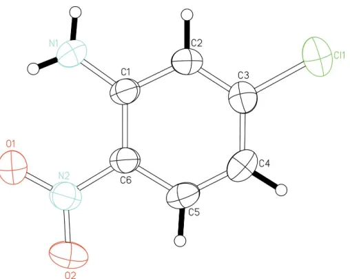
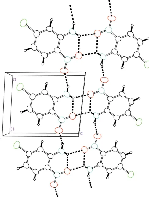
The title compound, C6H5ClN2O2, exists as a planar molecule; adjacent molecules are linked by N O hydrogen bonds into ribbons.
Comment
Chloronitroanilines possess non-linear optical properties. The crystal structure of 5-chloro-2-aniline, (I), was presented at a conference (Gerbiet al., 1988), but it has not been abstracted into the Cambridge Structural Database (Version 5.26; Allen, 2002). The first chloronitroaniline to be authenticated was 2-chloro-4-nitroaniline (McPhail & Sim, 1965); chloronitro-anilines are generally not well studied, and 4,5-dichloro-2-nitroaniline (Doyle, 1999) represents a rare example. The title compound (Fig. 1) exists as a planar molecule; adjacent mol-ecules are linked by hydrogen bonds (Table 1) into a ribbon (Fig. 2).
Experimental
The title compound was obtained commercially and was recrys-tallized from dimethyl sulfoxide.
[image:1.610.208.462.506.708.2]Received 16 June 2005 Accepted 22 June 2005 Online 30 June 2005
Figure 1
Crystal data
C6H5ClN2O2 Mr= 172.57
Triclinic,P1 a= 7.073 (3) A˚ b= 7.423 (3) A˚ c= 7.711 (3) A˚
= 83.87 (3)
= 81.98 (3)
= 62.24 (3)
V= 354.4 (3) A˚3
Z= 2
Dx= 1.617 Mg m 3 MoKradiation Cell parameters from 3038
reflections
= 3.1–27.5
= 0.48 mm1 T= 295 (2) K Plate, yellow
0.320.210.01 mm
Data collection
Rigaki R-AXIS RAPID IP diffractometer
!scans
Absorption correction: multi-scan (ABSCOR; Higashi, 1995) Tmin= 0.505,Tmax= 0.955 3525 measured reflections
1608 independent reflections 1299 reflections withI> 2(I) Rint= 0.023
max= 27.5 h=9!9 k=7!9 l=9!9
Refinement
Refinement onF2 R[F2> 2(F2)] = 0.041 wR(F2) = 0.122 S= 1.07 1608 reflections 120 parameters
All H-atom parameters refined w= 1/[2(F
o2) + (0.0843P)2] whereP= (Fo2+ 2Fc2)/3 (/)max= 0.001
max= 0.38 e A˚
3
min=0.21 e A˚
[image:2.610.318.562.72.395.2]3
Table 1
Hydrogen-bond geometry (A˚ ,).
D—H A D—H H A D A D—H A
N1—H11 O1 0.84 (1) 2.07 (2) 2.633 (2) 124 (2) N1—H11 O1i
0.84 (1) 2.45 (2) 3.152 (2) 141 (2) N1—H12 O2ii
0.85 (1) 2.35 (1) 3.113 (2) 150 (2)
Symmetry codes: (i)xþ1;y;zþ2; (ii)xþ1;y;z.
H atoms were located in difference Fourier maps and were refined with distance restraints of N—H = 0.85 (1) A˚ and C—H = 0.95 (1) A˚. Data collection: RAPID-AUTO(Rigaku, 1998); cell refinement: RAPID-AUTO; data reduction: CrystalStructure (Rigaku/MSC, 2002); program(s) used to solve structure: SHELXS97 (Sheldrick, 1997); program(s) used to refine structure:SHELXL97(Sheldrick, 1997); molecular graphics:ORTEPII(Johnson, 1976); software used to prepare material for publication:SHELXL97.
The author thanks Heilongjiang University for the diffrac-tion measurements and the University of Malaya for supporting this work.
References
Allen, F. H. (2002).Acta Cryst.B58, 380–388. Doyle, B. (1999).Acta Cryst.C55, IUC9900116.
Gerbi, D. J., Boyd, G. T., Ender, D. A. & Leung, P. C. W. (1988).Mater. Res. Soc. Symp. Proc. (Non-linear Opt. Prop. Polym.),109, 331–338.
Higashi, T. (1995).ABSCOR. Rigaku Corporation, Tokyo, Japan.
Johnson, C. K. (1976).ORTEPII. Report ORNL-5138. Oak Ridge National Laboratory, Tennessee, USA.
McPhail, A. T. & Sim, G. A. (1965).J. Chem. Soc.pp. 227–236. Rigaku (1998).RAPID-AUTO. Rigaku Corporation, Tokyo, Japan. Rigaku/MSC (2002). CrystalStructure. Rigaku/MSC Inc., 9009 New Trails
Drive, The Woodlands, TX 77381-5209, USA.
Sheldrick, G. M. (1997). SHELXS97 and SHELXL97. University of Go¨ttingen, Germany.
Figure 2
supporting information
sup-1 Acta Cryst. (2005). E61, o2299–o2300
supporting information
Acta Cryst. (2005). E61, o2299–o2300 [https://doi.org/10.1107/S1600536805019707]
5-Chloro-2-nitroaniline
Seik Weng Ng
5-Chloro-2-nitroaniline
Crystal data
C6H5ClN2O2 Mr = 172.57
Triclinic, P1 Hall symbol: -P 1
a = 7.073 (3) Å
b = 7.423 (3) Å
c = 7.711 (3) Å
α = 83.87 (3)°
β = 81.98 (3)°
γ = 62.24 (3)°
V = 354.4 (3) Å3
Z = 2
F(000) = 176
Dx = 1.617 Mg m−3
Mo Kα radiation, λ = 0.71073 Å Cell parameters from 3038 reflections
θ = 3.1–27.5°
µ = 0.48 mm−1 T = 295 K Plate, yellow
0.32 × 0.21 × 0.01 mm
Data collection
Rigaki RAXIS-RAPID IP diffractometer
Radiation source: fine-focus sealed tube Graphite monochromator
ω scans
Absorption correction: multi-scan (ABSCOR; Higashi, 1995)
Tmin = 0.505, Tmax = 0.955
3525 measured reflections 1608 independent reflections 1299 reflections with I > 2σ(I)
Rint = 0.023
θmax = 27.5°, θmin = 3.1° h = −9→9
k = −7→9
l = −9→9
Refinement
Refinement on F2
Least-squares matrix: full
R[F2 > 2σ(F2)] = 0.041 wR(F2) = 0.122 S = 1.07 1608 reflections 120 parameters 5 restraints
Primary atom site location: structure-invariant direct methods
Secondary atom site location: difference Fourier map
Hydrogen site location: difference Fourier map All H-atom parameters refined
w = 1/[σ2(F
o2) + (0.0843P)2]
where P = (Fo2 + 2Fc2)/3
(Δ/σ)max = 0.001
Δρmax = 0.38 e Å−3
Δρmin = −0.21 e Å−3
Fractional atomic coordinates and isotropic or equivalent isotropic displacement parameters (Å2)
x y z Uiso*/Ueq
Cl1 0.80361 (7) 0.31629 (7) 0.13860 (5) 0.0635 (2)
O2 0.0645 (2) 0.2492 (2) 0.7162 (2) 0.0667 (4)
N1 0.6846 (2) 0.1401 (3) 0.7783 (2) 0.0540 (4)
N2 0.2476 (2) 0.2088 (2) 0.7433 (2) 0.0441 (3)
C1 0.5971 (2) 0.1984 (2) 0.6259 (2) 0.0364 (3)
C2 0.7202 (2) 0.2283 (2) 0.4758 (2) 0.0404 (3)
C3 0.6407 (2) 0.2864 (2) 0.3167 (2) 0.0412 (3)
C4 0.4340 (3) 0.3203 (3) 0.2930 (2) 0.0468 (4)
C5 0.3114 (2) 0.2935 (2) 0.4360 (2) 0.0425 (4)
C6 0.3882 (2) 0.2334 (2) 0.6008 (2) 0.0362 (3)
H11 0.622 (3) 0.111 (3) 0.869 (2) 0.067 (6)*
H12 0.810 (2) 0.127 (3) 0.785 (3) 0.077 (7)*
H2 0.861 (2) 0.204 (3) 0.493 (2) 0.054 (5)*
H4 0.379 (3) 0.364 (3) 0.182 (2) 0.064 (5)*
H5 0.170 (2) 0.317 (3) 0.427 (3) 0.057 (5)*
Atomic displacement parameters (Å2)
U11 U22 U33 U12 U13 U23
Cl1 0.0644 (3) 0.0810 (4) 0.0433 (3) −0.0359 (3) 0.0065 (2) 0.0008 (2) O1 0.0550 (7) 0.0859 (9) 0.0463 (7) −0.0379 (7) −0.0092 (5) 0.0182 (6) O2 0.0370 (6) 0.095 (1) 0.0745 (9) −0.0370 (6) −0.0093 (6) 0.0079 (7) N1 0.0463 (7) 0.082 (1) 0.0436 (8) −0.0376 (7) −0.0187 (6) 0.0151 (7) N2 0.0380 (6) 0.0471 (7) 0.0499 (7) −0.0222 (5) −0.0062 (5) 0.0027 (5) C1 0.0335 (6) 0.0382 (7) 0.0396 (7) −0.0171 (6) −0.0102 (5) 0.0010 (5) C2 0.0330 (7) 0.0459 (8) 0.0441 (8) −0.0190 (6) −0.0069 (6) −0.0003 (6) C3 0.0429 (7) 0.0415 (7) 0.0375 (7) −0.0186 (6) −0.0008 (6) −0.0021 (6) C4 0.0496 (8) 0.0507 (8) 0.0392 (8) −0.0203 (7) −0.0161 (6) 0.0037 (6) C5 0.0355 (7) 0.0450 (8) 0.0482 (8) −0.0171 (6) −0.0155 (6) 0.0013 (6) C6 0.0319 (7) 0.0371 (7) 0.0419 (8) −0.0171 (6) −0.0080 (5) 0.0005 (6)
Geometric parameters (Å, º)
Cl1—C3 1.730 (2) C3—C4 1.399 (2)
O1—N2 1.231 (2) C4—C5 1.362 (2)
O2—N2 1.229 (2) C5—C6 1.398 (2)
N1—C1 1.340 (2) N1—H11 0.84 (1)
N2—C6 1.432 (2) N1—H12 0.85 (1)
C1—C2 1.411 (2) C2—H2 0.95 (1)
C1—C6 1.414 (2) C4—H4 0.95 (1)
C2—C3 1.364 (2) C5—H5 0.94 (1)
O2—N2—O1 121.5 (1) C5—C6—C1 121.2 (1)
O2—N2—C6 118.7 (1) C5—C6—N2 117.6 (1)
O1—N2—C6 119.8 (1) C1—C6—N2 121.2 (1)
N1—C1—C2 118.6 (1) C1—N1—H11 122 (2)
N1—C1—C6 125.5 (1) C1—N1—H12 121 (2)
C2—C1—C6 116.0 (1) H11—N1—H12 117 (2)
supporting information
sup-3 Acta Cryst. (2005). E61, o2299–o2300
C2—C3—C4 122.3 (1) C1—C2—H2 116 (1)
C2—C3—Cl1 118.5 (1) C3—C4—H4 122 (1)
C4—C3—Cl1 119.3 (1) C5—C4—H4 121 (1)
C5—C4—C3 117.5 (1) C4—C5—H5 121 (1)
C4—C5—C6 121.6 (1) C6—C5—H5 118 (1)
N1—C1—C2—C3 179.6 (1) N1—C1—C6—C5 −179.8 (2)
C6—C1—C2—C3 −0.3 (2) C2—C1—C6—C5 0.1 (2)
C1—C2—C3—C4 0.2 (2) N1—C1—C6—N2 0.3 (2)
C1—C2—C3—Cl1 −179.1 (1) C2—C1—C6—N2 −179.8 (1)
C2—C3—C4—C5 0.1 (2) O2—N2—C6—C5 −2.9 (2)
Cl1—C3—C4—C5 179.4 (1) O1—N2—C6—C5 176.8 (1)
C3—C4—C5—C6 −0.3 (2) O2—N2—C6—C1 177.0 (1)
C4—C5—C6—C1 0.2 (2) O1—N2—C6—C1 −3.3 (2)
C4—C5—C6—N2 −179.9 (1)
Hydrogen-bond geometry (Å, º)
D—H···A D—H H···A D···A D—H···A
N1—H11···O1 0.84 (1) 2.07 (2) 2.633 (2) 124 (2)
N1—H11···O1i 0.84 (1) 2.45 (2) 3.152 (2) 141 (2)
N1—H12···O2ii 0.85 (1) 2.35 (1) 3.113 (2) 150 (2)