
bacterial responses to aspartate
LACEY, M. <http://orcid.org/0000-0003-0997-0217>, AGASING, A., LOWRY,
R. and GREEN, J.
Available from Sheffield Hallam University Research Archive (SHURA) at:
http://shura.shu.ac.uk/7271/
This document is the author deposited version. You are advised to consult the
publisher's version if you wish to cite from it.
Published version
LACEY, M., AGASING, A., LOWRY, R. and GREEN, J. (2013). Identification of the
YfgF MASE1 domain as a modulator of bacterial responses to aspartate. Open
Biology, 3 (6), p. 130046.
Copyright and re-use policy
See
http://shura.shu.ac.uk/information.html
Sheffield Hallam University Research Archive
, 130046, published 5 June 2013
3
2013
Open Biol.
Melissa Lacey, Agnieshka Agasing, Rebecca Lowry and Jeffrey Green
bacterial responses to aspartate
Identification of the YfgF MASE1 domain as a modulator of
References
http://rsob.royalsocietypublishing.org/content/3/6/130046.full.html#ref-list-1
This article cites 25 articles, 9 of which can be accessed free
any medium, provided the original work is properly cited.
Attribution License, which permits unrestricted use, distribution, and reproduction in
This is an open-access article distributed under the terms of the Creative Commons
Subject collections
(25 articles)
microbiology
Articles on similar topics can be found in the following collections
rsob.royalsocietypublishing.org
Research
Cite this article:
Lacey M, Agasing A, Lowry
R, Green J. 2013 Identification of the YfgF
MASE1 domain as a modulator of bacterial
responses to aspartate. Open Biol 3: 130046.
http://dx.doi.org/10.1098/rsob.130046
Received: 8 March 2013
Accepted: 10 May 2013
Subject Area:
microbiology
Keywords:
chemotaxis, cyclic-di-GMP, MASE1 domain,
oxidative stress,
Salmonella
Author for correspondence:
Melissa Lacey
e-mail: [email protected]
†
Present address: Department of Biosciences,
Sheffield Hallam University, City Campus,
Howard Street, Sheffield S1 1WB, UK.
Identification of the YfgF MASE1
domain as a modulator of
bacterial responses to aspartate
Melissa Lacey
†
, Agnieshka Agasing, Rebecca Lowry
and Jeffrey Green
Department of Molecular Biology and Biotechnology, The Krebs Institute, University of
Sheffield, Sheffield S10 2TN, UK
1. Summary
Complex 30
-50
-cyclic diguanylic acid (c-di-GMP) responsive regulatory net-works that are modulated by the action of multiple diguanylate cyclases (DGC; GGDEF domain proteins) and phosphodiesterases (PDE; EAL domain proteins) have evolved in many bacteria. YfgF proteins possess a membrane-anchoring domain (MASE1), a catalytically inactive GGDEF domain and a catalytically active EAL domain. Here, sustained expression of the Salmonella entericaspp. Enterica ser. Enteritidis YfgF protein is shown to mediate inhibition of the formation of the aspartate chemotactic ring on motility agar under aerobic conditions. This phenomenon was c-di-GMP-independent because it occurred in aSalmonellastrain that lacked the ability to synthesize c-di-GMP and also when PDE activity was abolished by site-directed mutagenesis of the EAL domain. YfgF-mediated inhibition of aspartate chemotactic ring formation was impaired in the altered redox environment generated by exogenousp-benzoquinone. This ability of YfgF to inhibit the response to aspartate required a motif,213
Lys-Lys-Glu215, in the predicted cytoplasmic loop between trans-membrane regions 5 and 6 of the MASE1 domain. Thus, for the first time the function of a MASE1 domain as a redox-responsive regulator of bacterial responses to aspartate has been shown.
2. Introduction
30
-50
-cyclic diguanylic acid (c-di-GMP) is a bacterial second messenger that regulates diverse cellular functions including the cell cycle, synthesis of extra-cellular polysaccharide and virulence factors, and transitions between sessility and motility [1]. Generally, motility and the production of virulence factors are promoted by low intracellular concentrations of c-di-GMP, whereas high intra-cellular concentrations promote biofilm formation and a sessile lifestyle [2]. In many bacteria, c-di-GMP concentrations are controlled by complex networks of diguanylate cyclases (DGC; GGDEF domain proteins) and phosphodiesterases (PDE; EAL or HD-GYP domain proteins). Coordination of the opposing DGC and PDE activities that form these networks is required for optimal bacterial responses to environmental change [1]. This is achieved, at least in part, via signal perception and/or signal transduction domains, such as BluF (blue-light sensory domain), HAMP (histidine kinases–adenylyl cyclases–methyl accepting chemotaxis proteins–phosphatase domain), GAF (cGMP PDE–adenylyl cyclase– FhlA domain), PAS (Per-Amt-Sim domain), as well as DNA-binding domains and as yet uncharacterized domains, that are known to, or thought to, control the DGC and PDE activities of the corresponding proteins [1].
Facultative anaerobic enterobacteria such as Salmonella entericaspp. Enterica ser. Enteritidis (S. Enteritidis) experience transitions between aerobic and anaerobic environments during their pathogenic and commensal lifestyles. In these bac-teria, theyfgFgene encodes a c-di-GMP PDE that is maximally expressed under anaerobic conditions inEscherichia coli,with expression under microaerobic conditions (2.5% oxygen atmosphere) being only slightly reduced compared with that observed under anaerobic conditions [3]. TheyfgFpromoter ofS. Enteritidis is very similar to that for E. coli, retaining both the FNR box at 241.5 (TTGAT– –ATCAA in E. coli;
TTGAC– –ATCAA in S. Enteritidis) and the 210 element
(TAGAAT in E. coli; TACAAT in S. Enteritidis), suggesting the presence of a Class II FNR-dependent promoter in both bacteria [3].SalmonellaEnteritidis YfgF has three domains: a trans-membrane MASE1 (membrane-associated sensor 1) domain; a degenerate (inactive) GGDEF domain; and an active EAL domain [3]. While GGDEF and EAL domains are associated with well-defined functions, MASE1 domains are poorly characterized. The MASE1 domain was identified as a trans-membrane domain of unknown function often associated with histidine kinase, GGDEF, GGDEF-EAL and PAS domains [4], implying that MASE1 domains act as sensors that modulate the activity of various output domains.
The starting point for this work was the hypothesis that the MASE1 membrane anchor acts as a receptor for a signal that modulates YfgF function. It is shown that: (i) the PDE activity of S. Enteritidis YfgF (STM2503 in S. enterica ser. Typhimurium) restores motility to a c-di-GMP PDE-deficient strain of E. coli (yhjHmutant); (ii) sustained expression of
yfgFunder aerobic conditions specifically inhibits formation of theS. Enteritidis aspartate chemotactic ring independently of c-di-GMP and PDE activity; (iii) YfgF-mediated inhibition of the bacterial response to aspartate is relieved under anaerobic conditions and by the presence of exogenous
p-benzoquinone under aerobic conditions; and (iv) amino acid residues Lys-213, Lys-214 and Glu-215 of a predicted cytoplasmic loop located between trans-membrane regions 5 and 6 of the MASE1 domain are required for YfgF function. Thus, it was concluded that YfgF affects bacterial behaviour in c-di-GMP-dependent (through the action of its PDE activity to modulate motility) and -independent (through MASE1 domain-mediated changes in the response to aspar-tate) mechanisms.
3. Material and methods
3.1. Bacterial strains, plasmids and growth conditions
The prototrophS. Enteritidis 3934 and a derivativeSalmonella DXII which has deletions of all 12 genes encoding GGDEF proteins [5] were used in the investigation of c-di-GMP-indepen-dent regulation of aspartate chemotaxis by YfgF. Sustained expression of theS. Enteritidis YfgF protein (and site-directed variants thereof) was achieved by expression of the correspond-ing genes under the control of the pBAD promoter in the indicated plasmids (all YfgF proteins were expressed with a C-terminal FLAG-tag for immunoassays). The pGS2421 plas-mid that encoded the wild-typeyfgFgene was the template for site-directed mutagenesis using the Quikchange (Stratagene) protocol with appropriate oligonucleotides to express the YfgF variants studied here. The prototroph Escherichia coli K-12
MG1655 and an isogenic yhjH mutant (JRG5963/HSG139) [6] were used to demonstrate that the plasmid-encodedyfgF
genes were expressed and resulted in the synthesis of folded functional proteins. Bacteria were routinely cultured in L broth [7] supplemented with ampicillin (100mg ml21) when
necess-ary. DNA was isolated and manipulated by conventional methods [8].
3.2. Motility assays for chemotaxis
Motility was determined using motility agar (0.5% tryptone, 0.5% NaCl, 0.3% agar) as described previously [3], or minimal semi-solid agar [9] using Evans defined medium [10] containing 100 mM chemoattractant, as indicated. Motility in aspartate gradients generated independently of nutrient consumption was measured by spotting bacterial suspensions grown under aerobic or anaerobic conditions onto Evans minimal medium without a carbon source approximately 20 mm from a 6 mm diameter filter disc loaded with 10ml of 1 M aspartate. The plates were then incubated under aerobic or anaerobic conditions, as indicated. Ampicillin (100mg ml21), arabinose
(0.0002% or 0.002% w/v forE. coli andS. Enteritidis plates, respectively) and p-benzoquinone (200mg ml21) were added
as required. Motility plates were inoculated with a stab from agar stock plate and incubated at 378C (E. coli) or 288C (S. Enteritidis). Identification of the aspartate ring by pertur-bation was achieved by adding 2ml of 1 M aspartate 5 mm ahead of the motility halo on a motility plate, which was then incubated for 8 h [11].
3.3. Immunoblotting
Samples were collected from cultures (50 ml) grown at 288C in motility broth (0.5% tryptone, 0.5% NaCl) to an optical density at 600 mm of 0.1–0.2. Where indicated,p -benzoqui-none (200mg ml21) was then added and incubated for 1 h
at 288C. The cell pellets were boiled in reducing Laemmli sample buffer with 8 M urea, before electrophoresis on sodium dodecylsulphate-polyacrylamide gels [12] followed by electroblotting onto Hybond-C membrane (Amersham). Proteins were detected using FLAG primary anti-body raised in rabbit (1 : 30 000) followed by anti-rabbit immunoglobulin-horseradish peroxidase conjugate (1 : 10 000 dilution; Amersham). Bound antibody was visualized by chemiluminescence (ECL detection kit, Amersham).
4. Results
4.1. YfgF inhibits the development of the aspartate
chemotactic ring in
S. Enteritidis independently of
c-di-GMP under aerobic conditions
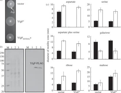
When S. Enteritidis transformed with the vector control ( pGS2390) was incubated on motility agar plates, two chemo-tactic rings were observed (figure 1a). The innermost ring was assigned as the aspartate ring (arrowed in figure 1a) and the outermost ring as the serine ring [11,13]. However, the aspar-tate chemotactic ring was not observed whenS. Enteritidis was transformed by plasmid pGS2421 to permit sustained expression of YfgF (with a C-terminal FLAG tag; figure 1a). Expression of YfgF from pGS2421 in the S. Enteritidis
rsob.r
oy
alsocietypublishing.org
Open
Biol
3:
130046
derivativeSalmonella DXII, which lacks all GGDEF proteins and contains no detectable c-di-GMP [5], also inhibited the development of the aspartate chemotactic ring (not shown).
TheSalmonellaDXIIstrain lacks GGDEF domain proteins and detectable c-di-GMP, and thus the PDE activity of YfgF is unlikely to account for the inhibition of aspartate chemo-taxis. Accordingly, a YfgF variant, YfgF(E526A), that lacks
PDE activity was still able to prevent the appearance of the aspartate chemotactic ring in both wild-type S. Enteritidis (figure 1a) andSalmonellaDXII(not shown). The abolition of PDE activity by the E526A substitution was demonstrated by the inability of YfgF(E526A)to degrade c-di-GMPin vitro and
its failure to complement the motility defect of anE. coli yhjH
mutant in vivo (not shown). Western blotting using anti-FLAG-tag antibodies indicated that the YfgF-FLAG and YfgF(E526A)-FLAG proteins were expressed at similar levels
in the transformed bacteria (figure 1b); note that, like many other membrane proteins, YfgF-FLAG (molecular mass 88.5 kDa) migrates aberrantly on denaturing polyacrylamide gels (Mr≏65 kDa).
The extent of the influence of YfgF on chemotaxis was measured using minimal media containing either a single chemoattractant or a combination of two chemoattractants such that consumption of the substrate(s) generates concen-tration gradients. Expression of YfgF-FLAG inS. Enteritidis
10 (c) (a)
(b)
aspartate
aspartate plus serine
diameter of motility zone (mm)
ribose
vector vector
Asp
YfgF+ YfgF+
YfgF-FLAG M
250 150
100 75
50
37
25
20
1 2 3 M 1 2 3
YfgF(E526A)+
vector YfgF+ maltose
galactose serine
8
6
4
2
0
16
12
8
0 4
16
12
8
0 4
25
20
15
10
0 5 20
15
10
5
0
12
8
4
[image:5.595.93.504.48.361.2]0
Figure 1.
S
. Enteritidis YfgF modulates aerobic aspartate chemotaxis via a c-di-GMP-independent mechanism. (
a
) Swimming motility of
S
. Enteritidis transformed
with plasmids expressing no YfgF (vector control),
S
. Enteritidis YfgF and
S
. Enteritidis YfgF
(E526A). The aspartate (Asp) chemotactic ring is arrowed. (
b
) Coomassie
blue-stained SDS polyacrylamide gel (left) and Western blot developed with anti-FLAG antibodies (right) for
S
. Enteritidis transformed with pGS2390 (vector control,
lanes 1), pGS2421 (YfgF
þ, lane 2), or pGS2460 (YfgF
(E526A), lane 3). The Coomassie blue-stained gel shows the protein loading for each lane. The locations of the
YfgF protein and the protein markers to calibrate the gel (lane M; molecular weights in kDa) are indicated. (
c
) Effects of sustained expression of YfgF. The
che-motactic response of
S
. Enteritidis was assessed on media containing the indicated chemoeffector(s) under aerobic (black bars) and anaerobic (white bars) conditions.
Each experiment was performed a minimum of three times; the mean values and standard deviations are shown.
vector
A
A
YfgF+
(a)
(b)
Figure 2.
Sustained expression of YfgF inhibits the migration of
S
. Enteritidis
towards a source of aspartate. An aspartate concentration gradient was
cre-ated by placing a filter disc (marked as A in the figure) loaded with 10
m
l of
1 M aspartate in the centre of a minimal medium agar plate lacking other
carbon sources. Suspensions of
S
. Enteritis transformed with pGS2390 (vector
control) or pGS2421 (YfgF
þ) were pipetted onto the agar, and the plates
were incubated under (
a
) aerobic and (
b
) anaerobic conditions at 28
8
C for
16 h. Chemotaxis was visible as the crescent formed as the bacteria migrated
from the point of inoculation towards the source of aspartate.
rsob.r
oy
alsocietypublishing.org
Open
Biol
3:
130046
[image:5.595.324.541.504.682.2]under aerobic conditions abolished colony expansion in the presence of aspartate, but did not affect the responses to serine, galactose, ribose or maltose (figure 1c, black bars). Furthermore, YfgF did not impair the chemotactic response to serine even in the presence of aspartate (figure 1c). Under anaerobic conditions both strains responded to all the chemoeffectors tested (figure 1c, white bars), including a positive response to aspartate. Expression of YfgF-FLAG inS. Enteritidis under aerobic conditions had no effect on the response to the repellants nickel and cobalt (data not shown). Together these data suggest that YfgF modulates colony expansion of S. Enteritidis in response to aspartate under aerobic conditions independently of c-di-GMP.
4.2. Sustained expression of YfgF inhibits bacterial
migration toward a source of aspartate under
aerobic conditions
In the experiments described in §4.1, the formation of the aspartate chemotactic ring is dependent upon the ability of the bacteria to consume aspartate and thereby create a con-centration gradient. In such experiments YfgF might impair bacterial migration by inhibiting aspartate utilization rather than by inhibiting aspartate chemotaxis. Therefore, the effects
of sustained YfgF-FLAG expression on the ability ofS. Enter-itidis to migrate in an aspartate gradient was tested on motility agar in which a concentration gradient was formed by diffusion from a filter disc loaded with aspartate placed on an agar plate. The experiment showed thatS. Enteritidis transformed with the vector ( pGS2390) was able to migrate towards the aspartate-loaded disc, whereas S. Enteritidis expressing YfgF-FLAG failed to do so under aerobic con-ditions (figure 2a). In contrast, under anaerobic conditions the inhibition of aspartate chemotaxis by YfgF-FLAG was not observed (figure 2b), confirming that YfgF is an inhibitor of bacterial migration in response to aspartate only under aerobic conditions (figure 1c). This experiment indicates that the metabolic generation of aspartate gradients is not necessary for the aerobic inhibition of aspartate chemotactic ring formation by YfgF.
4.3. The ability of YfgF to inhibit the formation of the
aspartate chemotactic ring is modulated by
exogenous
p-benzoquinone
In E. coli K-12 yfgF expression is activated by the oxygen-responsive transcription factor FNR and maximal under anaerobic conditions [3]. Analysis of the DNA sequence of
250
8
6
4
2
0
6
4
2
0
aspartate
diameter of motility zone (mm)
aspartate plus p-benzoquinone
M
vector (c)
(d) (a)
(b)
1 2 1 2
YfgF-FLAG
YfgF+ vector
Asp
YfgF+
vector YfgF+
150
100
75
50
37
25
[image:6.595.108.490.44.346.2]20
Figure 3.
YfgF-mediated inhibition of aspartate chemotaxis is relieved by exogenous
p
-benzoquinone. (
a
) Swimming motility of
S
. Enteritidis transformed with
plasmids expressing no YfgF (vector control) and
S
. Enteritidis YfgF in the presence of
p
-benzoquinone (200
m
g ml
21) under aerobic conditions. The aspartate (Asp)
chemotactic ring (arrowed) is apparent in the absence (left) and presence (right) of YfgF. (
b
) The motility of
S
. Enteritidis was assessed on medium containing
aspartate in the presence and absence of
p
-benzoquinone under aerobic conditions. In all cases, each experiment was performed a minimum of three times (mean
values with standard deviation are shown). (
c
) Coomassie blue-stained SDS polyacrylamide gel (left) and Western blot developed with anti-FLAG antibodies (right)
for
S
. Enteritidis transformed with pGS2421 (YfgF
þ) in the absence (lane 1) and presence (lane 2) of
p
-benzoquinone (200
m
g ml
21). The Coomassie blue-stained
gel shows the protein loading for each lane. The locations of the YfgF protein and the protein markers to calibrate the gel (lane M; molecular weights in kDa) are
indicated. (
d
) Motility of
E. coli yhjH
mutant transformed with pGS2390 (vector control) or pGS2421 (YfgF
þ) in the presence of
p
-benzoquinone (200
m
g ml
21).
Plates were incubated under aerobic conditions for 20 h at 37
8
C.
rsob.r
oy
alsocietypublishing.org
Open
Biol
3:
130046
theS. EnteritidisyfgFpromoter region indicated that the FNR regulatory element is conserved. DespiteE. coli yfgFexpression being maximal under anaerobic conditions, theyfgF mutant exhibits an oxidative stress phenotype [3]. Furthermore, the YfgF-mediated inhibition of aspartate chemotaxis observed here was apparent under aerobic but not anaerobic conditions (figures 1cand 2). Therefore, the effect of modulating the redox environment on YfgF function was investigated. Addition of increasing concentrations ofp-benzoquinone (which inhibits respiration, generates reactive oxygen species and lowers the proton motive force [13–15]) to motility plates resulted in restoration of the aspartate chemotactic ring under aerobic conditions (figure 3a). The identity of the aspartate ring was confirmed by spotting aspartate onto motility plates and observing colony morphology (not shown). Furthermore, aerobic colony expansion on minimal agar containing aspartate was restored in the presence ofp-benzoquinone (figure 3b). Western blotting using antibodies to the FLAG-tag showed that the level of YfgF protein in the presence and absence ofp-benzoquinone were similar (figure 3c) and the YfgF-FLAG protein was functional in the presence of
p-benzoquinone as judged by the ability to complement the motility defect of anE. coli yhjHmutant (figure 3d). There-fore, it was concluded that a redox signal, generated by the presence of exogenous p-benzoquinone in the experiments described here, is perceived by YfgF to restore the aspartate chemotactic ring.
4.4. A motif (Lys-Lys-Glu) linking trans-membrane
regions 5 and 6 is required for function of the
YfgF MASE1 domain
The E. coli YfeA protein has a similar domain structure to YfgF (MASE1-GGDEF-EAL), including a catalytically inac-tive GGDEF domain and catalytically acinac-tive EAL domain. Expression of YfeA inS. Enteritidis did not affect the bacterial response to aspartate; in contrast, expression ofE. coli YfgF (73% identical and 87% similar to S. Enteritidis YfgF over 733 amino acids) resulted in the same phenotypes as were observed for S. Enteritidis YfgF (not shown). These obser-vations and the experiments described above suggested a possible function for the YfgF MASE1 domain as a redox sensor. Three cysteine residues (131, 183 and Cys-231) within membrane spanning regions 4, 5 and 6 of YfgF were initially targeted for site-directed mutagenesis as they could mediate a redox signal by changes in oxida-tion state; however, the phenotypes of bacteria expressing these YfgF variants was the same as those expressing unal-tered YfgF (i.e. aspartate chemotactic ring formation was inhibited; not shown). Alignment of the amino acid sequence ofS. Enteritidis YfgF with theE. coliYfgF and YfeA MASE1 domains allowed the identification of residues that are con-served inS. Enteritidis andE. coliYfgF but are not in YfeA. Therefore, selected conserved charged/polar amino acids
(a)
(b)
diameter of motility zone (mm)
(c)
(d) vector
Asp
aspartate
aspartate plus serine 8
6 4 2 0
8 6 4 2 0
YfgF+ vector
vector
YfgF+
YfgF+
YfgF(KKE-AAA)+
YfgF(KKE-AAA)+
YfgF-FLAG YfgF(KKE-AAA)+
250 150 100 75 50
37
25 20
[image:7.595.127.477.41.338.2]M 1 2 3 M 1 2 3
Figure 4.
Replacement of Lys-Lys-Glu (amino acids 213 – 215) by Ala-Ala-Ala in the YfgF MASE1 domain permits formation of the aspartate chemotactic ring and
S
. Enteritidis colony expansion. (
a
) Swimming motility of
S
. Enteritidis transformed with plasmids expressing no YfgF (vector control, left),
S
. Enteritidis YfgF
þ(right)
and
S
. Enteritidis YfgF
(KKE-AAA)þ
(bottom). (
b
) Motility of
S
. Enteritidis on medium containing aspartate or aspartate plus serine was measured under aerobic conditions
as described above. (
c
) YfgF
(KKE-AAA)complements the motility defect of an
E. coli
K-12
yhjH
mutant. Swimming motility of
E. coli yhjH
transformed with pGS2390
(vector), pGS2421 (YfgF) and pGS2426 (YfgF
(KKE-AAA)). Plates were incubated for 20 h at 37
8
C under aerobic conditions. (
d
) Coomassie blue-stained SDS
polyacryl-amide gel (left) and Western blot developed with anti-FLAG antibodies (right) for
S
. Enteritidis transformed with pGS2390 (vector control, lane 1), pGS2421 (YfgF
þ,
lane 2), or pGS2426 (YfgF
(KKE-AAA), lane 3). The Coomassie blue-stained gel shows the protein loading for each lane. Lane M contains protein markers to calibrate the
gel (molecular weights in kDa are indicated). The location of the YfgF protein is also indicated.
rsob.r
oy
alsocietypublishing.org
Open
Biol
3:
130046
within membrane spanning regions 3, 7, 8 and 9 (Tyr-83, Glu-84, Thr-243, Asn-244, Thr-246, Leu-247, Ser-248, Arg-260, Ser-299, Ser-300, Ser-301, Tyr-302) were replaced, but these variants also exhibited wild-type behaviour (not shown). Thus, a third phase of site-directed mutagenesis was targeted towards conserved residues in the loops linking the trans-membrane regions. Replacement of Arg-109, Arg-110, His-115, Asp-117, Ser-151, Arg-191, Asn-192, Arg-197, Tyr-199, Tyr-200, Gln-202, Leu-203, 204, Gln-206, Asp-208, Lys-264, His-279 showed that these residues were not required for YfgF-mediated inhibition of the response to aspartate; the phenotypes of all these variants were similar to wild-type YfgF (not shown). However, replacement of a motif Lys-Lys-Glu (positions 213–215, located in the cytoplasmic loop between trans-membrane regions 5 and 6; pGS2426) by Ala-Ala-Ala abolished the ability of YfgF to inhibit the formation of the aspartate chemotactic ring (figure 4a) and permitted colony expansion on aspartate-containing medium without affecting the response to serine (figure 4b). These data indicated that the integrity of the predicted cytoplasmic loop located between trans-membrane regions 5 and 6 was required for YfgF MASE1 function. Replacement of the Lys-Lys-Glu motif did not impair the ability of YfgF to complement the motility defect of anE. coli yhjH mutant, indicating that the overall fold and PDE activity of the mutant protein were retained (figure 4c). Furthermore, immunoblotting using anti-FLAG antibodies indicated that the expression of YfgF and YfgF(KKE-AAA) were similar, and hence the inability of the
latter to mediate a response to aspartate was not caused by inadequate expression (figure 4d).
5. Discussion
Here, it is shown that S. Enteritidis YfgF controls bacterial motility by c-di-GMP-dependent and c-di-GMP-independent mechanisms, as shown by PDE-dependent restoration of moti-lity to anE. coli yhjHmutant, and PDE-independent, MASE1 domain-mediated inhibition of bacterial migration in response to aspartate. The results presented here used YfgF protein with a C-terminal FLAG-tag, but it should be noted that similar results were obtained with YfgF carrying a C-terminal His-tag (not shown), suggesting that the presence of the C-terminal tag does not impair YfgF function.
The initial experimental conditions required substrate (aspartate) utilization to create a concentration gradient and showed that YfgF specifically inhibited the formation of the aspartate chemotactic ring and expansion of bacterial colonies on aspartate medium without affecting the response to serine or sugar molecules under aerobic conditions (figure 1). When exposed to aspartate gradients generated independently of aspartate metabolism bacteria expressing yfgF were still unable to move along the concentration gradient (figure 2). Therefore, YfgF specifically inhibited S. Enteritidis colony expansion in response to aspartate in a c-di-GMP-independent manner under aerobic, but not anaerobic, conditions.
The ability of YfgF to inhibit aspartate chemotaxis was modulated by exogenous p-benzoquinone (figure 3), sug-gesting that YfgF activity responds to a redox signal. Site-directed mutagenesis identified a motif Lys-Lys-Glu (pos-ition 213–215) in the MASE1 domain that was required for YfgF-mediated inhibition of aspartate taxis (figure 4). This motif is likely to represent a component of a redox sensing
and/or effector region of YfgF. TheE. coliserine chemoreceptor Tsr is able to sense changes in the proton motive force, and it has been suggested that this might involve charged amino acids located on either side of the membrane spanning regions of the protein [16,17]. It is possible that the MASE1 amino acid motif identified here could form part of a similar sensory mech-anism. However, it is not known whether the apparent inhibition of aspartate taxis by the YfgF MASE1 domain is direct or indirect. For direct regulation, it is not obvious how the cytoplasmic loop linking trans-membrane regions 5 and 6 of YfgF could effectively inhibit the periplasmic interactions between Tar and its effector, aspartate. However, apparent inhibition of aspartate taxis would be observed if YfgF acted to promote aspartate excretion from the bacterium. The excreted aspartate could then act as an attractant, and thereby oppose the outward migration of the colony. Such apparent inhibition of aspartate chemotaxis has been observed pre-viously. Thus, some of the complex symmetrical patterns formed by motileE. colicells on soft-agar plates were found to be associated with the response of Tar to aspartate excreted by the bacteria triggered in part by oxidative stress [18–20].
Asp
(a)
(b)
Asp
YfgF
O2
Asp Isuc
Isuc H2O2
[image:8.595.316.546.43.222.2]Suc Suc Frd ETC AspA AspA Fum Fum c-di-GMP c-di-GMP pGpG pGpG NadB NadB Asp chemotaxis Asp uptake Asp excretion Asp Asp Frd redox perturbation ETC Asp chemotaxis Asp Asp ? Asp uptake Asp excretion YfgF
Figure 5.
YfgF modulates aspartate chemotaxis to lessen oxidative damage
when anaerobic bacteria are first exposed to molecular oxygen (O
2). (
a
) In
the absence of O
2, expression of
yfgF
is maximal [3] and the proton motive
force is low [24]. Under these conditions aspartate chemotaxis (in the presence
of an appropriate energy source), aspartate uptake and metabolism are
sup-ported (solid arrows). Aspartase (AspA) converts Asp to fumarate (Fum),
which in turn is reduced to succinate (Suc) by fumarate reductase (Frd)
using reducing equivalents from the electron transport chain (ETC). Aspartate
is also the substrate for NadB, which converts Asp to iminosuccinate (Isuc),
which is required for NAD
þbiosynthesis, with Fum acting as the electron
acceptor [25]. (
b
) If left unchecked, when anaerobic bacteria are exposed to
O
2NadB and Frd use O
2as an electron acceptor [22,23], generating the reactive
oxygen species hydrogen peroxide (H
2O
2) (broken arrows), resulting in oxidative
stress and redox perturbation. It is suggested (denoted by question mark) that
the MASE1 domain of YfgF perceives the onset of this stress and either inhibits
(
?
) aspartate chemotaxis or promotes () aspartate excretion to decrease Asp
utilization and thereby limit H
2O
2generation until the bacteria have adapted to
the new aerobic conditions. The presence of O
2switches off the transcription
factor FNR, thereby minimizing
yfgF
expression (
?
). After adaptation growth
and protein turnover removes YfgF from the now aerobic bacteria, restoring
aspartate utilization and observable aspartate chemotaxis. Sustained expression
of
yfgF
in the experimental system used here has allowed this normally
tran-sient behaviour to be revealed. Hydrolysis of c-di-GMP by YfgF promotes
motility under anaerobic and aerobic conditions.
Subsequently, E. coli aer(aerotaxis signal transducer) mutants supplied with succinate failed to migrate from the point of inoculation on semi-solid agar, but this was not the case for the E. coli aer tar double mutant [21]. These observations were rationalized by suggesting that under these conditions the bacteria excrete aspartate, which acts as an attractant con-straining colony expansion in the absence of aerotaxis. Hence, it is possible that YfgF is the redox sensor that acts to promote aspartate excretion to counteract the potentially dele-terious use of O2as an electron acceptor by aspartate oxidase
and fumarate reductase, which are major sources of the reac-tive oxygen species H2O2 when E. coli is transferred from
anaerobic to aerobic conditions [22,23]. Thus, it is proposed that YfgF transiently inhibits aspartate taxis or promotes aspar-tate excretion to lower the production of reactive oxygen species when anaerobicSalmonellaare rapidly exposed to O2
(figure 5). Because theyfgF gene is under the control of a Class II FNR-dependent promoter, aerobic inhibition of aspar-tate taxis would not be readily observed in wild-type bacteria, and thus sustained expression ofyfgF has allowed this nor-mally transient behaviour to be revealed. Aerobic inhibition of aspartate chemotactic ring formation was observed when the nuo operon (encoding the proton translocating NADH dehydrogenase I of the ETC) ofE. coliwas disrupted [13]. How-ever, when supplied with glycerol or combined with a tsr
mutation thenuomutants did form the aspartate chemotactic ring, suggesting that simultaneous consumption of aspartate and another carbon source was necessary for the formation of the aspartate chemotactic ring [13]. The perturbation of cel-lular redox state by thenuomutation apparently prevented such conditions being met, resulting in inhibition of aspartate taxis [13]. Thus, the possibility that YfgF acts to inhibit the util-ization of an ancillary carbon source that is required for the
formation of the aspartate ring on semi-solid agar cannot be excluded, but this possibility is unlikely because YfgF inhibits migration towards a source of aspartate under aerobic conditions in the absence of other carbon sources (figure 2).
MASE1 domains are membrane spanning units that are present in 462 bacterial species, including 164 species of Enterobacteriaceae [26]. These domains are often linked to other domains involved in signalling pathways, including histi-dine kinases, PAS, GAF, GGDEF and EAL domains [4]. While most MASE1 domains are linked to signalling units, there are examples of ‘stand-alone’ MASE1 domains in 20 bacterial genera [26], suggesting that they can also function indepen-dently to sense and transduce signals. The widespread distribution of MASE1 domains suggests that they influence many aspects of bacterial physiology, but this is the first time to the authors’ knowledge that a function of a MASE1 domain has been assigned. Future work should be directed towards establishing the precise mechanisms of signal perception and transduction by the YfgF MASE1 domain.
In conclusion, analysis of YfgF shows that it regulates bacterial motility by c-di-GMP-dependent and -independent mechanisms. Cyclic-di-GMP-dependent YfgF activity requires a catalytically active EAL domain, and thus YfgF is an important component in the complex array of interacting DGCs and PDEs that control several aspects of bacterial behaviour. Cyclic-di-GMP-independent YfgF activity is mediated by its MASE1 domain, revealing for the first time the function of a MASE1 domain as a redox-responsive regulator of aspartate taxis.
Acknowledgements. The authors thank Prof. In˜ igo Lasa (Instituto de Agrobiotecnologia, Universidad Pu´blica de Navarra-CSIC-Gobierno de Navarra) for useful discussions and the gift of bacterial strains. This work was supported by the Biotechnology and Biological Sciences Research Council UK (grant no. BB/G006024/1).
References
1. Mills E, Pultz IS, Kulasekara HD, Miller SI. 2011 The bacterial second messenger c-di-GMP: mechanisms of signalling.Cell Microbiol.13, 1122 – 1129. (doi:10.1111/j.1462-5822.2011.01619.x)
2. Pesavento C, Hengge R. 2009 Bacterial nucleotide-based second messengers.Curr. Opin. Microbiol.12, 170 – 176. (doi:10.1016/j.mib.2009.01.007) 3. Lacey MM, Partridge JD, Green J. 2010Escherichia coli
K-12 YfgF is an anaerobic cyclic di-GMP phosphodiesterase with roles in cell surface re-modelling and the oxidative stress response.Microbiology156, 2873–2888. (doi:10. 1099/mic.0.037887-0)
4. Nikolskaya AN, Mulkidjanian AY, Beech IB, Galperin MY. 2003 MASE1 and MASE2: two novel integral membrane sensory domains.J. Mol. Microbiol. Biotechnol.5, 11 – 16. (doi:10.1159/000068720) 5. Solano C, Garcı´a B, Latasa C, Toledo-Arana A,
Zorraquino V, Valle J, Casals J, Pedroso E, Lasa I. 2009 Genetic reductionist approach for dissecting individual roles of GGDEF proteins within the c-di-GMP signalling network inSalmonella.Proc. Natl Acad. Sci. USA101, 16 322 – 16 327. (doi:10.1073/ pnas.0812573106)
6. Girgis HS, Liu Y, Ryu WS, Tavazoie S. 2007 A comprehensive genetic characterization of bacterial
motility.PLoS Genet.3, 1644 – 1660. (doi:10.1371/ journal.pgen.0030154)
7. Lennox ES. 1955 Transduction of linked genetic characters of the host by bacteriophage P1. Virology1, 190 – 206. (doi:10.1016/0042-6822(55)90016-7)
8. Sambrook JW, Russell DW. 2001Molecular cloning: a laboratory manual, 3rd edn. Cold Spring Harbor, NY: Cold Spring Harbor Press.
9. Umemura T, Matsumoto Y, Ohnishi K, Homma M, Kawagishi I. 2002 Sensing of cytoplasmic pH by bacterial chemoreceptors involves the linker region that connects the membrane-spanning and the signal-modulating helices.J. Biol. Chem.277, 1593 – 1598. (doi:10.1074/jbc.M109930200) 10. Evans CGT, Herbert D, Tempest DW. 1970 The
continuous cultivation of micro-organisms.Methods Microbiol.2, 278 – 327.
11. Adler J. 1966 Chemotaxis in bacteria.Science153, 708 – 716. (doi:10.1126/science.153.3737.708) 12. Laemmli UK. 1970 Cleavage of structural proteins
during the assembly of the head of bacteriophage T4.Nature227, 680 – 685. (doi:10.1038/227680a0) 13. Pru¨ss BM, Nelms JM, Park C, Wolfe AJ. 1994
Mutations in NADH : ubiquinone oxidoreductase of
Escherichia coliaffect growth on mixed amino acids. J. Bacteriol.176, 2143 – 2150.
14. Bespalov VA, Zhulin IB, Taylor BL. 1996 Behavioral responses ofEscherichia colito changes in redox potential.Proc. Natl Acad. Sci. USA93, 10 084 – 10 089. (doi:10.1073/pnas.93.19.10084) 15. Sato-Watanabe M, Mogi T, Miyoshi H, Iwamura H,
Matsushita K, Adachi O, Anraku Y. 1994 Structure – function studies on the ubiquinol oxidation site of the cytochromebocomplex fromEscherichia coli usingp-benzoquinones and substituted phenols. J. Biol. Chem.269, 28 899 – 28 907.
16. Rebbapragada A, Johnson MS, Harding GP, Zuccarelli AJ, Fletcher HM, Zhulin IB, Taylor BL. 1997 The Aer protein and the serine chemoreceptor Tsr independently sense intracellular energy levels and transducer oxygen, redox, and energy signals for Escherichia colibehaviour.Proc. Natl Acad. Sci. USA94, 10 541–10 546. (doi:10.1073/pnas.94.20.10541) 17. Edwards JC, Johnson MS, Taylor BL. 2006
18. Budrene EO, Berg H. 1991 Complex patterns formed by motile cells ofEscherichia coli.Nature349, 630 – 633. (doi:10.1038/349630a0)
19. Budrene EO, Berg H. 1995 Dynamics of formation of symmetrical patterns by
chemotactic bacteria.Nature376, 49–53. (doi:10. 1038/376049a0)
20. Woodward DE, Tyson R, Myerscough MR, Murray JD, Budrene EO, Berg H. 1995 Spatio-temporal patterns generated bySalmonella typhimurium. Biophys. J.68, 2181–2189. (doi:10.1016/S0006-3495(95)80400-5)
21. Biblikov SI, Barnes LA, Gitin Y, Parkinson JS. 2000 Domain organizationand flavin dinucleotide-binding
determinants in the aerotaxis signal transducer Aer ofEscherichia coli.Proc. Natl Acad. Sci. USA97, 5830 – 5835. (doi:10.1073/pnas. 100118697)
22. Korshunov S, Imlay JA. 2010 Two sources of endogenous hydrogen peroxide inEscherichia coli. Mol. Microbiol.75, 1389 – 1401. (doi:10.1111/j. 1365-2958.2010.07059.x)
23. Messner KR, Imlay JA. 2002 Mechanism of superoxide and hydrogen peroxide formation by fumarate reductase, succinate dehydrogenase, and aspartate oxidase.J. Biol. Chem.
277, 42 563 – 42 571. (doi:10.1074/jbc. M204958200)
24. Kashkert ER. 1981 Effects of aerobiosis and nitrogen source on the proton motive force in growing Escherichia coliandKlebsiella pneumoniae cells. J. Bacteriol.146, 377 – 384.
25. Tedeschi G, Negri A, Mortarino M, Ceciliani F, Simonic T, Faotto L, Ronchi S. 1996L-Aspartate
oxidase fromEscherichia coli: II. Interaction with C4dicarboxylic acids and identification of a novelL
-aspartate:fumarate oxidoreductase activity. Eur. J. Biochem.239, 427 – 433. (doi:10.1111/j. 1432-1033.1996.0427u.x)
26. Finn RDet al. 2010 The Pfam protein families database.Nucl. Acids Res.38, D211 – D222. (doi:10. 1093/nar/gkp985)
rsob.r
oy
alsocietypublishing.org
Open
Biol
3:
130046


![Figure 5. YfgF modulates aspartate chemotaxis to lessen oxidative damagethe absence of Oacceptor [25]](https://thumb-us.123doks.com/thumbv2/123dok_us/743456.579342/8.595.316.546.43.222/figure-modulates-aspartate-chemotaxis-oxidative-damagethe-absence-oacceptor.webp)
