5-HT1A receptor expression : Studies in postmortem tissue
and characterisation of a model system.
BUNN, Lindsey J.
Available from Sheffield Hallam University Research Archive (SHURA) at:
http://shura.shu.ac.uk/19409/
This document is the author deposited version. You are advised to consult the
publisher's version if you wish to cite from it.
Published version
BUNN, Lindsey J. (2008). 5-HT1A receptor expression : Studies in postmortem
tissue and characterisation of a model system. Doctoral, Sheffield Hallam University
(United Kingdom)..
Copyright and re-use policy
c ''V/i. d-o Hailem University Lev ">?• T' Services Adset -. Cam; .'Ivy Campus
-Sheffield SI 1WB
ProQuest Number: 10694290
All rights reserved
INFORMATION TO ALL USERS
The qua lity of this reproduction is d e p e n d e n t upon the qua lity of the copy subm itted.
In the unlikely e v e n t that the author did not send a c o m p le te m anuscript and there are missing pages, these will be noted. Also, if m aterial had to be rem oved,
a n o te will in d ica te the deletion.
uest
ProQuest 10694290
Published by ProQuest LLC(2017). C o pyright of the Dissertation is held by the Author.
All rights reserved.
This work is protected against unauthorized copying under Title 17, United States C o d e M icroform Edition © ProQuest LLC.
ProQuest LLC.
789 East Eisenhower Parkway P.O. Box 1346
5-HT
ia
re
ceptor expression; studies in
postmortem tissue and
characterisation of a model system
Lindsey Janice Bunn
A thesis submitted in partial fulfilment of the requirements
of Sheffield Hallam University
for the degree of Doctor of Philosophy
Abstract
Serotonin (5-HT) neurotransmission is involved in the psychopharmacology of several psychiatric disorders including, depression, anxiety disorders and schizophrenia. The release of HT in neurons is mediated by somatodendritic 5-HTia autoreceptors. The presence of 5-HTia receptor is thought to be increased
in depressed patients, producing a reduction in the synthesis of 5-HT. A common single nucleotide polymorphism in the promoter region of 5HT1A receptor C-1019G is also associated with depression and suicide. The nuclear DEAF-1 related (NUDR) protein represses the 5-HTiA promoter region hence regulating
both 5-HT1A transcription and receptor expression.
The project involved undertaking a post-mortem study to determine any association between the HT1A promoter polymorphism and the expression of 5-HTia receptor mRNA and receptor density in control human hippocampal brain
tissue. This was achieved by genotyping human brain tissue for the 5-HTiA
receptor polymorphism C-1019G and 5-HTiA receptor mRNA levels were
quantified using real-time PCR. Radioligand binding was used to determine Bmax and Kd quantifying 5-HT1A receptor density.
The SHSY-5Y neuroblastoma cell line is a well characterised cell line model used in neurotransmitter studies when differentiated. The 5-HT1A receptor couples to Gj proteins inhibiting AC activity and hence mediating a variety of intracellular changes such as, decreasing cAMP leading to decreased Ca2+ levels. The SH-SY5Y cell line study investigated whether the 5-HTiA agonist 8-OH-DPAT, inhibited forskolin stimulated Ca2+ release in the SHSY-5Y cell line and whether the 5-HT1A antagonist p-MPPI reversed this effect using flow cytometry.
The post-mortem study showed that the G-1019 allele had significantly higher 5-HT1a expression compared to the C allele in control hippocampal tissue. Radioligand binding data demonstrated that control samples with a GG or G/C genotype had a significantly higher 5-HT1A receptor density compared to samples with a CC genotype. SH-SY5Y cells differentiated with RA for 5 days or NGF and aphidicolin for 10 days had significantly increased 5-HT1A receptor mRNA levels compared to undifferentiated cells. Western blots and immunocytochemistry confirmed the presence of the 5-HT1A receptor in this cell line. An increase in NUDR expression was observed at the same time there is an increase in 5-HTiA receptor expression in SH-SY5Y cells treated with RA or NGF and aphidicolin. Flow cytometry showed that 8-OH-DPAT efficiently diminished forskolin-stimulated increase in intracellular Ca2+ in RA differentiated cells. HT also a 5-HT1A agonist had a similar effect. SH-SY5Y cells treated with both p-MPPI and 8 -OH-DPAT demonstrated that cells treated with p-MPPI at higher concentrations significantly increased forskolin-stimulated intracellular Ca2+ levels and therefore effectively reversed the agonistic effect of 8-OH-DPAT.
The findings presented in the post-mortem study are novel and the SH-SY5Y cell line study demonstrates that this cell line when differentiated with either RA or NGF and aphidicolin is a useful cell-line model system for studying the 5-HT1A receptor.
Abbreviations
AC Adenylyl cyclase
ACTH Adrenocorticotropic hormone
APS Ammonium Persulfate
ARMS Amplification refractory mutation system
ASO Allele-specific oligonucleotides
BCA Bicinchonic acid
Bmax Binding site density
BP Binding potential
BSA Bovine serum albumin
CA Cornu ammonis
cAMP Cyclic AMP
CAMK Calmodulin-dependent protein kinase
cDNA Single-stranded complementary DNA synthesised from RNA template
CNS Central nervous system
CRF Corticotrophin-releasing factor
CRH Corticotrophin-releasing hormone
CSF Cerebrospinal fluid
ct
Threshold cycleDAPI 4'6-diamino-2-phenylindole
DMEM Dulbecco's modified Eagle's medium
DMSO Dimethyl sulfoxide
DR Dorsal raphe area
DRE Dual repressor element
dsDNA Double stranded DNA
ERK Extracellular signal related kinases
FCS Foetal bovine (calf) serum
FRET Fluorescence resonance energy transfer
GABA y-Aminobutyric acid
GDP Inactive guanine diphosphate
GPCR'S G-protein coupled receptor superfamily
GR Glucocorticoid receptor
GTP Activated guanine triphosphate
HPA Hypothalamic pituitary adrenal axis
ICC Immunocytochemistry
Kd Measure of the concentration of radioactive ligand that is required to occupy 50 percent of receptors
LC Locus coeruleus
MGB Minor groove binding
MAOI's Monoamine oxidase inhibitors
MAP Mitogen-activated protein kinases
MDD Major depressive disorder
mRNA Messenger RNA
MR Median raphe area
MR Mineralocorticoid receptor
MTC Mesiotemporal cortex
NA Noradrenaline
NGF Nerve growth factor
NFQ Non-fluorescent quencher
NRI's Norepinephrine selective reuptake inhibitors
NUDR Human nuclear deformed epidermal autoregulatory factor
PBST PBS and Tween 20
PCR Polymerase chain reaction
PET Position emission tomography
PKA Protein kinase A
PMI Post-mortem delay
p-MPPI 4(2-methoxy-phenyl)-1 -1 [2'-(n-2"-pyridinyl)-p-lodobenzamido]- ethyl- piperazine
PNS Peripheral nervous system
PRS Polymorphism ratio sequence
PTSD Panic disorder
PVDF Polyvinylidene fluoride
PVN Paraventricular nucleus of hypothalamus
RA Retinoic acid
RARE Retinoic acid response element
Rn Normalised intensities
RNA Ribonucleic acid
RT-PCR Real-Time PCR
SDS Sodium dodecyl sulfate
SDS-PAGE Sodium dodecyl sulfate polyacrylamide gel electrophoresis
SNP Single oligonucleotide polymorphism
SSCP Single stranded conformational polymorphism
ssDNA Single stranded DNA
SSRI's Selective serotonin reuptake inhibitors TEMED KN .N '.N ’-Tetramethylethylenediamine
TCAs Tricyclic antidepressants
Tm Melting temperature
TPA 12-O-tetradecanoylphorbol
TPH Tryptophan hydroxylase
WAY100635 (N-[2-[4-(2-methoxyphenyl) 1 -piperaziny!]ethyl]n-(2- pyridinyl)cyclohexanecarboxamidetrihydrochloride
5-HT Serotonin
5-HIAA 5-hydroxyindoleacetic acid
5-HT1A Serotonin 1A receptor
8-OH-DPAT 8-hydroxy-2-di-n-propylaminotetralin
Acknowledgements
I would like to thank Dr Caroline Dalton and Dr Adrian Hall for all their supervision, guidance and support. I have very much appreciated this throughout my studies.
I would also like to thank Professor Gavin Reynolds and his research group for providing the post-mortem tissue used in this study and for their advice and help with radioligand binding.
I would like to express all my love to my family for their love, support and encouragement throughout my life. Special thanks to Robert for his love and for always being there for me and for making me laugh and smile through the ups and downs.
Publications
Bunn LJ et al (2007) The 5-HTiA agonist 8-O H-DPAT inhibits forskolin
m ediated intracellular C a2+ release in differentiated SHSY-5Y cells. Journal of Psychopharm acology abstract supplem ent, 21, 7, A54.
Bunn LJ et al (2007) Induction of 5-HTiA receptor and NUDR m RNA in
differentiated SHSY-5Y neuroblastom a cells. Poster presented at Lifesciences conference, Glasgow, UK
Bunn LJ et al (2006) Induction of 5-HT1Areceptor m RNA in differentiated SHSY-5Y neuroblastom a cells. Proceedings of the British Pharm acological Society at http://w w w .pA 2online.org/abstractsA /ol4lssue2abst038P .pdf
Bunn LJ et al (2006) A 5-H TiA receptor prom oter polym orphism has
effects on receptor density and m RNA expression. Journal of Psychopharm acology abstract supplem ent, 20, 5, A11.
Bunn LJ et al (2005) Expression of 5-HTiA receptor m RNA is influenced
by a com m on prom oter region polym orphism . Journal of Psychopharm acology abstract supplem ent, 19, 5, A35
Conferences attended
British Association for Psychopharm acology (BAP) Harrogate July, 2005
Collegium Internationale Neuro-Psychopharm acologicum (CINP) Belfast April, 2006
British Association for Psychopharm acology (BAP) Oxford July, 2006
British Pharm acological Society (BPS) W inter m eeting, 2006
Lifesciences conference Glasgow, 2007
British Association for Psychopharm acology (BAP) Harrogate July, 2007
Contents Page
Abstract... ...I Abbrevations... II Acknowledgements... Ill Publications... IV
Conferences attended... IV
List of figures...V List of tables...VI
C h a p te r o n e - Introduction...1
1.0- S e ro to n in (5 -H T )...2
1.1- Synthesis and m etabolism of serotonin...3
1.2- Serotonin release and uptake at a serotonergic syn apse...4
1.3- R eceptor subtype identification and classification...5
1.3.1- 5-HT, receptors...6
1.3.2- 5-HT2, 5-HT3 and 5-HT4 receptors...7
1.3.3- 5-ht5) 5-hte and 5-HT7 receptors... 7
1.4- Serotonergic system ...8
1.4.1- Anatomical localisation... 9
1.4.2- Anatomical localisation of 5-HT1A receptors...10
1.4.2.1- Raphe nuclei...10
1.4.2.2- Limbic system... 11
1.4.2.3- Hippocampus...11
1.4.2.4- Function of the hippocampus... 12
1.4.2.5- Serotonergic transmission in the hippocampus...13
1.4.2.6- Pre-synaptic mechanisms... 13
1.4.2.7- Post-synaptic mechanisms ... 14
1.4.2.8- Other limbic brain regions...14
1.4.2.9- Pre-frontal cortex (PFC)... 15
1.4.2.10- Hypothalamus...15
1.4.3.11-Amygdala.. ... 17
1.5- Non 5-HT Neurotransm itter system s... 18
1.5.1- GABAergic... 18
1.5.2- Noradrenergic system...18
1.5.3- Cholinergic system...20
1.5.4- Dopaminergic system...22
1.6- Depression... 24
1.6.1- Major depression (Unipolar)... 24
1.6.2- Bipolar depression (Manic depressive illness... 25
1.6.3- Stress-induced depression... 26
1.6.3.1- GR and MR receptors... 27
1.6.4- 5-HT receptor(s) involvement with depression... 28
1.6.5- Medical management of depression... 34
1.6.5.1- Monoamine oxidase inhibitors (MAOI's)... 35
1.6.5.2- Tricyclic antidepressants (TCA's)... 36
1.6.5.3- Selective reuptake inhibitors... 37
1.6.5.4- Selective serotonin reuptake inhibitors (SSRI's)... 37
1.7- 5-HTia receptor structure and pharmacology...39
1.7.1 - 5-HT 1A structure... 39
1.7.2- 5-HiA receptor polymorphisms... 41
1.7.3- 5-HT1A receptor ligand pharmacology... 43
1.7.3.1 - 5-HT 1A receptor agonists...43
1.7.3.2- 5-HT 1A receptor antagonists...45
1.7.4- 5-HT receptors and second messenger signalling pathways...47
1.7.4.1- 5-HT1A receptor signalling...47
1.7.4.2- Other 5-HT receptor signalling pathways...50
1.7.4.3- 5-HT1A signalling in depression...52
1.8- Summary... 55
1.9- Aims of thesis...57
C h a p te r tw o - Post-mortem tissue study of the 5-H TiA receptor... 58
2.0- A im s... 59
2.1- Introduction... ...60
2.1.1- Post-mortem tissue... 60
2.1.2- 5-HTiA polymorphism C-1019G... 61
2.1.3- Genotyping methods... 61
2.1.3.1- Real-time PCR... 62
2.1.3.2- PCR amplification phase... 63
2.1.3.3- Melt curve analysis... 63
2.1.3.4- Housekeeping genes... 64
2.1.3.5- Optimisation of PCR reaction... 65
2.1.3.6- Amplification efficiency of PCR primers... 65
2.1.3.7- Allele specific oligonucleotides (ASO)... 66
2.1.3.8- SNP real-time PCR genotyping... 67
2.1.3.9- TaqMan probe genotyping... 67
2.1.4- Radioligand binding... 68
2.2- M aterials and m ethods... 70
2.2.1- Ethical aspects... 70
2.2.2- Brain tissue samples... 70
2.2.3- Human post-mortem brain tissue genotyping...70
2.2.3.1- DNA extraction... 70
2.2.3.2- Allele specific oligonucleotide (ASO) PCR for 5-HT1A genotyping... 71
2.2.3.3- PCR reaction...71
2.2.3.4- PCR cycle conditions...72
2.2.4- SNP real-time PCR genotyping... 72
2.2.4.1- Primers... 72
2.2.4.2- PCR reaction... 73
2.2.4.3- PCR cycle conditions...73
2.2.5- TaqMan custom genotyping...73
2.2.5.1- PCR cycles... 73
2.2.6- RNA extraction... 74
2.2.6.1- Experion analysis of RNA...74
2.2.6.2- cDNA synthesis... 74
2.2.7- Real-time PC R... ... 75
2.2.7.1- Housekeeping gene validation... 75
2.2.7.2- Primer design... 75
2.2.7.4- PCR cycles... 77
2.2.7.5- Primer efficiency... 77
2.2.8- Radioligand binding of 5 - H T1A... 78
2.2.8.1- Sample preparation... 78
2.2.9- Data analysis... 79
2.2.9.1- Comparative Ct method... 79
2.2.9.2- Pfaffl method... 79
2.2.9.3- Primer efficiency calculation... 79
2.2.9.4- Bmax and Kd... 80
2.3- R e s u lts ... 81
2.3.1- ASO 5-HT,A genotyping... 81
2.3.2- Real-time PCR SNP genotyping... 82
2.3.3- TaqMan genotyping... 84
2.3.4- Real-time PCR gene expression results... 89
2.3.4.1- RNA analysis using the Experion system... 89
2.3.4.2- Housekeeping gene validation... 90
2.3.4.3- Efficiency of primers... 92
2.3.4.4- Post-mortem factors... 94
2.3.4.5- Real-time PCR data correlated to genotype... 96
2.3.5- Radioligand binding results... 98
2.3.5.1- Binding data correlated with genotype data... 100
2.4- D is c u s s io n ...102
2.4.1- 5-HT,A genotyping of human post-mortem brain tissue samples 102 2.4.2- Real-time gene expression PCR...104
2.4.2.1- RNA analysis... 104
2.4.2.2- Housekeeping gene validation... 105
2.4.2.3- Efficiency of primers... 105
2.4.2.4- Effect of age, Post-mortem interval (PMI) and sex on 5-HT,A receptor mRNA... 106
2.4.2.5- Real-time PCR correlated with 5-HT,A genotype data 107 2.4.3- Radioligand binding of 5-HT,A...107
C h a p te r th re e - SH -SY5Y cell line study...110
3.0- A im s ...111
3.1- In tro d u c tio n ...112
3.1.1- SH-SY5Y cell line...112
3.1.2- Human nuclear deformed epidermal autoregulatory factor (NUDR)... 114
3.1.3- Real-time PCR analysis of gene expression... 114
3.1.4- Immunocytochemistry... 115
3.1.5- Western blots...115
3.2- M a te ria ls and m e th o d s ...117
3.2.1- Cell culture... 117
3.2.2- Differentiation with Retinoic acid (RA), Nerve growth factor (NGF) and aphidicolin... 117
3.2.3- SH-SY5Y cell line genotyping... 117
3.2.4- Gene expression studies by real-time PCR... 117
3.2.4.1- Cell preparation before RNA extraction...118
3.2.4.2- RNA extraction...118
3.2.4.4- Primer design and Housekeeping gene validation... 118
3.2.4.5- PCR reaction... 118
3.2.4.6- PCR cycles... 119
3.2.4.7- Primer efficiency... 119
3.2.4- Im m unocytochem istry... 119
3.2.5- SH -SY5Y cell line western blot... 12°
3.2.5.1- Sample preparation... 120
3.2.5.2- Protein concentration using Amicon centricon centrifugal filter devices... 120
3.2.5.3- Bicinchonic acid (BCA) assay... 121
3.2.5.4- SDS-PAGE gel... 121
3.2.5.5- Blot...121
3.3- R esults...122
3.3.1- Time-points of SH-SY5Y RA differentiated cells... 123
3.3.2- Time-points of SH-SY5Y NGF and aphidicolin differentiated cells.123 3.3.3- SH-SY5Y cell line genotype... 123
3.3.4- Real-time PCR gene expression... 126
3.3.4.1- RNA...126
3.3.4.2- Housekeeping gene validation...126
3.3.4.3- Primer efficiency... 127
3.3.4.4- 5-HT-ia receptor expression in RA and NGF and 129 aphidicolin SH-SY5Y differentiated cells... 130
3.3.4.5- NUDR mRNA expression in RA and NGF and aphidicolin SH-SY5Y differentiated cells...133
3.3.5- Immunocytochemistry...134
3.3.6- Western blot...136
3.4- D iscussion...137
3.4.1- Time-points of retinoic acid differentiated SH-SY5Y cells... 137
3.4.2- Time-points of NGF and aphidicolin differentiated SH-SY5Y cells...137
3.4.3- Real-time PCR...138
3.4.3.1- RNA...138
3.4.3.2- Housekeeping gene validation...138
3.4.3.3- Efficiency of primers... 139
3.4.3.4- 5-HT1A receptor mRNA expression in RA or NGF and aphidicolin differentiated SH-SY5Y cells... 139
3.4.3.5- NUDR mRNA expression in RA or NGF and aphidicolin differentiated SH-SY5Y cells... ...141
3.4.4- Immunocytochemistry... 142
3.4.5- Western blots...142
3.4.6- Conclusion... 143
C h a p te r fo u r- 5-H Tia second messenger signalling...144
4.0- A im ... 145
4.1- Introduction...146
4.1.2- Calcium signalling and 5-HT1A receptor...14®
4.1.3- Measurements of intracellular calcium... 147
4.1.4- Techniques for measuring calcium...149
4.2- M aterials and m ethods...150
4.2.1- Cell culture... 150
4.2.2- Plate based assay...150
4.3- R esults... 152
4.3.1- Plate based assay... 152
4.3.2- Flow cytometer... 153
4.3.3- Undifferentiated SH-SY5Y cells treated with and without 8-OH-DPAT... 154
4.3.4- Bar chart: Undifferentiated SH-SY5Y cells treated in the presence and absence of 8-OH-DPAT... 159
4.3.5- Bar Chart: RA differentiated SH-SY5Y cells treated in the presence and absence of 8-OH-DPAT... 160
4.3.6- 5-HT... 161
4.3.7- Bar chart: RA differentiated SH-SY5Y cells in the presence and absence of 5-HT...;... 163
4.3.8- p-MPPI...164
4.3.9- Bar chart: RA differentiated SH-SY5Y cells treated with p-MPPI... 166
4.4- D iscussion...170
C h a p te r 5 - F in al D is c u s s io n ... 172
5.0- Final Discussion and conclusions... 173
5.1- Hum an-post m ortem study... 175
5.1.1- 5-HT1A receptor genotype and expression... I75 5.1.2- 5-HT-iA receptor density... I 7® 5.1.3- Conclusions of post-mortem study... I 7® 5.2- SH -SY5Y cell line...I77 5.2.1- Differentiation of the SH-SY5Y cell line... I77 5.2.2- mRNA and protein expression of the 5-HT1A receptor in differentiated SH-SY5Y... I77 5.2.3- NUDR mRNA expression in differentiated SH-SY5Y cells... 178
5.2.4- Second messenger signalling of the 5-HT1A receptor...179
5.2.5- SH-SY5Y cell line conclusions...180
5.3- Future w o rk...181
List of Figures
Chapter one
1.1- Biosynthesis of 5-HT... 3
1.2- 5-HT neurotransmission... 5
1.3- The serotonergic system... 9
1.4- Schematic representation of the hippocampus... 12
1.5- Diagram of the cross section of the hypothalamus... 16
1.6- Cross-section of the Amygdala... 17
1.7- The noradrenergic system... 20
1.8- Cholinergic system... 21
1.9- Dopaminergic system... 22
1.10- Summary of neural circuitry of the brain..:... 23
1.11- HPA axis...27
1.12- Transcriptional regulatory elements of the human 5-HT 1A gene...30
1.13- Actions of the C-1019G 5-HT 1A polymorphism in 5-HT neurons... 31
1.14- Mechanism of action of monamine oxidase inhibitors antidepressants 35
1.15- Tricyclic antidepressants (TCA) mode of action on 5-HT...36
1.16- Mechanism of action of SSRI’s...38
1.17- Noradrenaline selective reuptake inhibitors (NRI’s) mode of action on NA... 39
1.18- Diagrammatic representation of the structure of the 5-HT 1A receptor 41 1.19- Regulation of MAPK by 5-HT1A receptors... 50
1.20- Schematic of 5-HT2 receptor second messenger signalling ...51
1.21- Schematic of 5-HT4, 5-ht6 and 5-HT7 receptor second messenger signalling... 52
1.22- 5-HT 1A receptor activated transduction pathways...54
1.23- Overview of 5-HT neurotransmission...56
Chapter two
2.1- Diagrammatic representation of the PCR phases... 632.2- Melt curve analysis... 64
2.3- Schematic of ASO genotyping method... 66
2.4- Schematic of TaqMan probe genotyping method... 68
2.5- An example agarose gel of 5-HT1A receptor genotypes... . 81
2.6- Example data of real-time PCR SNP genotyping ... 83
2.7- Example of TaqMan allelic discrimination plot... 85
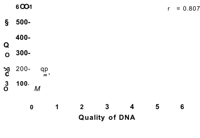
2.8- Correlation between quality and quantity of DNA... 87
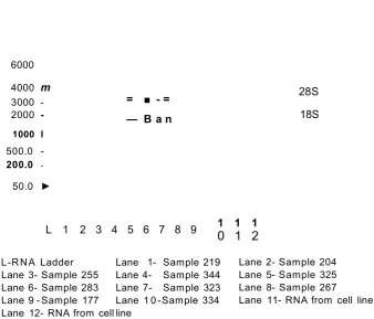
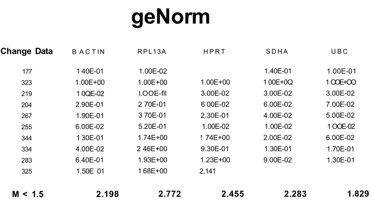
2.9- Experion gel data RNA from human post-mortem brain tissues samples 89 2.10- Example of a RNA sample electrogram from the experion system...go 2.11- geNorm data of 5 housekeeping genes...91
2.12- geNorm data of 2 most stable housekeeping genes... 92
2.13- Efficiency of the housekeeping primers SDHA, UBC and 5-HT1A...93
2.14- Correlation between age and relative...94
2.15- Correlation between gender of subject and relative expression ratio 95 2.16- Correlation between post-mortem delay and relative expression ratio...96
2.17- Log of relative expression correlated with genotype...97
2.18- Log of relative gene expression of G-allele versus the C-allele...97
2.19- Radioligand binding of a human post-mortem tissue sample with the 5-HTiA silent antagonist WAY100635...99
C h a p te r th re e
3.1- Time-points of SH-SY5Y RA differentiated cells... .124
3.2- Time-points of SH-SY5Y NGF and aphidicolin differentiated cells...125
3.3- 5-HT1A receptor genotype in SH-SY5Ycell line... 126
3.4- RNA agarose gel ...127
3.5- geNorm data of 5 housekeeping genes... 128
3.6- geNorm data of the two most stable housekeeping genes...128
3.7- Efficiency of the primers UBC, YWHAZ, 5-HT 1A and NUDR...129
3.8- 5-HT1A receptor mRNA expression in SH-SY5Y cells differentiated with NGF (100pg/ml) for different lengths of time (0-14days)...131
3.9- 5-HT1A receptor mRNA expression in SH-SY5Y cells differentiated with RA (10‘5M) for different lengths of time (0-14days)... 132
3.10- NUDR mRNA expression in SH-SY5Y cells differentiated with NGF (100pg/ml) for different lengths of time (0-14days)... 133
3.11- NUDR mRNA expression in SH-SY5Y cells differentiated with RA (10'5M) for different lengths of time (0-14days)... 134
3.12- 5-HT1A expression in SH-SY5Y cells using immunocytochemistry...135
3.13- Western blots of RA and NGF and aphidicolin differentiated SH-SY5Y cells for (0-14 days)... 136
C h a p te r fo u r 4.1- Schematic of intracellular calcium assay...148
4.2- Effects of 8-OH-DPAT on intracellular Ca2+ in SH-SY5Y cells...152
4.3- Forward and side scatter dot plot... 153
4.4- Undifferentiated SH-SY5Y cells treated in the absence of 8-OH-DPAT...155
4.5- Undifferentiated SH-SY5Y cells treated in the presence of 8-OH-DPAT 156 4.6- SH-SY5Y RA differentiated cells treated in the absence of 8-OH-DPAT... 157
4.7- RA differentiated SH-SY5Y cells treated in the presence of 8-OH-DPAT...158
4.8- Bar chart: Undifferentiated SH-SY5Y cells treated in the presence and absence of 8-OH-DPAT... 159
4.9- Bar chart: RA differentiated SH-SY5Y cells treated in the presence and absence of 8-OH-DPAT... 160
4.10- SH-SY5Y RA differentiated cells treated in the absence of 5-HT... 161
4.11- SH-SY5Y RA differentiated cells treated in the presence of 5-HT...162
4.12- Bar chart: RA differentiated SH-SY5Y cells treated in the presence and absence of 5-HT...163
4.13- RA differentiated SH-SY5Y cells treated with forskolin (OpM), p-MPPI (OpM, 10pM and 100 pM) and 8-OH-DPAT (2pM)... 164
4.14- SH-SY5Y RA differentiated cells treated with forskolin (20pM), p-MPPI (OpM, 10pM and 100 pM) and 8-OH-DPAT (2pM)... 165
4.15- Bar chart: RA differentiated SH-SY5Y cells treated with no forskolin, p-MPPI (OpM, 10pM and 100 pM) and 8-OH-DPAT...166
4.16- Bar chart: RA differentiated SH-SY5Y cells treated with 20pM forskolin, p-MPPI (OpM, 10pM and 100 pM) and 8-OH-DPAT... 167
4.17- RA differentiated SH-SY5Y cells treated with forskolin (50pM), p-MPPI (0pM,10pM and 100 pM) and 8-OH-DPAT (2pM)... .168
4.18- Bar chart: RA differentiated SH-SY5Y cells treated with 50pM forskolin, p-MPPI (0pM,1OpM and 100 pM) and 8-OH-DPAT...169
List of tables
Chapter one
1.1- 5-HT receptor nomenclature...8
Chapter two
2 .1 - ASO primer sequences... ...71 2 .2 - Oligonucleotide primers for real-time PCR SNP genotyping...72 2.3- Oligonucleotide primer sequences for housekeeping genes used
for real-time PCR... 73 2 .4 - Oligonucleotide primer sequences for 5-HT1A receptor gene... 76 2 .5 - Genotype of human hippocampal post-mortem brain tissue samples
using ASO method... ... 82 2 .6 - Genotype of human hippocampal brain tissue samples using
real-time PCR SNP genotyping method... 84 2 .7 - Genotype of human post-mortem tissue samples using custom
TaqMan probes... 86
2 .8 - Final genotype of assigned to samples... 88
2 .9 - Relative expression values of human post-mortem tissue samples 98
2.1 0- The calculated Bmax and Kd, and receptor density (fmol) values with
concurrent genotype of each post-mortem brain tissue sample...101
2 .1 1 - Summary table of 5-HT1A receptor density and genotype ... 101
Chapter three
3 .1- Oligonucleotide primer sequences for the 5-HT1A gene ... 110
Chapter 1
1.0- Serotonin (5-HT)
The isolation and characterisation of serotonin (5-HT) and its final identification as serotonin took place between 1940 and 1949. However, back in 1868 it was already widely acknowledged that blood contained a vasoconstrictive substance that was released in serum during platelet breakdown (Green, 2006). This substance proved to be a problem for Irvine Page in his studies on malignant hypertension due to the substance's ability to elicit large pressor responses and, depending on dose administered, this substance could act as either a vasoconstrictor or vasodilator (Rapport, Green and Page, 1948b). The substance was isolated and characterised and in 1949 it was finally identified by Maurice Rapport as serotonin, named after its vasoconstrictor properties (Rapport, Green and Page, 1948a).
One year later in 1950 Gaddum observed that serotonin was present in the brain; he also showed that the action of 5-HT in the gut was antagonised by the hallucinogen, lysergic acid diethylamide (LSD) (Gaddum and Hameed, 1954). Erspamer in the 1950’s demonstrated that “enteramine” a substance now known to be serotonin was distributed widely and involved in smooth muscle contraction. Erspamer named serotonin “enteramine” as large amounts of this substance were stored in enterochromaffin cells of the gastrointestinal tract (Erspamer, 1963).
Also in the 1950’s, Wooley and Shaw suggested that there was a role for serotonin in mental illness. This was hypothesised due to the knowledge of pyschotomimetic activity of serotonin analogues and of LSD. It was not until the 1970’s that a role for serotonin in depression was established (Clarke et al, 1975). Underlying this theory was work by a group of Scandinavian scientists who developed selective inhibitors of serotonin uptake and showed these
inhibitors to be successful antidepressants (Carlsson et al, 1969).
development. Presently, there is a substantial amount of information on the neuropharmacology of serotonin (5-HT) which implicates the serotonin system as an important modulator in a variety of central nervous system processes (Green, 2006). These processes include: anxiety, fear, depression and aggression; control of sleep and modulation of ingestive behaviours and the cardiovascular system (Gingrich and Hen, 2001; Hoyer et al, 2002).
1.1- Synthesis and metabolism of serotonin
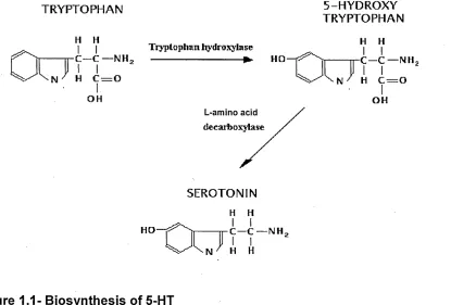
The precursor amino acid utilised in the biosynthesis of 5-HT is tryptophan. Tryptophan hydroxylase (TPH) catalyzes the hydroxylation of L-tryptophan via the oxidation of tetrahydrobiopterin in the presence of the reductive incorporation of molecular oxygen (Kappock and Caradonna, 1996). This is the first step in the biosynthesis of the indoleamines (serotonin (5-HT) and melatonin) (Martinez, Knappskog and Haavik, 2001), (Figure 1.1).
Figure 1.1- Biosynthesis of 5-HT
The conversion of tryptophan to 5-hydroxytryptophan, occurs in the chromaffin cells and neurons. The second step is the decarboxylation of 5- hydroxytryptophan to 5-HT by the aromatic L-amino acid decarboxylase.
TRYPTOPHAN 5-HYDROXY
TRYPTOPHAN
H H Tryptophan hydroxylase H H
OH OH
L-amino decarbo:
SEROTONIN
[image:20.612.107.522.340.621.2]In mammalian metabolism of 5-HT, the reaction catalysed by TPH preceding a-decarboxylation is thought to be the rate limiting step in the production of 5-HT (Lovenberg, Jequier and Sjoerdsma, 1967; Jequier, Lovenberg and Sjoerdsma,
1967).
Once 5-HT has been synthesised, 5-HT is stored in secretory vesicles, as the free compound can be rapidly oxidised to 5-hydroxyindoleacetic acid (5-HIAA) through the actions of the enzymes monoamine oxidase and aldehyde dehydrogenase. Stored 5-HT is then released in response to mechanical and neuronal stimuli (Boadle-Biber, 1993).
1.2- Serotonin release and reuptake at a serotonergic synapse
5-HT as previously mentioned is taken up into and stored in storage vesicles, where the neurotransmitter is released into the synaptic cleft only when an action potential occurs at the pre-synaptic neuron. 5-HT then diffuses across the synaptic cleft and can bind with any of the 5-HT receptor classes (1,2,3,4,5,6 and 7) on the post-synaptic neuron.
S*r<tfC*1in revaptons
Serotonin
Postsynaptic neuron Presynaptic neuron
Serotonin reuptake transporter
Synapse
Vesicle containing serotonin
Figure 1.2- 5-HT neurotransm ission
An action potential initiates the release of 5-HT from synaptic vesicles into the synaptic cleft. 5-HT binds to 5-HT receptors present on the postsynaptic neuron. Excess 5-HT is reuptaken by 5-HTT or degraded by MAO.
1.3- Receptor subtype identification and classification
The first attem pt to categorise 5-HT receptor subtypes was by G addum and Picarelli in 1957 although it had been previously published that 5-HT possessed two receptor subtypes by Gaddum and Hadeem in 1954. G addum ’s classification of 5-HT receptors divided them into D and M receptor subtypes. The naming of these receptors was based on the selectivity of the receptor subtypes to dibenzyline and m orphine as blockers. However, G addum ’s proposal was generally accepted to be relevant to peripheral receptors only and not those present in the brain (Gaddum and Picarelli, 1957).
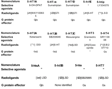
The current classification of 5-HT receptors and their subtypes are based on their molecular structure, signal transduction pathway and operational properties (Kelly, 1995). There are seven classes of 5-HT receptors 5-HT-I-5-HT7, with some of these receptor classes being further subdivided into subtypes such as 5-HT1A, B, D, E and F (Hoyer and Martin, 1997).
In accordance with the recommendations of NU-IUPHAR (the main IUPHAR nomenclature committee), newly described recombinant receptors are described in lower case i.e. 5-htn and described in uppercase only when operational and transductional information are available.
1.3.1- 5-HT-j receptors
The 5-HT1 receptor class is subdivided into five receptor subtypes (5-HT1A,1B, 1D, 1E and 1F), which share 40-63% overall sequence identity in humans and couple preferentially, although not exclusively to, Gj/oto inhibit cAMP formation.
The 5-H Tia receptor is widely distributed throughout the CNS in particular in the
raphe nuclei and limbic structures including the hippocampus. The 5-H Tia
1.3.2- 5-HT2, 5-HT3 and 5-HT4 receptors
The 5-HT2 class comprises of the 5-HT2A, 2B and 2C subtypes which exhibit 46-50% overall sequence identity and this receptor class preferentially couple to Gq/n to increase the hydrolysis of inositol phosphates and elevate cytosolic calcium. The 5-H T2a receptor is widely distributed in peripheral and central tissues with particularly high expression found in cortical regions (Eison and Mullins, 1996). 5-HT2b receptor expression has been found in organs including vascular smooth muscle (Ullmer et al, 1995) spinal cord (Helton and Colbert, 1994) and the brain (Choi and Maroteaux, 1996). 5-H T2C receptors are found in the choroid plexus (Pazos, Hoyer and Palacios, 1984), the cortex, basal ganglia, hippocampus and hypothalamus (Molineaux et al, 1989).
5-H T3 receptors can be found on neurones of both central and peripheral origin, the CAI pyramidal cell layer in the hippocampus and the dorsal motor nucleus (Hoyer and Martin, 1997). The 5-HT4 receptor is widely distributed within the CNS and peripheral tissues where it is thought to play an important role in the function of several organ responses including the alimentary tract, urinary blader, heart and adrenal glands (Hedge and Eglen, 1996).
1.3.3- 5-ht5, 5-ht6and 5-HT7 receptors
Nomenclature 5-HT1A 5-HT-ib 5-HT1D 5-ht1E 5-htip Selective
agonists 8-OH-DPAT Sumatriptan Sumatriptan " LY334370 Radioligands [JH]WAY10063
5 [ IZ3I]GTI [1Z5I]GTI [JH]5-HT ['"l]L S D
G protein
effector Gj/o Gj/o Gj/o Gj/o Gj/o
Nomenclature 5-HT2A 5-HT2B 5-HT2C 5-HT3 5-HT4 Selective
agonists Ketanserin SB200646 Mesuiergine Granisetron GR113808
Radioligands ['" I] DOI [JH]5-HT [1tol]LSD [JH](S)zac
opride [^ l]S B 207710
G protein
effector Gq/|| Gq/|| Gq/|| " Gs
Nomenclature 5-htsA 5-ht5B 5-hte 5-HT7
Selective agonists ” “ “ “
Radioligands [lzol] LSD [1Z0I]LSD [1ZDI]SB25885 [1Z0I]LSD
G protein effector Gj/o None identified Gs Gs
Table 1.1- 5-HT receptor nom enclature
Abbreviations: Gq/n- activates phospholipase C, Gi/0- Inhibits adenylyl cyclase, Gs- Stimulatory G-protein activates adenylyl cyclise. Modified from Hoyer and Martin (1997).
1.4 - Serotonergic system
[image:25.612.100.483.49.352.2]Basal ganglia
Thalamus Neocortex
Hypothalamus
Temporal lobe
Cerebellum Raphe nuclei
To spinal cord
Figure 1.3- The serotonergic system
Ascending projections arise from the dorsal and median raphe nuclei and travel through to target regions including the limbic system (hippocampus, hypothalamus and amygdala), striatrum and cerebral cortex. The descending projections arise from the raphe magnus and project towards the spinal cord (Bear, Connors and Paradiso, 2001).
1.4.1-Anatomical localisation
5-HT secreting neurons are distributed and contained throughout the brain and the gastrointestinal tract.
pathophysiology of a number of clinical entities such as functional gut disorders including irritable bowel syndrome.
5-HT acts as a mucosal signalling molecule for mucosal enterochromaffin cells which act as sensory transducers that respond to mechanical pressure (Bulbring and Crema, 1959) or nutrients (Kim, Cooke and Javed, 2001; Raybould et al, 2003; Fukumoto, Takewaki and Yamada, 2003) to secrete 5-HT into the wall of the bowel and initiate peristaltic (Grider, Kuemmerle and Jin, 1996) and secretory reflexes (Cooke, 2000).
The central nervous system accounts for 5 percent of the 5-HT present in the body with 5-HT being present within several areas of the midbrain including the hippocampus, frontal cortex, limbic system and the hypothalamus (Tork, 1990). Within the central nervous system (CNS) 5-HT acts as a neurotransmitter and high concentrations of 5-HT can be predominantly found in localised nerve projections of the mid-brain particularly in several large clusters of cells referred to as Raphe nuclei (Rang and Ritter, 1999), (Figure 1.3).
1.4.2- Anatomical localisation of 5-HT1A receptors
1.4.2.1 - Raphe nuclei
influence on 5-HT neuronal firing (Nestler et al, 2001; Blier and Ward, 2003; Stahl, 1996; Ou e t a l , 2000).
In the raphe area the activation of 5-H Tia autoreceptors reduces the release of
5-HT at the level of terminals in the hippocampus (Sharp and Hjorth, 1990; Blier, Serrano and Scatton, 1990). It is therefore assumed that changes in the firing rate of 5-HT neurons which is induced by drugs acting at 5-H TiA receptors can
alter the level of activation of post-synaptic 5-HT receptors in the brain (Mongeau, Blier and de Montigny, 1997).
1.4.2.2 - Lim bic system
The limbic system is made from several heavily interconnected nuclei and several regions of the cerebral cortex. The limbic system including the hypothalamus, cingulated gyrus and the hippocampus and their interconnections comprise a harmonious mechanism, which involve the function of central emotion as well as participating in emotional expression (Morgane et al, 2005; Papez, 1937).
1.4.2.3 - Hippocam pus
The hippocampal formation is one of the most complex and vulnerable brain structures which is recognised as a crucial brain area subserving the human long-term memory (Henke et al, 1999). The hippocampal formation consists of the dentate gyrus and the cornu ammonis (CA) plus the subiculum collectively known as the hippocampus.
5-HT stim ulates 5-H Tia receptors which are localised on both excitatory pyram idal and granule neurons (Gulyas, Acsadi and Freund, 1999) resulting in neuronal hyperpolarisation and inhibition of neuronal activity in the hippocam pus (Schm itz et al, 1998). 5-HT acting via its 5 -H Tia receptor has
been im plicated in rhythm ic slow activity, which has been associated with learning and m em ory processes in the hippocam pus (Vanderwolf and Barker, 1986; M cEntree and Crook, 1992 and Pom peiano et al, 1992).
Fimbria
Alveus
Den fate
gyrus CA3 CAl
Subiculum
Figure 1.4- Schem atic representation of the hippocam pus
The hippocampal pyramidal neurons (CA1-3) and the dentate gyrus granule cells (DG) receive a direct cortical input via the perforant pathway from the entorhinal cortex (EC). Hippocampal pyramidal neurons (CA1) and the sibiculum are involved in hippocampal cortical output to associated limbic cortices. 5-HT1A receptors are located on both pyramidal CA1-3 neurons and on granule neurones present in the DG (Nolte and Angevine, 2000).
1.4.2.4- Function of the Hippocam pus
lobe where information about relationships, combinations, and conjunctions among and between stimuli is processed (Riedel and Micheau, 2001). Both the medial temporal lobe and the hippocampus (CA, DG, and subiculum) are the place for either the temporary storage of to-be consolidated information (Squire, 1992) or as the locus of permanent information storage through multiple memory traces (Nadel and Moscovitch, 1997).
Specific encoding of new information requires the dentate gyrus granule cells and processes of memory consolidation, either short-term or long-term, by contrast, should depend on the network activity of the hippocampus proper (CA1-CA3), (Riedel and Micheau, 2001).
1.4.2.5 - Serotonergic transm ission in the hippocam pus 1.4.2.6- Pre-synaptic m echanism s in the hippocam pus
5-HT neuron firing activity is credited to a pacemaker cycle that involves a calcium dependent potassium current. The discharge rate of these neurons is mediated by 5-H TiA autoreceptors localised in the somatodendritic region on
the presynaptic neuron (Mongeau, Blier and de Montigny, 1997). The activation of 5-HT-ia autoreceptors is regulated by an hyperpolarisation of the membrane
occurring by the opening of potassium channels (Aghajanian and Lakoski, 1984; Sprouse and Aghajanian, 1987; Williams, Henderson and North, 1985).
Contrary to somatodendritic autoreceptors of the raphe area, terminal presynaptic 5-HT autoreceptors of the hippocampus are not of the 5-H Tia subtype and control the release of 5-HT without interfering with the propagation of action potentials (Starke, Gothert and Kilbinger, 1989). Terminal 5-HT autoreceptors in mammalian species excluding rodent are of the 5-HT-id subtype (Mongeau, Blier and de Montigny, 1997).
1
.4.2.7 - Post-synaptic mechanisms in the hippocampus
Activation of postsynaptic 5-H TiA receptors, results in an inhibition of the activity
of neurons of the limbic system (Sprouse and Aghajanian, 1988).
There are two subsets of post-synaptic 5-H Tia receptors in the hippocampus
that differentially couple to G-proteins that suppress pyramidal cell firing. Extrasynaptic 5-HT-jA receptors are located on the soma of hippocampal pyramidal cells, which can be activated by microiontophoretic application of agonists and are inactivated by pertussis toxin (Blier, de Montigny and Lista, 1993). However, the other subset intrasynaptic 5-H T1A receptors are localised on dendrites of hippocampal pyramidal cells and activated by endogenous 5-HT. Intrasynaptic 5-H TiA receptors are un-affected by pertussis toxin (Blier, de
Montigny and Lista, 1993). It has therefore been hypothesised that extrasynaptic 5-H TiA receptors are coupled with G j /0 proteins, whereas
intrasynaptic 5-H T1A receptors are not.
1.4.2.8 - Other Limbic brain regions
1
.4.2.9 - Prefrontal Cortex (PFC)
The prefrontal cortex can be divided into the ventromedial and dorsolateral regions, each of which is associated with posterior and subcortical brain regions (Wood and Grafman, 2003).
The ventral medial PFC has been strongly implicated in the expression of behavioural, neuroendocrine and autonomic responses to emotionally relevant stimuli. Recent imaging studies have indicated that abnormalities in structure and function of this region is present in patients with mood disorders (Drevets et al, 1997; Kennedy et al, 2001). Tracing studies have shown that 5-HT pathways ascend from the midbrain dorsal (DRN) and median raphe nuclei (MRN) which project extensively in to the ventral mPFC (O'Hearn and Molliver, 1984; Steinbusch, 1981). This region has also been shown to contain a high density of 5-HT transporter sites (Battaglia et al, 1991; Herbert et al, 2001) and 5-HT receptors including 5 -H Tia and 5 -H T2a (Pazos and Palacios, 1985; Pompeiano et al, 1992). The DRN and MRN are thought to receive projections from the ventral mPFC (Hajos et al, 1998; Peyron et al, 1998; Sesack et al, 1989; Varga et al, 2001) activation of which has been shown to mediate 5-HT neuronal activity (Celada et al, 2001; Hajos et al, 1998; Varga et al, 2001).
From anatomical and electrophysiological observations it has been suggested that there is an excitatory mPFC-DRN projection that brings about inhibition of 5-HT neurones in the DRN through the activation of local raphe GABA neurones (Hajos et al, 1998; Varga et al, 2001).
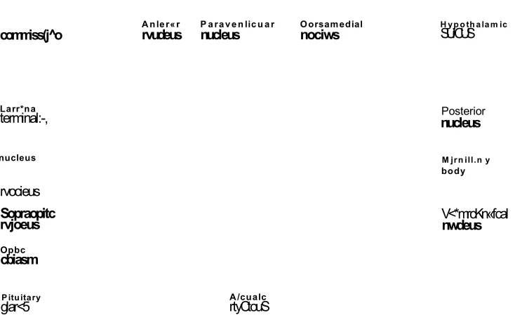
1.4.2.10-Hypothalamus
It has been shown that serotonergic m echanism s exert an excitatory influence on the HPA axis (Chaouloff, 1993) and 5-HT has also been shown to elicit the release of ACTH release directly from the pituitary (Spinedi and Negro-Vilar,
1983) by activation of 5 -H Tia and 5 -H T2a receptors (Calogero et al, 1990;
Ritten-House et al, 1994).
The 5-HT system acting through 5 -H Tia receptors may be able to m ediate the negative feedback control of the HPA axis.
commiss(j^o A nler«r rvudeus nucleus P a raven licu ar Oorsamedialnociws H yp othalam icSUiCUS
Larr*na
terminal:-,
nucleus
rvocieus
Posterior nucleus
M jrn ill.n y body
Sopraopitc
rvjoeus V<*mrcKn«fcalnwdeus
Opbc
cbiasm
Pituitary
[image:33.619.120.487.234.472.2]glar<5 rtyCtouSA/cualc
Figure 1.5 - Diagram of the cross section of the hypothalamus
1
.4.2.11 Amygdala
Several studies have shown the role of the am ygdala to be associated with conditioned fear (Davis, 1998; Catill et al, 1999; Le Doux, 2000). The am ygdala is thought to m ediate the ability of previously non-threatening stimuli, when associated with naturally frightening stim uli (including the exposure to severe stress) to educe a wide range of stress responses (Nestler et al, 2002).
Postsynaptic 5-H T1A receptors can be found in the am ygdala m ainly in the central nucleus (Pazos and Palacios, 1985; O huoha et al, 1993), (Figure 1.6). Recent data has shown that som atodendritic 5-H T1A autoreceptors in the claudal linear raphe nucleus are capable of m odulating extracellular 5-HT levels in this area by reducing 5-HT cell firing by an as yet unknown pathway (Bosker, Klom pm akers and W estenberg, 1997).
Lateral nucleus
L*
Central
nucleus 6asalnucleus Medial nucleus
Insular cortex
Cortical nucleus
Figure 1.6- Cross- section of the Amygdala
1.5- Non 5-HT Neurotransmitter systems
Serotonergic neurons in the central nervous system impinge on many other neurons and modulate their neurotransmitter release. Serotonergic neurons interact with GABAergic, noradrenergic, cholinergic and dopaminergic neurons.
1.5.1- GABAergic
y-Aminobutyric acid (GABA), a neurotransmitter that is inhibitory, is present in the CNS and is distributed across all brain regions (Zachmann, Tocci and Nyhan, 1966).
In the majority of brain regions the release of GABA is controlled by inhibitory presynaptic 5-HT-ia receptors that are present at GABAergic nerve terminals.
This control of GABA release is thought to be caused by the inhibition of adenylyl cyclase and cAMP signal transduction pathway. This pathway directly acts on the GABA release process in independent K+ and Ca2+ channels (Koyama et al, 1999). Hence, this pathway has a negative effect on GABA release.
The hypothesised involvement of GABAergic dysfunction in mood disorders came from a study by Enrich et al (1980). The study looked at the mood stabiliser valproate which is used as an effective treatment for bipolar patients. The pathophysiology of mood disorders has been linked with a GABAergic deficiency (Enrich et al, 1980). Preclinical animal studies have also shown that GABA levels may be decreased in animal models of depression and clinical studies have reported low plasma and CSF GABA levels in mood disorder patients (Bambilla et al, 2003).
1.5.2- Noradrenergic system
the brain norepinephrine functions as a general regulator of mood responses to stimuli including stress (Wang et al, 1999).
In the hippocampus NA terminals are thought to originate exclusively from the locus coeruleus (LC) (Haring and Davis, 1985; Jones and Moore, 1977). It is generally accepted that an increase in the availability of NA in the biophase of adrenoceptors in this brain region is involved in the mechanism of action of antidepressants. It has also been suggested that 5-HT receptor ligands that enhance NA release may be expected to positively influence mood disorders (Fink and Gothert, 2007).
Inhibitory presynaptic 5 -H Tib/id receptors have been identified on the noradrenergic axon terminals of the cardiovascular system of various species both
in vitro
andin vivo
(Charlton et al, 1986; Gothert et al, 1986; Medhurst et al, 1997 and Harris et al, 2002). Therefore, it has been hypothesised that noradrenergic nerve terminals in the CNS could also have inhibitory presynaptic 5-H T-ib/id receptors present (Taube et al, 1977; Schlicker et al, 1983).Neocortex
Thalamus
H ypothalam us
Temporal lobe Locus coeruleus
^ Cerebellum
To spinal cord
F
igure 1.7- The noradrenergic system
T h e n oradrenergic system exten d s from th e locus co eru leu s sending projections to w ards th e neocortex, th alam u s, h ypo th alam u s and cereb ellu m (B e a r, C o n n o rs and P arad iso , 2 0 0 1 ).
1.5.3- Cholinergic system
The cholinergic system is im portant for the role of m em ory and cognition (Cassel and Jeltsch, 1995; Steckler and Sahgal, 1995; Feuerstein and Seeger, 1997; Ruotsalainen et al, 1998), an observed increase in acetylcholine concentration in the synaptic cleft represents a therapeutic option in dem entia which is associated with A lzheim er’s disease.
Im m unohistochem ical double staining has dem onstrated that 5 -H Tia receptors
occur on cholinergic cell bodies in the septum and project to the hippocam pus and neocortical areas (Kia et al, 1996). In the presence of 5 -H Tia receptor
serotonergic neurons are activated. The activation of these autoreceptors inhibits the activity of serotonergic neurons and therefore, there is an assum ed decrease in the stim ulation of inhibitory G ABAergic interneurons, which, in turn leads to the disinhibition of cholinergic neurons (Fink and Gothert, 2007).
Neocortex
Thalamus
Basal nucleus of Meynert
Hippocampus
Pontomesencephaio- tegmental complex Medial
septal nuclei
F
igure 1.8- Cholinergic system
1
.5.4- Dopaminergic system
The dopam inergic system in the brain arises from the midbrain and in particular from a group of cells in this region and from the hypothalam us (Figure 1.9), (Kapur and Mann, 1992). The rat and m ouse brain have been used to investigate the m echanism in which 5-HT receptors regulate dopam ine (DA) release.
5-H T1A receptors are thought to increase DA release and it is likely that the 5-
H Tia receptors are located as pre-and/or postsynaptic 5-H TiA receptors on
inhibitory G ABAergic interneurons which exert an inhibitory tone on the activity of the dopam inergic neurons (Fink and Gothert, 2007).
Dopamine system
Frontal lobe
\
Ventral tegmental area
Striatum
Substantia
Figure 1.9- Dopaminergic system
The serotonergic system as previously m entioned interacts with several non- serotonergic neurons within the CNS and these neurotransm itter system s are present within the limbic region of the brain i.e. the hypothalam us, am ygdala and the hippocam pus with projections expanding to the dorsal raphe (DR), the locus coeruleus and the pre-frontal cortex (PFC) as sum m arised in Figure 1.10. The figure shows a sim plified sum m ary of a series of neurotransm itter neurons in the brain, which interact with the afore-m entioned brain regions and thus contribute to depressive sym ptom s.
VTA
To
— GABAergic — Dopaminergic
NAergic/5HTergic
Figure 1.10-Summary of the neural circuitary of the brain
S c h e m a tic rep resen tatio n show ing th e interconnections of neural circuitary in th e brain. Abbrevations: NAc- nucleus a ccu m b en s, VTA- ventral te g m e n ta l a re a . M o d ified from
N astier fit al 2002.
Hypo thalamus
Amygdala
Hippocampus
1.6- Depression
Depressive disorders are amongst the most common psychiatric diseases with prevalence estimates ranging from 5 percent to a maximum of 20 percent (Hamet and Tremblay, 2005). Less severe forms of depression may affect an additional 10 percent of the American population. The interaction between genes and the environment have been acknowledged to play a role in the pathophysiology of depression (Hamet and Tremblay, 2005).
Depression is recurrent and tends to have chronic course, and can often be comorbid in nature. It is thought that depression is a clinically heterogeneous disorder thought to result from an interaction of multiple genes cooperating with environmental and developmental epigenetic components (Hamet and Tremblay, 2005).
There are two etiologically different forms of depression, bipolar disorder (manic depression) and unipolar disorder (Lesch, 2004). There are also many different symptoms of depression including disturbance of mood, thinking, sleep, appetite, and motor activity, with suicidal thoughts or attempts that occur to different degrees (American Psychiatric Association, 1994).
1.6.1- Major depression (Unipolar)
Major depression (unipolar) is a serious medical condition. The risk of developing major depression is thought to be approximately one in ten of the population at some time in their lives and twice as great among women than amongst men in almost all cultures studied (Elliot, 1998). Major depression is. characterised by sad mood, loss of interest, sleep disturbances and recurrent thoughts of death and suicide (Rajkowska, 2003; Lucki, 1998). In the United
Q to fo c o n H \A/orlrl\A/irlQ m o in r rlftn ro o c lA n So rJ ^ 11 r
wvwtkww UI IV4 V»VI iViMiMW) i i ivijs/i Owwivi i iw Li iv iOuviii WUUOO Ol
Many individuals suffering from panic disorder (PTSD) or other anxiety related disorders tend to develop m ajor depression. This observation has lead to the thought that there may be overlapping neural circuitry involved in the pathophysiology of depression and that of certain anxiety disorders.
1
.6.2- Bipolar depression (Manic Depressive illness)
Bipolar depression is a m ajor public health problem . It is estim ated that there is a 0.3-1.5 percent worldwide lifetime prevalence of bipolar depression (W eissm an, et al, 1996). Bipolar depression has also been associated with a m ortality risk; approxim ately 25 percent of patients attem pt suicide at some point during their lives and 11 percent of patients die by suicide (Prien and Potter, 1990). Bipolar depression is also characterised by fam ilial transm ission the incidence of bipolar depression among first-degree relatives of affected individuals is 8-25 percent.
This type of depression is often characterised by episodes of mania, with or without distinct episodes of depression. Mania is characterised by euphoria or irritability, increased energy, and a decreased need for sleep (Am erican Psychiatric Association, 1993).
Both m anic and depressive states of this disorder are thought to be due to low serotonergic function through defective dam pening of other neurotransm itters such as, norepinephrine and dopam ine (Hilty, Brady and Hales, 1999). Lesions in the frontal and tem poral lobes are linked with bipolar disorder. Left-sided lesions are related with depression and right-sided lesions are associated with mania (Hilty, Brady and Hales, 1999).
1
.6.3- Stress-induced depression
Stress and the HPA axis have both been im plicated as a factor involved in the onset of depression (Taylor et al, 2004).
The release of stress horm ones, such as cortisol and also corticotrophin- releasing horm one (CRH), which are secreted from the hypothalam us; this occurs in m any individuals diagnosed with mood disorders and may result from hyperfunctioning of the am ygdala (which is known to activate the paraventricular nucleus of the hypothalam us (PVN)), or by the hypofunctioning of the hippocam pus (which exerts a potent inhibitory influence on the PVN), (Young, Lopez and M urphy-W einberg, 2003; M uller et al, 2002). The release of stress horm ones from the hypothalam us in turn stim ulates the release of glucocorticords from the adrenal cortex (Liberzon, Krstov and Young, 1997). Excessive am ounts of glucocorticoids can be dam aging and therefore the HPA axis is under tight regulation by a negative feedback system (Liberzon, Krstov and Young, 1997) which occurs m ainly through m ineralocorticoid and glucocorticoid receptors (Young, Lopez and M urphy-W einberg, 2003).
PVN Hippocampus Hippocampus — _j_
CRF Amygdala
Glucocorticoids Dexamethasone ACTH
Adrenal cortex
F
igure 1.11- HPA axis
T h e H P A axis receives pro m in en t neural inputs th at include excitatory a fferen ts from th e a m yg d ala. T h e H P A axis m ay contribute to d epressio n not only via th e a m y g d a la and inhibitory (postsynaptic) afferen ts from th e h ip p ocam pu s but also through e n h a n c e d C R F tra n s fe r (N e s tle r et al, 2 0 0 2 ).
1.6.3.1- Glucocorticoid receptor and Mineralocorticoid receptors
1
.6.4- 5-HT receptor(s) involvement with depression
Several 5-HT receptors have been linked with being involved in depression although exact roles for many of the 5-HT receptors have not yet been fully identified. The main 5-HT receptors of interest are the 5 -H T2a, 5-HTT and particularly the 5 -H Tia receptor.
Increases in 5 -H T 2a receptors are frequently mentioned in support of the hypothesis of alterations in serotonergic neurotransmission in suicide and affective disorders (Stockmeier, 2003). Various studies have investigated 5-H T 2a binding in platelets of depressed patients, these studies reported an increase in binding sites (Bmax) in both depressed and suicidal patients (Arora and Meltzer, 1989). Studies investigating 5-HT2A binding in post-mortem tissue have shown 5-H T2A receptors to be found in the frontal cortex suggesting a role in cognitive aspects of depression. In the hippocampus of suicide victims a significant decrease in the number of 5-HT2A receptors has been reported (Cheetham et al, 1988).
Two polymorphisms of the 5-HTT gene have been identified; a variable number of tandem repeats in the second intron (VNTR) (Lesch et al, 1994) and a 44bp insertion/deletion in the 5-HTT linked polymorphic region (long and short alleles) (Heils et al, 1996). An association between the short allele of the 5-HT transporter has been reported with both unipolar and bipolar patients (Collier et al, 1996). This functional polymorphism in the promoter region of the 5-HTT has also been found to moderate the influence of stressful life events on depression. It has been hypothesised that individuals possessing one or two copies of the short allele may exhibit more depressive symptoms and are more suicidal in relation to stressful life events than individuals who are homozygous for the long allele (Caspi et al, 2003).
5-H T1A receptor has a heterogenous localisation, presynaptically as an autoreceptor on the som a and dendrites found m ainly in the m edian and dorsal raphe nuclei and postsynaptically in the limbic regions of the brain (Jacobs and Azm itia, 1992). Som atodendritic receptors have an autoinhibitory function. It has been shown that on stim ulation a decrease in firing in serotonergic neurons, 5-HT synthesis and 5-HT release was observed (Blier, de-M ontigny, Chaput 1987; Sprouse and Aghajanian, 1987; Hjorth and M agnusson, 1988; Hutsen et al, 1989; M artin-R uiz and Ugedo, 2001).
Activation of postsynaptic 5-H T1A receptors, located in limbic areas (Pazos and Palacios, 1985; Li, Battagila and Van de Ker, 1997) results in an inhibition of the activity of neurons of the limbic system (Sprouse and Aghajanian, 1988; Blier, de m ontigny and Lista, 1993). W hen 5-H TiA receptors are activated by excess
am ounts of 5-HT they hyperpolarise the neuron, causing it to slow down its firing activity (Nestler et al, 2001; Blier and W ard, 2003; Stahl, 1996; Ou et al, 2000).
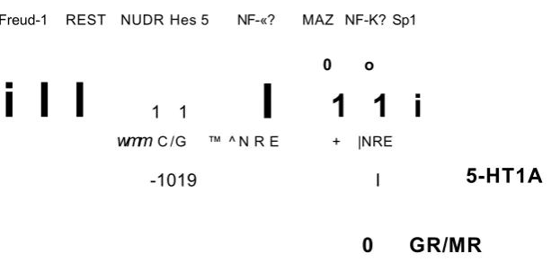
CAMK
Freud-1 REST NUDR Hes 5 NF-«? MAZ NF-K? Sp1
i l l
1 1I
0 o
1 1 i
wmm C /G ™ ^ N R E + |NRE
-1019 I
5-HT1A
0 GR/MR
Figure 1.12- Transcriptional regulatory elements of the human 5-HTiAgene.
H ighlighted in bold are th e key rep resso r e le m e n ts and proteins, activation is indicated by th e arrow s and blocked lines indicate repression; T h e C (-1 0 1 9 ) allele d e p e n d e n c e of N U D R /H e s 5 binding is indicated by the d ash ed lines. T h e re a re tw o ta n d e m copies of th e dual rep resso r e le m e n t (D R E ), and the R E -1 (w hich binds R E S T /N R S F ). A d a p te d from A lb ert and L e m o n d e (2 0 0 3 ).
It has been suggested from data obtained from transcriptional reporter assays, that Human nuclear deform ed epiderm al auto regulatory factor (NUDR) and Hes5 repress the transcription activity of the C (-1019) allele of the 5-H T1A promoter. W ith the G (-1019) allele, this transcriptional repression is significantly decreased (Lem onde et al, 2003). The G allele, unlike the C allele fails to bind and m ediate NUDR repression generating an overexpression of the 5-H TiA
[image:47.613.138.445.71.222.2]Normal
K K K K K K K K
5-HT
Depression/Suicide
n
i l
5 - H T
Figure 1.13- Actions of the C-1019G 5-HT1A polymorphism in 5-HT neurons
In non-depressed subjects those with a C allele, NUDR (red dot) binds and 5-HT1A receptor (yellow dots) expression is repressed leading to an increase in 5-HT firing rate ( a ). Whereas, the G-allele fails to bind to NUDR leading to an overexpression of 5-HT1A receptors and hence a decrease in 5-HT firing rate (Adapted from Albert and Lemonde, 2004).
In addition, under chronic stress conditions 5-H T1A receptor m RNA and bi