Crystal structure of methyl
4-(2-fluoro-
phenyl)-6-methyl-2-sulfanylidene-1,2,3,4-tetrahydropyrimidine-5-carboxylate
M. S. Krishnamurthy and Noor Shahina Begum*
Department of Studies in Chemistry, Bangalore University, Bangalore 560 001, Karnataka, India. *Correspondence e-mail: noorsb@rediffmail.com
Received 23 September 2015; accepted 7 October 2015
Edited by E. R. T. Tiekink, University of Malaya, Malaysia
In the title compound, C13H13FN2O2S, the pyrimidine ring adopts a twist-boat conformation with the MeCN and methine-C atoms displaced by 0.0938 (6) and 0.2739 (3) A˚ , respectively, from the mean plane through the other four atoms of the ring. The 2-fluorobenzene ring is positioned axially and forms a dihedral angle of 89.13 (4)with the mean
plane through the pyrimidine ring. The crystal structure features N—H O, N—H S and C—H O hydrogen bonds that link molecules into supramolecular chains along thebaxis. These chains are linked into a layer parallel to (101) by C—H interactions; layers stack with no specific interactions between them.
Keywords:crystal structure; pyrimidine derivative; hydrogen bonding,C— H interactions.
CCDC reference:1430034
1. Related literature
For the bioactivity of organo-fluorine compounds, see: Guru Row, (1999); Yamazakiet al., (2009). For biological activity of pyrimidine derivatives, see: Kappe (2000) and of dihydro-pyrimidines (DHPMs) and their derivatives, see; Jauk et al.
(2000); Kappe (1998); Mayer et al. (1999). For the Biginelli reaction, see: Biginelli (1893). For bond length data, see: Qin
et al. (2006). For related structures, see: Krishnamurthy & Begum (2015a,b).
2. Experimental
2.1. Crystal data
C13H13FN2O2S Mr= 280.31
Monoclinic,P21=n a= 13.3298 (15) A˚ b= 7.1509 (8) A˚ c= 14.5703 (17) A˚
= 109.854 (4)
V= 1306.3 (3) A˚3 Z= 4
MoKradiation
= 0.26 mm1 T= 100 K
0.240.220.18 mm
2.2. Data collection
Bruker SMART APEX CCD diffractometer
Absorption correction: multi-scan (SADABS; Bruker, 1998) Tmin= 0.955,Tmax= 0.960
9937 measured reflections 2296 independent reflections 1340 reflections withI> 2(I) Rint= 0.112
2.3. Refinement
R[F2> 2(F2)] = 0.064 wR(F2) = 0.133 S= 0.95 2296 reflections
174 parameters
H-atom parameters constrained
max= 0.42 e A˚
3
min=0.26 e A˚
[image:1.610.314.565.526.581.2]3
Table 1
Hydrogen-bond geometry (A˚ ,).
Cgis the centroid of the C8–C13 ring.
D—H A D—H H A D A D—H A
N1—H1 O1i
0.88 2.14 2.977 (4) 159
N2—H2 S1ii
0.88 2.55 3.386 (2) 159
C1—H1B O1i
0.98 2.52 3.262 (5) 133
C10—H10 Cgiii 0.95 2.86 3.648 (2) 141
Symmetry codes: (i)x;yþ1;z; (ii)xþ2;yþ1;z; (iii)x;yþ1;z.
Data collection: SMART (Bruker, 1998); cell refinement: SAINT-Plus(Bruker,1998); data reduction:SAINT-Plus; program(s) used to solve structure: SHELXS97(Sheldrick, 2008); program(s) used to refine structure:SHELXL97(Sheldrick, 2008); molecular graphics: ORTEP-3 for Windows(Farrugia, 2012) andCAMERON(Watkinet al., 1996); software used to prepare material for publication:WinGX (Farrugia, 2012).
Acknowledgements
MSK thanks the University Grants Commission (UGC), India, for a UGC–BSR Meritorious Fellowship.
data reports
Supporting information for this paper is available from the IUCr electronic archives (Reference: TK5392).
References
Biginelli, P. (1893).Gazz. Chim. Ital.23, 360–413.
Bruker. (1998). SMART, SAINT-Plus and SADABS. Bruker Axs Inc., Madison, Wisconsin, USA.
Farrugia, L. J. (2012).J. Appl. Cryst.45, 849–854. Guru Row, T. N. (1999).Chem. Rev.183, 81–100.
Jauk, B., Pernat, T. & Kappe, C. O. (2000).Molecules,5, 227–239. Kappe, C. O. (1998).Molecules,3, 1–9.
Krishnamurthy, M. S. & Begum, N. S. (2015a).Acta Cryst.E71, o268–o269. Krishnamurthy, M. S. & Begum, N. S. (2015b).Acta Cryst.E71, o699–o700. Mayer, T. U., Kapoor, T. M., Haggarty, S. J., King, R. W., Schreiber, S. I. &
Mitchison, T. J. (1999).Science,286, 971–974.
Qin, Y.-Q., Ren, X.-Y., Liang, T.-L. & Jian, F.-F. (2006).Acta Cryst.E62, o5215–o5216.
Sheldrick, G. M. (2008).Acta Cryst.A64, 112–122.
Watkin, D. J., Prout, C. K. & Pearce, L. J. (1996).CAMERON.Chemical Crystallography Laboratory, University of Oxford, England.
supporting information
sup-1 Acta Cryst. (2015). E71, o838–o839
supporting information
Acta Cryst. (2015). E71, o838–o839 [https://doi.org/10.1107/S2056989015018873]
Crystal structure of methyl
4-(2-fluorophenyl)-6-methyl-2-sulfanylidene-1,2,3,4-tetrahydropyrimidine-5-carboxylate
M. S. Krishnamurthy and Noor Shahina Begum
S1. Comment
In recent years, dihydropyrimidines (DHPMs) and their derivatives have attracted considerable attention in synthetic
organic chemistry because of their wide range of biological activities (Kappe et al., 2000), such as antibacterial, antiviral,
antitumor and anti-inflammatory activities (Mayer et al., 1999). The Biginelli reaction (Biginelli et al., 1893), a one-pot
condensation of aldehyde, acetoacetate and urea under strongly acidic conditions, is one of the most useful
multicomponent reactions (MCRs), gaining increasing importance in organic and medicinal chemistry because of its
capacity to generate multifunctionalized products including 3,4-dihydropyrimidin-2-ones, their thione analogs, and other
related heterocyclic compounds. They are also noteworthy as calcium channel modulators (Kappe, 1998; Jauk et al.,
2000). The presence of a fluorine atom in the molecule can have profound and unexpected results on the biological
activity of the compound (Guru Row, 1999; Yamazaki et al., 2009). Herein, we report the crystal structure of the title
compound. It is one of the analogue of our previously reported fluoro-DHPMs (Krishnamurthy & Begum, 2015a;
Krishnamurthy & Begum, 2015b). The bond lengths and angles in the title compound are in good agreement with the
corresponding bond distances and angles reported in closely related structures (Quin et al., 2006).
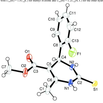
In the title compound, Fig. 1, the 2-fluorobenzene ring at chiral carbon atom C4 is positioned axially and bisects the
pyrimidine ring with a dihedral angle of 89.13 (4)°. The pyrimidine ring adopts a twist-boat conformation with atoms C4
and N1 displaced by 0.2739 (3) Å and 0.0938 (6) Å from the mean plane of the other four atoms (C5/C6/C2/N2)
respectively. The carbonyl group of the exocyclic ester at C5 adopts a trans orientation with respect to C5=C6 double
bond. The 2-fluorobenzene ring shows an anti periplanar conformation with respect to C4—H4 bond of the pyrimidine
ring. The molecular structure is stabilized by intermolecular C1—H1B···O1 and N1—H1···O1 interactions generating
bifurcated bonds from two donor atoms C1 and N1, to the same acceptor O1 to form an R2
2(6) ring motif, which are in
turn linked to form a molecular chain along crystallographic b axis. The packing is further stabilized by intermolecular N
—H···S hydrogen bonds (N2—H2···S1) resulting in a centrosymmetric head to head dimer with graph set R2
2(8) notation
(Table 1; Fig. 2). In addition, the crystal structure is stabilized by C10—H10···Cg (Cg is the centroid of aryl ring C8—
C13) interaction (Table 1).
S2. Experimental
The title compound was synthesized by the reaction of 2-fluorobenzaldehyde (1.24 g, 10 mmol), methylacetoacetate
(1.38 g, 12 mmol) and thiourea (1.14 g, 15 mmol) in 15 ml ethanol. The solution was refluxed for 6 h in the presence of
concentrated hydrochloric acid as a catalyst. The reaction was monitored with TLC and the reaction medium was
quenched in ice cold water. The precipitate obtained was filtered and dried. The compound was recrystallized from
ethanol solvent by slow evaporation method, yielding colorless blocks suitable for X-ray diffraction studies (yield 72%;
The H atoms were placed at calculated positions in the riding-model approximation with N—H = 0.86 Å and C—H =
[image:4.610.130.493.110.472.2]0.93–0.96 Å, and with Uiso(H) = 1.5Ueq(C) for methyl H atoms and Uiso(H) = 1.2Ueq(N, C) for the other hydrogen atoms.
Figure 1
The molecular structure of the title compound with the atom-numbering scheme. Displacement ellipsoids are drawn at
supporting information
[image:5.610.146.466.71.492.2]sup-3 Acta Cryst. (2015). E71, o838–o839
Figure 2
Unit cell packing of the title compound showing intermolecular C—H···O, N—H···O and N—H···S interactions as dotted
lines. H atoms not involved in hydrogen bonding have been excluded.
4-(2-Fluorophenyl)-6-methyl-2-sulfanylidene-1,2,3,4-tetrahydropyrimidine-5-carboxylate
Crystal data
C13H13FN2O2S
Mr = 280.31
Monoclinic, P21/n
Hall symbol: -P 2yn
a = 13.3298 (15) Å
b = 7.1509 (8) Å
c = 14.5703 (17) Å
β = 109.854 (4)°
V = 1306.3 (3) Å3
Z = 4
F(000) = 584
Dx = 1.425 Mg m−3
Mo Kα radiation, λ = 0.71073 Å
Cell parameters from 2296 reflections
θ = 3.0–25.0°
µ = 0.26 mm−1
T = 100 K
Bruker SMART APEX CCD diffractometer
Radiation source: fine-focus sealed tube Graphite monochromator
ω scans
Absorption correction: multi-scan (SADABS; Bruker, 1998)
Tmin = 0.955, Tmax = 0.960
9937 measured reflections 2296 independent reflections 1340 reflections with I > 2σ(I)
Rint = 0.112
θmax = 25.0°, θmin = 3.0°
h = −15→15
k = −8→8
l = −15→17
Refinement
Refinement on F2
Least-squares matrix: full R[F2 > 2σ(F2)] = 0.064
wR(F2) = 0.133
S = 0.95
2296 reflections 174 parameters 0 restraints
Primary atom site location: structure-invariant direct methods
Secondary atom site location: difference Fourier map
Hydrogen site location: inferred from neighbouring sites
H-atom parameters constrained w = 1/[σ2(F
o2) + (0.0421P)2 + 1.6067P]
where P = (Fo2 + 2Fc2)/3
(Δ/σ)max < 0.001
Δρmax = 0.42 e Å−3
Δρmin = −0.26 e Å−3
Special details
Geometry. All s.u.'s (except the s.u. in the dihedral angle between two l.s. planes) are estimated using the full covariance matrix. The cell s.u.'s are taken into account individually in the estimation of s.u.'s in distances, angles and torsion angles; correlations between s.u.'s in cell parameters are only used when they are defined by crystal symmetry. An approximate (isotropic) treatment of cell s.u.'s is used for estimating s.u.'s involving l.s. planes.
Refinement. Refinement of F2 against ALL reflections. The weighted R-factor wR and goodness of fit S are based on F2,
conventional R-factors R are based on F, with F set to zero for negative F2. The threshold expression of F2 > 2σ(F2) is
used only for calculating R-factors(gt) etc. and is not relevant to the choice of reflections for refinement. R-factors based
on F2 are statistically about twice as large as those based on F, and R- factors based on ALL data will be even larger.
Fractional atomic coordinates and isotropic or equivalent isotropic displacement parameters (Å2)
x y z Uiso*/Ueq
S1 0.95357 (8) 0.77963 (14) 0.05365 (8) 0.0312 (3)
O1 0.6807 (2) 0.0312 (4) 0.0894 (2) 0.0452 (9)
O2 0.5913 (2) 0.2422 (3) 0.1440 (2) 0.0460 (8)
N1 0.7803 (2) 0.6600 (4) 0.0856 (2) 0.0310 (9)
H1 0.7685 0.7788 0.0941 0.037*
N2 0.8688 (2) 0.4407 (4) 0.0289 (2) 0.0243 (8)
H2 0.9267 0.4070 0.0170 0.029*
F1 0.64843 (18) 0.5589 (3) −0.10951 (18) 0.0491 (7)
C1 0.6436 (4) 0.6093 (5) 0.1568 (4) 0.0500 (14)
H1A 0.5696 0.6128 0.1121 0.075*
H1B 0.6672 0.7365 0.1789 0.075*
H1C 0.6481 0.5311 0.2133 0.075*
C2 0.8628 (3) 0.6175 (5) 0.0542 (3) 0.0257 (9)
C3 0.6629 (3) 0.1929 (5) 0.1031 (3) 0.0305 (10)
C4 0.7879 (3) 0.2971 (5) 0.0189 (3) 0.0229 (9)
supporting information
sup-5 Acta Cryst. (2015). E71, o838–o839
C5 0.7196 (3) 0.3493 (5) 0.0792 (3) 0.0251 (9)
C6 0.7138 (3) 0.5290 (5) 0.1050 (3) 0.0306 (10)
C7 0.5324 (4) 0.0920 (6) 0.1694 (4) 0.0523 (14)
H7A 0.4917 0.0232 0.1103 0.079*
H7B 0.4833 0.1446 0.1997 0.079*
H7C 0.5824 0.0068 0.2154 0.079*
C8 0.7244 (3) 0.2591 (5) −0.0870 (3) 0.0237 (9)
C9 0.7339 (3) 0.0877 (6) −0.1298 (3) 0.0323 (10)
H9 0.7803 −0.0052 −0.0914 0.039*
C10 0.6779 (3) 0.0514 (7) −0.2255 (3) 0.0429 (12)
H10 0.6851 −0.0666 −0.2525 0.052*
C11 0.6117 (4) 0.1832 (8) −0.2826 (3) 0.0550 (15)
H11 0.5737 0.1571 −0.3494 0.066*
C12 0.5999 (3) 0.3552 (8) −0.2436 (3) 0.0509 (14)
H12 0.5536 0.4478 −0.2824 0.061*
C13 0.6574 (3) 0.3874 (6) −0.1470 (3) 0.0343 (10)
Atomic displacement parameters (Å2)
U11 U22 U33 U12 U13 U23
S1 0.0345 (6) 0.0216 (5) 0.0415 (6) −0.0051 (5) 0.0183 (5) −0.0047 (5)
O1 0.072 (2) 0.0159 (16) 0.070 (2) 0.0026 (15) 0.0524 (19) −0.0029 (15)
O2 0.058 (2) 0.0187 (16) 0.085 (2) −0.0037 (14) 0.0553 (19) −0.0002 (15)
N1 0.043 (2) 0.0139 (17) 0.048 (2) 0.0005 (16) 0.0300 (19) 0.0032 (15)
N2 0.0213 (18) 0.0183 (18) 0.036 (2) 0.0019 (15) 0.0136 (15) −0.0013 (15)
F1 0.0443 (16) 0.0364 (15) 0.0604 (17) 0.0111 (13) 0.0097 (13) 0.0185 (13)
C1 0.075 (4) 0.014 (2) 0.089 (4) −0.003 (2) 0.065 (3) −0.004 (2)
C2 0.030 (2) 0.024 (2) 0.026 (2) 0.0024 (19) 0.013 (2) 0.0033 (18)
C3 0.039 (3) 0.022 (2) 0.039 (2) 0.001 (2) 0.023 (2) 0.002 (2)
C4 0.027 (2) 0.0146 (19) 0.029 (2) 0.0040 (18) 0.0123 (18) 0.0056 (18)
C5 0.031 (2) 0.014 (2) 0.034 (2) 0.0049 (18) 0.017 (2) 0.0040 (17)
C6 0.041 (3) 0.019 (2) 0.042 (3) 0.001 (2) 0.027 (2) 0.0048 (19)
C7 0.067 (3) 0.024 (2) 0.095 (4) −0.009 (2) 0.065 (3) −0.002 (3)
C8 0.023 (2) 0.024 (2) 0.028 (2) −0.0047 (19) 0.0144 (18) 0.0040 (19)
C9 0.031 (3) 0.038 (3) 0.033 (3) −0.006 (2) 0.017 (2) −0.006 (2)
C10 0.034 (3) 0.060 (3) 0.041 (3) −0.019 (3) 0.022 (2) −0.014 (3)
C11 0.045 (3) 0.090 (5) 0.030 (3) −0.031 (3) 0.013 (3) −0.012 (3)
C12 0.027 (3) 0.076 (4) 0.042 (3) −0.008 (3) 0.000 (2) 0.024 (3)
C13 0.029 (2) 0.030 (3) 0.045 (3) −0.003 (2) 0.015 (2) 0.002 (2)
Geometric parameters (Å, º)
S1—C2 1.677 (4) C4—C5 1.512 (5)
O1—C3 1.211 (4) C4—H4 1.0000
O2—C3 1.332 (4) C5—C6 1.348 (5)
O2—C7 1.451 (4) C7—H7A 0.9800
N1—C2 1.362 (4) C7—H7B 0.9800
N2—C2 1.327 (4) C8—C9 1.400 (5)
N2—C4 1.460 (4) C9—C10 1.364 (5)
N2—H2 0.8800 C9—H9 0.9500
F1—C13 1.364 (5) C10—C11 1.365 (7)
C1—C6 1.502 (5) C10—H10 0.9500
C1—H1A 0.9800 C11—C12 1.386 (7)
C1—H1B 0.9800 C11—H11 0.9500
C1—H1C 0.9800 C12—C13 1.374 (6)
C3—C5 1.457 (5) C12—H12 0.9500
C4—C8 1.511 (5)
C3—O2—C7 116.8 (3) C5—C6—N1 119.1 (3)
C2—N1—C6 124.4 (3) C5—C6—C1 127.5 (4)
C2—N1—H1 117.8 N1—C6—C1 113.3 (3)
C6—N1—H1 117.8 O2—C7—H7A 109.5
C2—N2—C4 125.8 (3) O2—C7—H7B 109.5
C2—N2—H2 117.1 H7A—C7—H7B 109.5
C4—N2—H2 117.1 O2—C7—H7C 109.5
C6—C1—H1A 109.5 H7A—C7—H7C 109.5
C6—C1—H1B 109.5 H7B—C7—H7C 109.5
H1A—C1—H1B 109.5 C13—C8—C9 116.1 (4)
C6—C1—H1C 109.5 C13—C8—C4 123.3 (3)
H1A—C1—H1C 109.5 C9—C8—C4 120.5 (3)
H1B—C1—H1C 109.5 C10—C9—C8 121.4 (4)
N2—C2—N1 116.0 (3) C10—C9—H9 119.3
N2—C2—S1 123.1 (3) C8—C9—H9 119.3
N1—C2—S1 120.9 (3) C9—C10—C11 120.6 (5)
O1—C3—O2 122.4 (3) C9—C10—H10 119.7
O1—C3—C5 123.2 (4) C11—C10—H10 119.7
O2—C3—C5 114.3 (3) C10—C11—C12 120.1 (4)
N2—C4—C8 111.5 (3) C10—C11—H11 119.9
N2—C4—C5 109.8 (3) C12—C11—H11 119.9
C8—C4—C5 113.5 (3) C13—C12—C11 117.9 (4)
N2—C4—H4 107.2 C13—C12—H12 121.1
C8—C4—H4 107.2 C11—C12—H12 121.1
C5—C4—H4 107.2 F1—C13—C8 118.3 (4)
C6—C5—C3 125.5 (3) F1—C13—C12 117.8 (4)
C6—C5—C4 120.1 (3) C8—C13—C12 123.9 (4)
C3—C5—C4 114.4 (3)
C4—N2—C2—N1 9.1 (5) C4—C5—C6—C1 174.3 (4)
C4—N2—C2—S1 −173.8 (3) C2—N1—C6—C5 −9.7 (6)
C6—N1—C2—N2 9.3 (5) C2—N1—C6—C1 168.7 (4)
C6—N1—C2—S1 −167.8 (3) N2—C4—C8—C13 −66.5 (4)
C7—O2—C3—O1 −1.7 (6) C5—C4—C8—C13 58.1 (5)
C7—O2—C3—C5 −180.0 (4) N2—C4—C8—C9 111.8 (4)
supporting information
sup-7 Acta Cryst. (2015). E71, o838–o839
C2—N2—C4—C5 −23.3 (5) C13—C8—C9—C10 −1.0 (5)
O1—C3—C5—C6 −170.2 (4) C4—C8—C9—C10 −179.4 (3)
O2—C3—C5—C6 8.1 (6) C8—C9—C10—C11 0.8 (6)
O1—C3—C5—C4 10.8 (6) C9—C10—C11—C12 −0.7 (6)
O2—C3—C5—C4 −170.9 (3) C10—C11—C12—C13 0.6 (6)
N2—C4—C5—C6 21.7 (5) C9—C8—C13—F1 −177.7 (3)
C8—C4—C5—C6 −103.9 (4) C4—C8—C13—F1 0.7 (5)
N2—C4—C5—C3 −159.3 (3) C9—C8—C13—C12 1.0 (6)
C8—C4—C5—C3 75.1 (4) C4—C8—C13—C12 179.4 (4)
C3—C5—C6—N1 173.6 (4) C11—C12—C13—F1 177.9 (4)
C4—C5—C6—N1 −7.5 (6) C11—C12—C13—C8 −0.8 (6)
C3—C5—C6—C1 −4.6 (7)
Hydrogen-bond geometry (Å, º)
Cg is the centroid of the C8–C13 ring.
D—H···A D—H H···A D···A D—H···A
N1—H1···O1i 0.88 2.14 2.977 (4) 159
N2—H2···S1ii 0.88 2.55 3.386 (2) 159
C1—H1B···O1i 0.98 2.52 3.262 (5) 133
C10—H10···Cgiii 0.95 2.86 3.648 (2) 141