Age and sex-related changes in superoxide dismutase
activity in bovine tissues
M. Giergiel, M. Kankofer
Department of Biochemistry, Faculty of Veterinary Medicine, University of Life Sciences, Lublin, Poland
ABSTRACT:The influence of age, gender, and type of tissue on superoxide dismutase (SOD) activity in bovine organs and tissues was investigated. The investigated material consisted of fragments of tissues and organs (liver, heart, lung, kidney, skeletal muscles, and diaphragm) from healthy cows (n = 15), bulls (n = 15), and female calves (n = 12) collected immediately after slaughter at the slaughterhouse. The total SOD activity was measured in tissue and organ homogenates by spectrophotometric method. PAGE electrophoresis and Western blotting technique with specific anti-SOD antibodies as well as zymography confirmed the presence of enzymatic protein and SOD isoenzymes in examined tissues. The study revealed significant differences in SOD activity between organs and tissues, which might be connected with different metabolic rate. Age-related changes were also observed. SOD activity was twice higher in kidneys, lungs, heart of calves compared to other groups of animals. It was also noticed that SOD activity was higher in younger animals and decreased with ageing among the group of bulls. Gender-related differences were observed in lungs and diaphragm. It was concluded that SOD activity depends on sex steroids, which is reflected in age and gender discrepancies, as well as metabolic rate of tissues.
Keywords: antioxidative enzymes; cows; calves; bulls; age; sex
INTRODUCTION
Imbalance between production and neutralization of reactive oxygen species (ROS) results in the oc-currence of oxidative stress. Elevated concentrations of ROS are harmful for cell structures and cause lipid and protein peroxidation. Accumulation of lipid and protein peroxidation products might be the reason of aging and also some diseases. ROS may change cell metabolism and lead to cell death by alterations in cell membrane permeability. The hypothesis is confirmed by a range of diseases like atherosclerosis, cardiovascular diseases, diabetes mellitus or cancer occurring in older age. Moreover there are gender-related differences, e.g. Alzheimer’s disease occurs more frequently in women while Parkinson’s disease in men (Giustarini et al. 2009). The body is protected against ROS and their toxic products by a wide range of defense mechanisms. Among them there are antioxidative enzymes as
well as nonenzymatic antioxidants. One of the main antioxidative enzymes which is involved in the defense against ROS is superoxide dismutase (SOD), which catalyzes the dismutation of super-oxide anion to hydrogen persuper-oxide and oxygen. SOD occurs in three isoforms differing from each other in the localization within the cells and tissues, quaternary structure, ions in the active centre, susceptibility to inhibitors, as well as the level of expression among species.
an increase in enzymatic activity in older animals, but also a decrease in erythrocytes with age. No effect of age on SOD activity was noticed in the mouse brain (Hamilton et al. 2001), in the liver of mice (Sverko et al. 2004) as well as in the rat muscles (Chen et al. 2008).
The age related changes and their intensity are different depending on the tissue (Rikans and Hornbrook 1997) due to the metabolic rate of particular tissue and the presence of different isoforms of enzymes. There are significant differ-ences between pre- and post-mitotic tissues. There are some data indicating the impact of gender on SOD activity (Giergiel et al. 2012). Sobocanec et al. (2003) found significantly higher SOD activity in the brain of female mice than in males. The dif-ferences between the sexes in SOD activity can be associated with the regulation of the enzyme by sex steroid hormones (Pajovic and Saicic 2008), which is reinforced in particular by the effect of age. It is known that estrogens may exert their antioxidant properties via genomic and non-genomic ways of action (Pfeilschifter et al. 2002).
Available literature describes age and gender-re-lated changes in SOD activity in laboratory animals and humans but there is still lack of information about SOD activity in bovine tissues. This infor-mation could be helpful in protection from oxi-dative stress and could improve the maintenance of animal welfare in milk and meat production.
The aim of the study was to determine and to describe age and gender related changes in SOD activity in particular bovine tissues differing from metabolic rate.
MATERIAL AND METHODS
The experimental protocol was approved by the Ethic Committee of the University of Life Sciences in Lublin (decision No. 60/2011).
Experimental design. Samples of liver, kidneys, lungs, heart, skeletal muscles, and diaphragm were collected from cows aged between 14 and 27 months (n = 15), female calves aged between 2 weeks and 2 months (n = 12), and bulls aged between 13 and 27 months (n = 15).
Cows and bulls were additionally divided into 3 subgroups as follows: cows: 14–16 months (n = 4), 17–19 months (n = 7), 20–27 months (n = 4); bulls: 13–16 months (n = 4), 17–19 months (n = 4), 20–27 months (n = 7).
All animals were healthy, of Holstein-Friesian breed, fed in accordance with available standards. Tissues were collected immediately after slaugh-ter of animals, washed in 0.9% NaCl, portioned, and frozen. Tissues were homogenized in phos-phate buffer (0.05 mol/dm3, pH 7.2), centrifuged at 6000 g (20 min, 4°C). The obtained supernatants were subjected to further determinations.
SOD activity was measured according to the method of Sun and Zigman (1978) which is based on the measurement of percentage of inhibition of spontaneous autooxidation of adrenaline to adrenochrom by the enzyme at 340 nm wavelength during a 10 min incubation.
The percentage of inhibition was converted into units of SOD activity (50% inhibition corresponded to one unit of SOD) and was expressed in units of SOD/mg protein in the supernatant.
Electrophoretic analysis. The supernatants were subjected to electrophoretic separation in 15% polyacrylamide gels with the addition of sodium dodecyl sulphate (SDS-PAGE) (Laem-mli 1970) by using Mighty Small SE 250 (Hoefer, Pharmacia, Uppsala, Sweden). The running buffer was 0.025 mol/dm3 Tris-Glycine (pH 8.3). The amount of protein added to each line was the same (10 μg). Electrophoresis was performed under reducing and non-reducing conditions using a constant voltage of 100 V. After electrophore-sis, gels were stained with a solution of 0.125% Coomassie brilliant blue R-250 (Merck, Warsaw, Poland). Obtained fractions were compared with the molecular weight standards (Fermentas, Inc., USA) (10, 15, 25, 35, 40, 50, 70, 100, 140, and 260 kDa, respectively).
Western blotting.Proteins separated by elec-trophoresis were transferred to Immun Blot PVDF membrane (Polyvinylidenedifluoride; BioRad, Warsaw, Poland). The process was carried out ac-cording to the procedure described by Towbin et al. (1979) at 4°C with the voltage of 100 V during 2 h.
The membrane was incubated for 1 h at 4% so-lution of fat-free cow’s milk in PBST to avoid non-specific immune response. The next step was the incubation of the membrane in a solution of sheep antibodies against bovine CuZn SOD in a concentration of 1 µg/ml (12 h, 4°C).
concentration of 0.25 µg/ml. Secondary antibod-ies were conjugated with alkaline phosphatase.
The positive response was the presence of dark bands in the location corresponding to proteins with a molecular weight of the search. In the nega-tive control the primary antibody was omitted.
Zymography.Zymography (also called positive staining) was used to confirm the activity of SOD and the presence of the enzyme protein in biologi-cal sample (Misra and Fridovich 1977).
For zymography, gels after electrophoretic sepa-ration in non-reducing conditions were used. They were washed in a solution of 2.5% Triton X-100 for 15 min and in distilled water for 15 min. The next step was the incubation of the gels in a solution con-taining 2 mmol/dm3 dianisidine and 0.1 mol/dm3 of riboflavin in 10 mmol/dm3 potassium phosphate buffer (pH 7.2) for 1 h at room temperature. After washing in distilled water, the gels were illumi-nated for 10 min. Fractions containing SOD were darker than the rest of the gel. In order to inhibit the enzyme activity potassium cyanide (KCN) in a concentration of 1.2 mmol/dm3 was used.
Determination of protein concentration.The concentration of total protein in the samples was measured by biuret method using a kit of reagents Cormay (Lublin, Poland) according to the manu-facturer’s instructions.
Statistical analysis.The effects of age and the type of tissue on enzyme activity in the group of cows and bulls was examined by Two-Way Analysis of Variance (ANOVA) while in the group of calves One-Way ANOVA was used.
To investigate the influence of the type of tis-sue on SOD activity as well as the effect of sex in the group of cows, calves, and bulls, the Tukey’s multiple comparison tests were used.
The effect of age on SOD activity was examined by One-Way ANOVA and Tukey’s multiple com-parison tests.
RESULTS
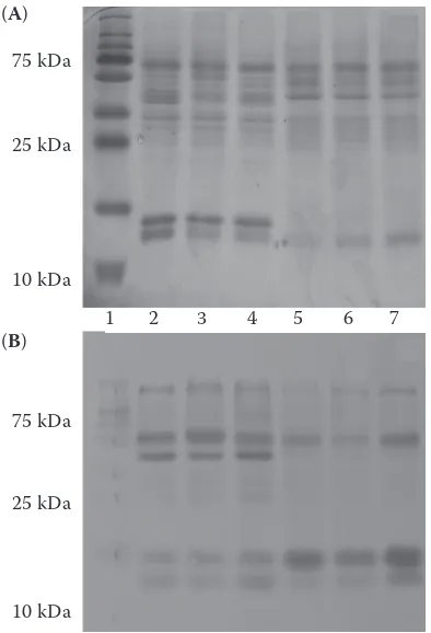
Electrophoresis, Western blotting, and zymog-raphy. The presence of enzymatic protein related to SOD isoforms in examined homogenates of tissues was confirmed by immunological reaction with appropriate antibodies (Figure 1). Zymography con-firmed that enzymatic protein was active (Figure 2).
[image:3.595.83.280.397.686.2]Effect of age on SOD activity in examined tis-sues. The influence of age on SOD activity was estimated by the comparison of enzyme activity
Figure 2. Zymographic analysis of chosen organs of cows, bulls, and calves; without KCN (A), with KCN (B)
[image:3.595.303.523.484.683.2]1 = calf heart, 2 = cow heart, 3 = bull heart, 4 = calf kidney, 5 = cow kidney, 6 = bull kidney, 7 = calf liver, 8 = cow liver, 9 = bull liver
Figure 1. Electrophoretic separation (A) and Western blotting (B) of chosen organs of cows, bulls, and calves
1 = standard mas (250, 150, 100, 75, 50, 37, 25, 20, 15, 10 kDa), 2 = bull heart, 3 = calf heart, 4 = cow heart, 5 = bull kidney, 6 = calf kidney, 7 = cow kidney
75 kDa
25 kDa
10 kDa
75 kDa
25 kDa
10 kDa
80 kDa
30 kDa
80 kDa
30 kDa 1 2 3 4 5 6 7
1 2 3 4 5 6 7 8 9 (A)
(B) (A)
in the same organs and tissues between cows, and female calves. The results are presented in Table 1.
In cows, the highest (P < 0.05) SOD activity was observed in homogenates of liver (4.59 ± 0.64 U/mg protein), but significantly the lowest in homogen-ates of the heart (2.40 ± 0.61) as compared to other examined tissues. In calves, the highest (P < 0.05) SOD activities were observed in the homogenates of kidney (6.61 ± 0.84 U/mg protein), but signifi-cantly the lowest in the skeletal muscles (2.17 ± 0.48) as compared to other organs and tissues.
SOD activity was significantly (P < 0.05) higher in the homogenates of heart, lung, and kidneys in calves than in cows, while in skeletal muscle the homogenates enzyme activity showed opposite relationship – 3.29 ± 0.68 U/mg protein vs 2.17 ± 0.48 (P < 0.05). There was no significant difference in the activity of SOD in the homogenates of liver and diaphragm between cows and calves.
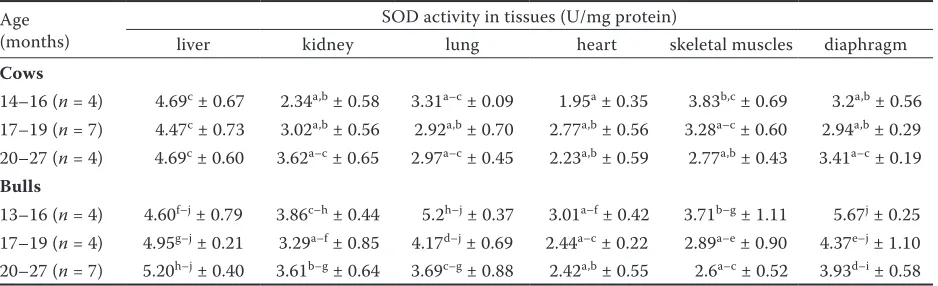
The activity of SOD was also compared within age subgroups of bulls and cows. The highest SOD activity was noticed in the homogenates of dia-phragm in young bulls (13–16 months) and reached 5.67 ± 0.25, while in older animals (20–27 months) it decreased significantly (P < 0.05) to the value of 3.93 ± 0.58. Similarly, in lung homogenates SOD
activity was higher in younger bulls at the age of 13–16 months (5.2 ± 0.37) whereas in bulls aged 20–27 months enzyme activity decreased signifi-cantly (P < 0.05) to values of 3.69 ± 0.88. In the other organs the differences in enzyme activity among age subgroups were also observed and showed the tendency (without statistical significance) to higher values of SOD activity in younger than in older bulls. In the group of cows, age related changes with statistical significance were not observed. The results are presented in Table 2.
Effect of gender on SOD activity. The impact of gender on SOD activity was examined by the comparison of enzyme activity in homogenates of various organs and tissues of cows, bulls, and calves (Table 1).
In the lung homogenates, the highest value of en-zyme activity was found in calves (5.35 ± 0.61 U/mg protein). Significantly (P < 0.05) lower values were observed in bulls (4.22 ± 0.94) and cows (3.04 ± 0.53).
The highest value of enzyme activity in homog-enates of diaphragm was observed in bulls (4.51 ± 0.99), while significantly (P < 0.05) lower in cows (3.13 ± 0.39) and calves (3.28 ± 0.67).
[image:4.595.64.531.113.185.2]There were no significant differences between bulls and cows in homogenates of skeletal muscles, Table 1. Influence of age, gender, and type of tissues on superoxide dismutase (SOD) activity
Animals SOD activity in tissues (U/mg protein)
liver kidney lung heart skeletal muscles diaphragm Cows 4.59AB ± 0.64 3B ± 0.73 3.04C ± 0.53 2.41B ± 0.61 3.29A ± 0.68 3.13B ± 0.39
Bulls 4.97A ± 0.52 3.59B ± 0.65 4.22B ± 0.94 2.58B ± 0.5 2.97A ± 0.88 4.51A ± 0.99
Calves 4.29B ± 0.4 6.61A ± 0.84 5.35A ± 0.61 4.13A ± 0.55 2.17B ± 0.48 3.28B ± 0.67
A–Cdifferent letters mean significant differences between results; P < 0.05 by ANOVA
Table. 2. Influence of age in the subgroups of cows and bulls
Age (months)
SOD activity in tissues (U/mg protein)
liver kidney lung heart skeletal muscles diaphragm
Cows
14–16 (n = 4) 4.69c ± 0.67 2.34a,b ± 0.58 3.31a–c ± 0.09 1.95a ± 0.35 3.83b,c ± 0.69 3.2a,b ± 0.56
17–19 (n = 7) 4.47c ± 0.73 3.02a,b ± 0.56 2.92a,b ± 0.70 2.77a,b ± 0.56 3.28a–c ± 0.60 2.94a,b ± 0.29
20–27 (n = 4) 4.69c ± 0.60 3.62a–c ± 0.65 2.97a–c ± 0.45 2.23a,b ± 0.59 2.77a,b ± 0.43 3.41a–c ± 0.19
Bulls
13–16 (n = 4) 4.60f–j ± 0.79 3.86c–h ± 0.44 5.2h–j ± 0.37 3.01a–f ± 0.42 3.71b–g ± 1.11 5.67j ± 0.25
17–19 (n = 4) 4.95g–j ± 0.21 3.29a–f ± 0.85 4.17d–j ± 0.69 2.44a–c ± 0.22 2.89a–e ± 0.90 4.37e–j ± 1.10
20–27 (n = 7) 5.20h–j ± 0.40 3.61b–g ± 0.64 3.69c–g ± 0.88 2.42a,b ± 0.55 2.6a–c ± 0.52 3.93d–i ± 0.58
SOD = sodium dismutase
[image:4.595.64.533.580.723.2]kidney, and heart. Significantly (P < 0.05) higher SOD activity in these organs was found in the group of calves apart from skeletal muscle, where enzyme activity was the lowest.
Significant (P < 0.05) differences in SOD activity were found in liver homogenates between calves (4.29 ± 0.4) and bulls (4.97 ± 0.52), however in cows the activity reached the value of 4.59 ± 0.64.
The influence of the type of tissues on SOD was observed in all groups of animals. The results confirm age-related changes in SOD activity while gender-related changes were observed only in some organs and tissues including lung and diaphragm.
DISCUSSION
The analysis of SOD protein in organs and tissues of cows, bulls, and female calves using electropho-retic methods as well as Western blotting and zy-mography confirmed the presence of enzyme in the examined tissues, and its physicochemical properties. Spectrophotometric determinations allowed for the quantitative analysis of enzymatic activity. The SOD activity showed the differences in the intensity of antioxidative processes which reflected the origin of the tissue enzyme and was dependent on age and gender of the examined animals.
Peroxidative processes are an inevitable part of aerobic metabolism. However, their intensity de-pends on the specific organs and cells. Proper func-tion of antioxidant protecfunc-tion is crucial for cells and tissues because it provides a balance between pro- and antioxidant processes. This balance en-sures correct permeability of cell membranes, and protects against the damage to macromolecules. SOD is one of the key elements of antioxidant protection and exists in three isoforms: cytoplas-mic SOD1 (CuZn-SOD), mitochondrial SOD2 (Mn-SOD), and extracellular EC-SOD (SOD3) (Fridovich 1997).
The liver is the organ having a particularly high metabolic activity. Antioxidant enzymes in the liver also show the highest activity in comparison to other tissues (Tian et al. 1998). However, the examination of the effect of age on the activity of antioxidant enzymes in the liver is difficult due to its role in detoxification of xenobiotics and metabolic functions. There are many factors that affect the metabolism of this organ.
The results obtained in the present study in the liver homogenates were confirmed by other authors
but in different species (Paynter and Caple 1984; Tian et al. 1998). There were no statistical differ-ences in the total enzyme activity in the group of older and younger females which may suggest no dependence on age in livers of these animals, which is also confirmed by other authors (Tian et al. 1998, Sverko et al. 2004). There are, however, others who observed increased SOD activity dur-ing agedur-ing in mice (Wozniak et al. 2004).
Kidneys, the same as the liver, are involved in the transformation of xenobiotics which is connected with higher metabolic rate. That is why they need appropriate antioxidant protection. In the present experiment, total SOD activity in homogenates of kidneys was twice higher in calves compared to other age groups of animals. There was no signifi-cant difference in activity between cows and bulls. This may suggest that SOD activity in kidneys decreased during ageing which was confirmed e.g. by Tian et al. (1998) and Meng et al. (2007). Uzun et al. (2013) also observed age and gender related changes in SOD activity which decreased with age but were higher in female old rats than in males while oxidative parameters increased dur-ing agedur-ing in both sexes. However, there are also opposite data describing increasing SOD activity during ageing (Paynter and Caple 1984).
The heart muscle is particularly vulnerable to oxidative stress, because it works constantly. It has a high oxygen partial pressure, and antioxidant protection is less effective than in other muscles. ROS are generated in mitochondria at high con-centrations, and therefore the Mn-SOD plays an important role in the overall antioxidant protec-tion and exhibits higher activity than CuZn-SOD. Xu et al. (2007) confirmed that myocardium as a post-mitotic tissue is less able to regulate anti-oxidant protection and repair damage caused by oxidative stress than rapidly dividing tissues and has the lowest activity and mRNA expression of CuZn-SOD, however, the highest activity of Mn-SOD isoform.
com-pared with other investigated tissues. A 3–5-fold lower activity of CuZn-SOD than of Mn- SOD isoform regardless of age has been observed. In contrast, the increase of total enzyme activity with age in rats was found by Gunduz et al. (2004).
Lung tissue is particularly exposed to inhaled contact with free radicals or molecules that cause local overproduction of ROS. Moreover, a relatively large area of the respiratory tract also enforces effec-tive antioxidant protection against excessive ROS. Paynter and Caple (1984) reported the increase of SOD activity in the lungs of lambs during ageing which may indicate the maturation of the antioxi-dant system with the growth of the organism. The present study has not confirmed this. Moreover, it showed sex-dependent alterations.
The metabolism of skeletal muscles depends on the types of fibres which predominate in their con-struction and are different in the concentration of myoglobin as well as resistance to fatigue. Skeletal muscles are particularly vulnerable to oxidative stress because they are constituted by post-mitotic cells consuming large amounts of oxygen and this greatly enhances the accumulation of oxidative damage. The effect of age on antioxidant system in the muscles may be quite different from that in the liver, kidney, brain, and heart.
Oh-Ishl et al. (1995) studied the effect of age on the activity and expression of mRNA SOD isoen-zymes (CuZn-SOD, Mn-SOD) in the skeletal muscles with various types of fibres in Fisher rats at the age of 4 and 24 months. The authors showed that the SOD activity in fibres type I was higher than in fib-ers type II. In addition, the increase of CuZn-SOD activity but no change in the activity of Mn-SOD isoenzyme with age was observed. These results were also confirmed by Capel et al. (2004) and Xu et al. (2007), who observed increased activity of CuZn-SOD as well as a higher expression of CuZn-CuZn-SOD mRNA with age. The increase of total SOD activity with age was observed in the present study.
Paynter and Caple (1984) did not observe any age-dependent increase in SOD activity in skeletal muscle in lambs. However, the enzyme activity in muscle was the lowest among other tissues (such as liver, heart, lungs, kidneys), what was confirmed in the present study, and Mn- SOD activity was 3–5 fold higher than CuZn-SOD activity.
The results indicate a close relationship between antioxidant enzymes and oxidative capacity in the muscle tissue. Another reason why muscles are
more susceptible to oxidative stress is the reduc-tion in intracellular glutathione (GSH) which may increase during the life.
The diaphragm is active constantly which means high consumption of oxygen and production of ROS. But when workload of the diaphragm is higher for example during exercise training or chronic obstructive lung disease, the production of ROS could increase which may lead to fatigue. The diaphragm, like other skeletal muscles, contains both slow and fast fibres.
There are significant differences between spe-cies in oxidative capacity and the type of fibre the diaphragm is composed of. Large animals have slow metabolism while small animals a relatively faster one and that is why oxidative capacity of their diaphragms can be higher. In the present study the SOD activity in diaphragm homogenates significantly decreased with age in the group of bulls. Furthermore, there was no significant differ-ence in enzymatic activity between the groups of cows and calves. However it is difficult to compare the effect of age on SOD activity in the diaphragm due to the lack of available literature.
The age-related changes in SOD activity in bovine tissues were confirmed in the present study. In pre-vious studies the activity of glutathione peroxidase (GSH-Px) was higher in calves than in cows but opposite relationship was observed in the total anti-oxidant capacity (Kankofer et al. 2013). Moreover, a previous examination of lipid and protein peroxida-tion in cows and calves showed that these processes were more intense in younger animals (Giergiel et al. 2014). Taking into consideration these facts we can suppose that the reason why SOD activity is higher in calves is the dynamic metabolic rate which causes the increase of oxidative stress products and leads to increased activity of antioxidant enzymes as a compensatory mechanism.
Additionally, the impact of sex steroid hormones on the regulation of antioxidant protection could be the part of the described discrepancies. The role of sex steroid hormones in the antioxidative system control is justified because of biological significance of these hormones and their changes with age (Pajovic et al. 2008).
A direct discussion of the obtained results is difficult due to scarce data from cattle. Some dis-crepancies in the SOD profile given in the present study as well as by the cited authors can be related with different SOD isoforms or total activity that were determined. Moreover, in many cases different age frames for comparisons were used. Differences related to species dependent specificity as well as metabolic rate of animals and tissues should be also taken into consideration.
The knowledge of the antioxidant status in live-stock is very important not only with respect to a proper function of organism and animal health, but also because of having an indirect impact on animal products (e.g. meat) quality (Bobcek et al. 2004; Lahucky et al. 2005).
CONCLUSION
The proper function of antioxidative protection is crucial for the health of animals. There are a lot of factors which may impact the antioxidant enzyme activities including SOD. Among them there are age, gender, and type of tissue, which was confirmed in the current study. Moreover, sex steroids can be involved in the regulation and control of enzyme activity. The obtained results could be helpful in the estimation of appropriate antioxidative supplementation which can be di-rected at particular age or sex groups of animals and improve welfare of older animals. But further investigation is needed to improve our knowledge on that topic in bovine species.
REFERENCES
Asha Devi S., Prathima S., Subramanyam M.V. (2003): Die-tary vitamin E and physical exercise: II. Antioxidant status and lipofuscin-like substances in aging rat heart. Experimental Gerontology, 38, 291–297.
Bobcek B., Lahucky R., Mrazova J., Bobcek R., Novotna K., Vasicek D. (2004): Effects of dietary organic selenium supplementation on selenium content, antioxidative sta-tus of muscles and meat quality of pigs. Czech Journal of Animal Science, 49, 411–417.
Capel F., Buffiere C., Mirand P.P., Mosoni L. (2004): Dif-ferential variation of mitochondrial H2O2 release during
aging in oxidative and glycolytic muscles in rats. Mecha-nisms of Ageing and Development, 125, 367–373. Chen C., Brown-Borg H.M., Rakoczy S.G., Thompson L.D.V.
(2008): Muscle disuse: adaptation of antioxidant systems
is age dependent. Journal of Gerontology: Biological Sci-ences, 63, 461–466.
Fridovich I. (1997): Superoxide anion radical (O2–),
super-oxide dismutases, and related matters. The Journal of Biological Chemistry, 272, 18515–18517.
Giergiel M., Lopucki M., Stachowicz N., Kankofer M. (2012): The influence of age and gender on antioxidant enzyme activities in humans and laboratory animals. Aging Clini-cal and Experimental Research, 24, 561–569.
Giergiel M., Zielinska A., Legutko K., Kankofer M. (2014): Protein and lipid peroxidation intensity in cows and fe-male calves. Acta Scientiae Veterinariae, 42, 1185. Giustarini D., Dalle-Donne I., Tsikas D., Milzani A., Rossi
R. (2009):Oxidative stress and human diseases: ori-gin, link, measurement, mechanisms, and biomarkers. Critical Reviews in Clinical Laboratory Sciences, 46, 241–281.
Gunduz F., Senturk U.K., Kuru O., Aktekin B., Aktekin M.R. (2004): The effect of one year’s swimming exercise on oxidant stress and antioxidant capacity in aged rats. Physiological Research, 53, 171–176.
Hamilton M.L., Van Remmen H., Drake J.A. (2001): Does oxidant damage to DNA increase with age? Proceedings of the National Academy of Sciences of the United States of America, 98, 10469–10474.
Kankofer M., Wawrzykowski J., Giergiel M. (2013): Sex and age dependent activity of glutathione peroxidase in reproductive organs in pre- and post-pubertal cattle in relation to total antioxidant capacity. Aging Clinical and Experimental Research, 25, 365–370.
Laemmli U.K. (1970): Cleavage of structural proteins during the assembly of the head of bacteriophage T4. Nature, 227, 680–685.
Lahucky R., Bahelka I., Novotna K., Vasickova K. (2005): Effects of dietary vitamin E and vitamin C supplementa-tion on the level of α-tocopherol and l-ascorbic acid in muscle and on the antioxidative status and meat quality of pigs. Czech Journal of Animal Science, 50, 175–184. Meng Q., Wong Y.T., Chen J., Ruan R. (2007): Age-related
changes in mitochondrial function and antioxidative en-zyme activity in Fischer 344 rats. Mechanisms of Ageing and Development, 128, 286–292.
Misra H.P., Fridovich I. (1977): Superoxide dismutase and peroxidase: a positive activity stain applicable to poly-acrylamide gel electropherograms. Archives of Biochem-istry and Biophysics, 183, 511–515.
Navarro-Arevalo A., Canavate C., Sanchez-del-Pino M.J. (1999): Myocardial and skeletal muscle aging and changes in oxidative stress in relationship to rigorous exercise training. Mechanisms of Ageing and Development, 108, 207–217. Oh-Ishl S., Kizaki T., Yamashita H., Nagatab N., Suzuki K.,
Taniguchi N., Ohno H. (1995): Alterations of superoxide dismutase iso-enzyme activity, content, and mRNA ex-pression with aging in rat skeletal muscle. Mechanisms of Ageing and Development, 84, 65–76.
Pajovic S.B., Saicic Z.S. (2008): Modulation of antioxidant enzyme activities by sexual steroid hormones. Physiologi-cal Research, 57, 801–811.
Paynter D.I., Caple I.W. (1984): Age-related changes in ac-tivities of the superoxide dismutase enzymes in tissues of the sheep and the effect of dietary copper and manganese on these changes. Journal of Nutrition, 114, 19089–19106. Pfeilschifter J., Koeditz R., Pfohl M., Schatz H. (2002): Chang-es in proinflammatory cytokine activity after menopause. Endocrine Reviews, 23, 90–119.
Rikans L.E., Hornbrook K.R. (1997): Lipid cooperation, an-tioxidant protection and ageing. Biochimica et Biophysica Acta, 1362, 116–127.
Rinaldi B., Corbi G., Boccuti S., Filippelli W., Rengo G., Leosco D., Rossi F., Filippelli A., Ferrara N. (2006): Ex-ercise training affects age-induced changes in SOD and heatshock protein expression in rat heart. Experimental Gerontology, 41, 764–770.
Sobocanec S., Balog T., Sverko V. (2003): Sex-dependent antioxidant enzyme activities and lipid peroxidation in ageing mouse brain. Free Radical Research, 37, 743–748.
Sun M., Zigman S. (1978): Determination of superoxide dismutase in erythrocytes using the method of adrenaline autooxidation. Analytical Biochemistry, 90, 81–89. Sverko V., Sobocanec S., Balog T., Marotti T. (2004): Age
and gender differences in antioxidant enzyme activity: potential relationship to liver carcinogenesis in male mice. Biogerontology, 5, 235–242.
Tian L., Cai Q., Wei H. (1998): Alteration of antioxidant enzymes and oxidative damage to macromolecules in different organs of rats during ageing. Free Radical Biol-ogy and Medicine, 24, 1477–1484.
Towbin H., Staechelin T., Gordon J. (1979): Electropho-retic transfer of proteins from polyacrylamide gels to nitrocellulose sheets: procedure and some applications. Proceedings of the National Academy of Sciences of the United States of America, 76, 4350–4355.
Uzun D., Korkmaz G.G., Sitar M.E., Cebe T., Yanar K., Cakatay U., Aydin S. (2013): Oxidative damage parameters in renal tissues of aged and young rats based on gender. Clinical Interventions in Aging, 8, 809–815.
Wozniak A., Drewa G., Wozniak B.,Schachtschabel D.O. (2004): Activity of antioxidant enzymes and concentration of lipid peroxidation products in selected tissues of mice of different ages, both healthy and melanoma-bearing. Zeitschrift für Gerontologie und Geriatrie, 37, 184–189. Xu C.L., Wang Y.Z., Guo J., Liu J.X., Feng J. (2007): Compari-son of age-related differences in expression of antioxidant enzyme mRNA and activity in various tissues of pigs. Comparative Biochemistry and Physiology, Biochemistry and Molecular Biology, 147, 445–451.
Received: 2014–05–13 Accepted after corrections: 2015–03–02
Corresponding Author
Marta Giergiel, Ph.D., University of Life Sciences, Faculty of Veterinary Medicine, Department of Biochemistry, Akademicka 12, 20-033 Lublin, Poland