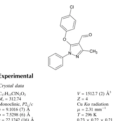
5-(4-Chlorophenoxy)-3-methyl-1-phenyl-1
H
-pyrazole-4-carbaldehyde
N. Vinutha,aS. Madan Kumar,aS. Shobhitha,b
B. Kalluraya,bN. K. Lokanathaand D. Revannasiddaiaha*
aDepartment of Studies in Physics, University of Mysore, Manasagangotri, Mysore 570 006, India, andbDepartment of Studies in Chemistry, Mangalore University, Mangalagangotri, Mangalore 574 199, India
Correspondence e-mail: [email protected]
Received 15 March 2014; accepted 9 April 2014
Key indicators: single-crystal X-ray study;T= 296 K; mean(C–C) = 0.003 A˚; Rfactor = 0.039;wRfactor = 0.105; data-to-parameter ratio = 12.5.
In the title compound, C17H13ClN2O2, the phenyl and
chlorobenzene rings are inclined to the central pyrazole ring at 40.84 (9) and 65.30 (9), respectively. In the crystal, pairs of
C—H interactions link the molecules into inversion dimers and C—H O hydrogen bonds link these dimers into columns extended in [010]. The crystal packing exhibits short intermolecular O Cl contacts of 3.0913 (16) A˚ .
Related literature
For biological properties and pharmocological applications of aryloxy pyrazole derivatives, see: Raiet al. (2008); Girishaet al.(2010); Islooret al.(2009, 2010); Shobhithaet al.(2013). For related structures, see: Shahani, Fun, Ragavan et al. (2011); Shahani, Fun, Shettyet al.(2011); Prasathet al.(2011).
Experimental
Crystal data
C17H13ClN2O2 Mr= 312.74 Monoclinic,P21=c a= 9.1016 (7) A˚
b= 7.5298 (6) A˚
c= 22.1242 (16) A˚
= 93.908 (3)
V= 1512.7 (2) A˚3
Z= 4
CuKradiation
= 2.31 mm1 T= 296 K
0.230.220.21 mm
Data collection
Bruker X8 Proteum diffractometer Absorption correction: multi-scan
(SADABS; Bruker, 2013)
Tmin= 0.619,Tmax= 0.643
9744 measured reflections 2501 independent reflections 2314 reflections withI> 2(I)
Rint= 0.041
Refinement
R[F2> 2(F2)] = 0.039 wR(F2) = 0.105 S= 1.03 2501 reflections
200 parameters
H-atom parameters constrained max= 0.25 e A˚
3
min=0.38 e A˚
[image:1.610.48.243.523.729.2]3
Table 1
Hydrogen-bond geometry (A˚ ,).
Cgis the centroid of the C11–C16 ring.
D—H A D—H H A D A D—H A
C6—H6 O2i
0.93 2.58 3.503 (2) 171
C2—H2 Cgii 0.93 2.63 3.476 (2) 152
Symmetry codes: (i)x;yþ1;z; (ii)xþ2;yþ1;zþ1.
Data collection:APEX2(Bruker, 2013); cell refinement:SAINT (Bruker, 2013); data reduction:SAINT; program(s) used to solve structure:SHELXS97(Sheldrick, 2008); program(s) used to refine structure: SHELXL97 (Sheldrick, 2008); molecular graphics: Mercury(Macraeet al., 2008); software used to prepare material for publication:PLATON(Spek, 2009).
The authors are thankful to the IOE and UPE, University of Mysore, for providing the single-crystal X-ray diffraction facility and for the financial support. VN is grateful to the UGC for the award of an RFSMS Fellowship. RD acknowl-edges the UGC, New Delhi, for financial support under the Major Research Project Scheme [UGC MRP No. F.41–882/ 2012 (SR) dated 01/07/2012].
Supporting information for this paper is available from the IUCr electronic archives (Reference: CV5447).
References
Bruker (2013).APEX2,SAINTandSADABS. Bruker AXS Inc., Madison, Wisconsin, USA.
Girisha, K. S., Kalluraya, B., Narayana, V. & Padmashree. (2010).Eur. J. Med. Chem.45, 4640–4644.
Isloor, A. M., Kalluraya, B. & Pai, S. K. (2010).Eur. J. Med. Chem.45, 825–830. Isloor, A. M., Kalluraya, B. & Shetty, P. (2009).Eur. J. Med. Chem.44, 3784–
3787.
Macrae, C. F., Bruno, I. J., Chisholm, J. A., Edgington, P. R., McCabe, P., Pidcock, E., Rodriguez-Monge, L., Taylor, R., van de Streek, J. & Wood, P. A. (2008).J. Appl. Cryst.41, 466–470.
Prasath, R., Bhavana, P., Ng, S. W. & Tiekink, E. R. T. (2011).Acta Cryst.E67, o2650.
Rai, N. S., Kalluraya, B., Lingappa, B., Shenoy, S. & Puranic, V. G. (2008).Eur. J. Med. Chem.43, 1715–1720.
Shahani, T., Fun, H.-K., Ragavan, R. V., Vijayakumar, V. & Venkatesh, M. (2011).Acta Cryst.E67, o475.
Shahani, T., Fun, H.-K., Shetty, S. & Kalluraya, B. (2011).Acta Cryst.E67, o2646.
Sheldrick, G. M. (2008).Acta Cryst.A64, 112–122.
Shobhitha, S., Kalluraya, B., Nithinchandra, B. M., Joshi, C. G., Joshi, H. & Nidavani, R. B. (2013).Indian J. Heterocycl. Chem.23, 33–38.
Spek, A. L. (2009).Acta Cryst.D65, 148–155. Acta Crystallographica Section E
Structure Reports
Online
supporting information
Acta Cryst. (2014). E70, o560 [doi:10.1107/S1600536814007879]
5-(4-Chlorophenoxy)-3-methyl-1-phenyl-1
H
-pyrazole-4-carbaldehyde
N. Vinutha, S. Madan Kumar, S. Shobhitha, B. Kalluraya, N. K. Lokanath and D.
Revannasiddaiah
S1. Comment
Aryloxy pyrazoles and their derivatives possess a significant pharmcological activities such as antimicrobial (Rai et al.
2008; Girisha et al., 2010), anti-inflammatory (Isloor et al., 2009) and analgesic activities (Shobhitha et al., 2013). The
title compound can serve as an intermediate in the synthesis of various pyrazole derivatives with significant
pharmacological activities (Isloor et al., 2010).
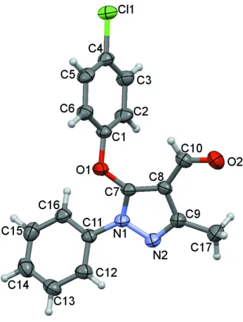
In the title compound (Fig.1), all bond lengths and angles are normal and correspond well to those observed in the
related compounds (Shahani, Fun, Ragavan et al., 2011; Shahani, Fun, Shetty et al., 2011; Prasath et al., 2011). The
pyrazole ring makes dihedral angles of 65.30 (9)° with chlorobenzene ring and 40.84 (9)° with benzene ring. The dihedral
angle between the chlorobenzene ring and benzene ring is 76.23 (9)°.
In the crystal, C–H···π interactions (Table 1) link the molecules into inversion dimers, and intermolecular C–H···O
hydrogen bonds (Table 1) link these dimers into columns extended in [010]. The crystal packing exhibits short
intermolecular O···Cl contacts of 3.0913 (16) Å.
S2. Experimental
The title compound was prepared by refluxing a mixture of 5-chloro-3-methyl-1-phenyl-1H-pyrazol-4-carboxaldehyde
(0.1 mol) and 4-chloro phenol (0.1 mol) in 10 ml of dimethyl sulfoxide. To this solution, 0.1 mol of potassium hydroxide
was added. The reaction mixture was refluxed for 3 hrs and then it was cooled to room temperature and poured to
crushed ice. The solid product that separated was filtered and dried. It was then recrystallized from ethanol. Crystals
suitable for X-ray analysis were obtained from slow evaporation of ethanol.
S3. Refinement
All the H atoms were fixed geometrically (C—H= 0.93–0.96 Å) and allowed to ride on their parent atoms with Uiso(H) =
Figure 1
The molecular structure of the title compound showing the atomic numbering and 50% probability displacement
ellipsoids.
5-(4-Chlorophenoxy)-3-methyl-1-phenyl-1H-pyrazole-4-carbaldehyde
Crystal data
C17H13ClN2O2
Mr = 312.74 Monoclinic, P21/c
Hall symbol: -P 2ybc
a = 9.1016 (7) Å
b = 7.5298 (6) Å
c = 22.1242 (16) Å
β = 93.908 (3)°
V = 1512.7 (2) Å3
Z = 4
F(000) = 648
Dx = 1.373 Mg m−3
Cu Kα radiation, λ = 1.54178 Å Cell parameters from 2501 reflections
θ = 4.0–64.4°
T = 296 K Block, brown
0.23 × 0.22 × 0.21 mm
Data collection
Bruker X8 Proteum diffractometer
Radiation source: Bruker MicroStar microfocus rotating anode
Helios multilayer optics monochromator Detector resolution: 18.4 pixels mm-1
φ and ω scans
Absorption correction: multi-scan
(SADABS; Bruker, 2013)
Tmin = 0.619, Tmax = 0.643
9744 measured reflections 2501 independent reflections 2314 reflections with I > 2σ(I)
Rint = 0.041
θmax = 64.5°, θmin = 4.0°
h = −10→10
k = −3→8
l = −25→25
Refinement
Refinement on F2
Least-squares matrix: full
R[F2 > 2σ(F2)] = 0.039
wR(F2) = 0.105
S = 1.03 2501 reflections 200 parameters 0 restraints
Primary atom site location: structure-invariant direct methods
Secondary atom site location: difference Fourier map
Hydrogen site location: inferred from neighbouring sites
H-atom parameters constrained
w = 1/[σ2(F
o2) + (0.0555P)2 + 0.5076P]
where P = (Fo2 + 2Fc2)/3
(Δ/σ)max < 0.001
Δρmax = 0.25 e Å−3
Δρmin = −0.38 e Å−3
Extinction correction: SHELXL97 (Sheldrick, 2008), FC*=KFC[1+0.001XFC2Λ3/SIN(2Θ)]-1/4
Extinction coefficient: 0.0171 (10)
Special details
Geometry. Bond distances, angles etc. have been calculated using the rounded fractional coordinates. All su's are estimated from the variances of the (full) variance-covariance matrix. The cell e.s.d.'s are taken into account in the estimation of distances, angles and torsion angles
Refinement. Refinement on F2 for ALL reflections except those flagged by the user for potential systematic errors.
Weighted R-factors wR and all goodnesses of fit S are based on F2, conventional R-factors R are based on F, with F set to
zero for negative F2. The observed criterion of F2 > σ(F2) is used only for calculating -R-factor-obs etc. and is not
relevant to the choice of reflections for refinement. R-factors based on F2 are statistically about twice as large as those
based on F, and R-factors based on ALL data will be even larger.
Fractional atomic coordinates and isotropic or equivalent isotropic displacement parameters (Å2)
x y z Uiso*/Ueq
C8 1.05021 (18) 0.3640 (2) 0.34914 (7) 0.0354 (5) C9 1.16849 (19) 0.2680 (2) 0.37941 (8) 0.0398 (5) C10 0.9588 (2) 0.3101 (3) 0.29611 (8) 0.0407 (6) C11 1.20153 (18) 0.6520 (2) 0.46814 (7) 0.0336 (5) C12 1.3510 (2) 0.6767 (2) 0.47974 (8) 0.0407 (6) C13 1.4008 (2) 0.8051 (3) 0.52092 (9) 0.0488 (6) C14 1.3028 (2) 0.9086 (3) 0.54945 (9) 0.0533 (7) C15 1.1529 (2) 0.8822 (3) 0.53819 (8) 0.0495 (6) C16 1.10095 (19) 0.7530 (2) 0.49751 (7) 0.0404 (5) C17 1.2243 (2) 0.0881 (3) 0.36473 (11) 0.0592 (7) H02A 1.30480 0.05740 0.39310 0.0890* H2 0.76800 0.44510 0.40090 0.0500* H02B 1.25740 0.08840 0.32440 0.0890* H3 0.51850 0.44700 0.37070 0.0500* H02C 1.14670 0.00270 0.36720 0.0890* H5 0.57430 0.89290 0.27670 0.0530* H6 0.82560 0.88770 0.30450 0.0480* H10 0.89020 0.39080 0.27940 0.0490* H12 1.41770 0.60760 0.46010 0.0490* H13 1.50150 0.82140 0.52930 0.0590* H14 1.33690 0.99660 0.57640 0.0640* H15 1.08660 0.95150 0.55800 0.0590* H16 1.00030 0.73440 0.49010 0.0480*
Atomic displacement parameters (Å2)
U11 U22 U33 U12 U13 U23
C17 0.0530 (12) 0.0482 (12) 0.0741 (13) 0.0116 (10) −0.0122 (10) −0.0135 (10)
Geometric parameters (Å, º)
Cl1—C4 1.7390 (18) C11—C16 1.386 (2)
O1—C1 1.403 (2) C12—C13 1.384 (3)
O1—C7 1.3490 (19) C13—C14 1.370 (3)
O2—C10 1.214 (3) C14—C15 1.384 (3)
N1—N2 1.384 (2) C15—C16 1.386 (3)
N1—C7 1.347 (2) C2—H2 0.9300
N1—C11 1.426 (2) C3—H3 0.9300
N2—C9 1.320 (2) C5—H5 0.9300
C1—C2 1.377 (3) C6—H6 0.9300
C1—C6 1.377 (2) C10—H10 0.9300
C2—C3 1.384 (2) C12—H12 0.9300
C3—C4 1.379 (3) C13—H13 0.9300
C4—C5 1.373 (2) C14—H14 0.9300
C5—C6 1.388 (3) C15—H15 0.9300
C7—C8 1.384 (2) C16—H16 0.9300
C8—C9 1.425 (2) C17—H02A 0.9600
C8—C10 1.449 (2) C17—H02B 0.9600
C9—C17 1.490 (3) C17—H02C 0.9600
C11—C12 1.380 (2)
O2—C10—C8 123.73 (18) C9—C17—H02B 109.00 N1—C11—C12 118.14 (14) C9—C17—H02C 109.00 N1—C11—C16 120.96 (14) H02A—C17—H02B 110.00 C12—C11—C16 120.89 (15) H02A—C17—H02C 109.00 C11—C12—C13 119.43 (16) H02B—C17—H02C 109.00 C12—C13—C14 120.43 (17)
C7—O1—C1—C2 36.1 (2) C2—C3—C4—C5 0.6 (3) C7—O1—C1—C6 −146.92 (15) Cl1—C4—C5—C6 −179.47 (13) C1—O1—C7—N1 −143.39 (14) C3—C4—C5—C6 0.3 (3) C1—O1—C7—C8 42.4 (2) C4—C5—C6—C1 −0.6 (2) C7—N1—N2—C9 −1.81 (18) O1—C7—C8—C9 174.01 (17) C11—N1—N2—C9 −175.88 (14) O1—C7—C8—C10 −3.9 (3) N2—N1—C7—O1 −174.08 (13) N1—C7—C8—C9 −0.60 (17) N2—N1—C7—C8 1.51 (18) N1—C7—C8—C10 −178.50 (16) C11—N1—C7—O1 −0.8 (2) C7—C8—C9—N2 −0.55 (19) C11—N1—C7—C8 174.78 (15) C7—C8—C9—C17 −179.41 (17) N2—N1—C11—C12 37.2 (2) C10—C8—C9—N2 177.37 (17) N2—N1—C11—C16 −141.54 (15) C10—C8—C9—C17 −1.5 (3) C7—N1—C11—C12 −135.63 (17) C7—C8—C10—O2 −177.79 (18) C7—N1—C11—C16 45.7 (2) C9—C8—C10—O2 4.8 (3) N1—N2—C9—C8 1.42 (19) N1—C11—C12—C13 −179.23 (16) N1—N2—C9—C17 −179.62 (16) C16—C11—C12—C13 −0.5 (2) O1—C1—C2—C3 177.88 (16) N1—C11—C16—C15 179.80 (15) C6—C1—C2—C3 1.0 (3) C12—C11—C16—C15 1.1 (2) O1—C1—C6—C5 −177.13 (15) C11—C12—C13—C14 −0.8 (3) C2—C1—C6—C5 −0.1 (3) C12—C13—C14—C15 1.5 (3) C1—C2—C3—C4 −1.3 (3) C13—C14—C15—C16 −0.9 (3) C2—C3—C4—Cl1 −179.59 (14) C14—C15—C16—C11 −0.4 (3)
Hydrogen-bond geometry (Å, º)
Cg is the centroid of the C11–C16 ring.
D—H···A D—H H···A D···A D—H···A
C6—H6···O2i 0.93 2.58 3.503 (2) 171
C2—H2···Cgii 0.93 2.63 3.476 (2) 152