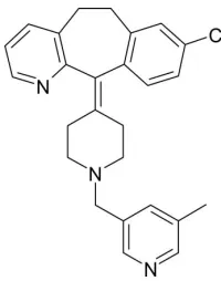
Rupatadine
Manpreet Kaur,aJerry P. Jasinski,b* Zane A. Luopa,b Neeraj Kumar,cNilesh G. Patel,cOmprakash Gudaparthic and H. S. Yathirajana
aDepartment of Studies in Chemistry, University of Mysore, Manasagangotri, Mysore
570 006, India,bDepartment of Chemistry, Keene State College, 229 Main Street,
Keene, NH 03435-2001, USA, andcCR & D, Cadila Pharmaceuticals Ltd, 1389,
Trasad Road, Dholka, Ahmedabad 387 810, Gujarat, India Correspondence e-mail: [email protected]
Received 21 May 2013; accepted 22 May 2013
Key indicators: single-crystal X-ray study;T= 173 K; mean(C–C) = 0.002 A˚; Rfactor = 0.041;wRfactor = 0.122; data-to-parameter ratio = 15.7.
In the title compound (systematic name: 8-chloro-11-{1-[(5- methylpyridin-3-yl)methyl]piperidin-4-ylidene}-6,11-dihydro-5H-benzo[5,6]cyclohepta[1,2-b]pyridine), C26H26ClN3, the
dihedral angle between the mean planes of the chlorophenyl and cyclohepta[1,2-b]pyridinyl rings fused to the cycloheptane ring is 56.6 (1). The mean planes of the cyclohepta[1,2-b
]-pyridinyl and 5-methylpyridin-3-yl rings are twisted by 64.9 (4). The central piperizene group is in a slightly distorted
chair configuration. A weak intramolecular C—H N inter-action is observed between the cyclohepta[1,2-b]pyridinyl and piperidin-4-ylidene moieties.
Related literature
For the pharmacological importance of rupatadine, see: Kean & Plosker (2007); Merlos et al. (1997); Mullol et al. (2008); Picado (2006). For the reported synthesis methodology of rupatadine, see: Agarwal et al. (2008). For standard bond lengths, see: Allenet al.(1987).
Crystal data
C26H26ClN3 Mr= 415.95
Monoclinic,P21=n a= 10.2655 (3) A˚ b= 11.3341 (4) A˚ c= 18.8111 (6) A˚
= 90.874 (3)
V= 2188.43 (11) A˚3 Z= 4
CuKradiation
= 1.67 mm 1 T= 173 K
0.420.380.22 mm
Data collection
Agilent Xcalibur (Eos, Gemini) diffractometer
Absorption correction: multi-scan (CrysAlis PROandCrysAlis RED; Agilent, 2012) Tmin= 0.673,Tmax= 1.000
13849 measured reflections 4281 independent reflections 3565 reflections withI> 2(I) Rint= 0.026
Refinement
R[F2> 2(F2)] = 0.041 wR(F2) = 0.122 S= 1.05 4281 reflections
273 parameters
H-atom parameters constrained
max= 0.22 e A˚ 3
[image:1.610.119.219.570.697.2]min= 0.29 e A˚ 3
Table 1
Hydrogen-bond geometry (A˚ ,).
D—H A D—H H A D A D—H A
C19—H19B N1 0.99 2.60 3.229 (2) 121
Data collection: CrysAlis PRO(Agilent, 2012); cell refinement: CrysAlis PRO; data reduction: CrysAlis RED (Agilent, 2012); program(s) used to solve structure: SHELXS97 (Sheldrick, 2008); program(s) used to refine structure:SHELXL2012(Sheldrick, 2008); molecular graphics:OLEX2(Dolomanovet al., 2009); software used to prepare material for publication:OLEX2.
HSY thanks the UOM for research facilities. JPJ acknowl-edges the NSF–MRI program (grant No. CHE-1039027) for funds to purchase the X-ray diffractometer.
Supplementary data and figures for this paper are available from the IUCr electronic archives (Reference: HG5317).
References
Agarwal, R., Bhirud, S. B., Bijukumar, G. & Khude, G. D. (2008).Synth. Commun.38, 122–127.
Agilent (2012). CrysAlis PRO and CrysAlis RED. Agilent Technologies, Yarnton, England.
Allen, F. H., Kennard, O., Watson, D. G., Brammer, L., Orpen, A. G. & Taylor, R. (1987).J. Chem. Soc. Perkin Trans. 2, pp. S1–19.
Dolomanov, O. V., Bourhis, L. J., Gildea, R. J., Howard, J. A. K. & Puschmann, H. (2009).J. Appl. Cryst.42, 339–341.
Kean, S. J. & Plosker, G. L. (2007).Drugs,67, 457–474.
Merlos, M., Giral, M., Balsa, D., Ferrando, R., Queralt, M., Puigdemont, A., Garcia-Rafanell, J. & Forn, J. (1997).J. Pharmacol. Exp. Ther.280, 114–121. Mullol, J., Bousquet, J., Bachert, C., Canonica, W. G., Gimenez-Arnau, A., Kowalski, M. L., Martı´-Guadan˜o, E., Maurer, M., Picado, C., Scadding, G. & Van Cauwenberge, P. (2008).Allergy,63, 5–28.
Picado, C. (2006).Expert Opin. Pharmacother.7, 1989–2001. Sheldrick, G. M. (2008).Acta Cryst.A64, 112–122. Structure Reports
Online
supporting information
supporting information
Acta Cryst. (2013). E69, o980 [doi:10.1107/S1600536813014256]
Rupatadine
Manpreet Kaur, Jerry P. Jasinski, Zane A. Luopa, Neeraj Kumar, Nilesh G. Patel, Omprakash
Gudaparthi and H. S. Yathirajan
S1. Comment
Rupatadine (IUPAC Name: 8-Chloro-6,11-dihydro-11-[1-[(5-methyl-3- pyridinyl)
methyl]-4-piperidinylidene]-5H-benzo[5,6]cyclohepta[1,2-b] pyridine) is a non-sedating antihistamine showing a rapid onset of action and a good safety
profile even in prolonged treatment periods of a year (Picado, 2006; Mullol et al., 2008). A review of its use in the
management of allergic disorders is published (Kean & Plosker, 2007). Rupatadine has shown as inhibition deregulation,
induced by the immunological and non-immunological stimulants and the inhibition of release of cytokines, particularly
the tumor necrosis factor alpha (TNF-alpha) in human mastocytes and monocytes (Picado, 2006). In vitro metabolism
studies indicate that rupatadine is metabolized mainly by the cytochrome P-450 in liver (Merlos et al., 1997). In view of
the importance of the title compound, (I), C26H26ClN3, we have synthesized rupatadine free base based on a reported
method (Agarwal et al., 2008) and its single crystal structure is reported herin.
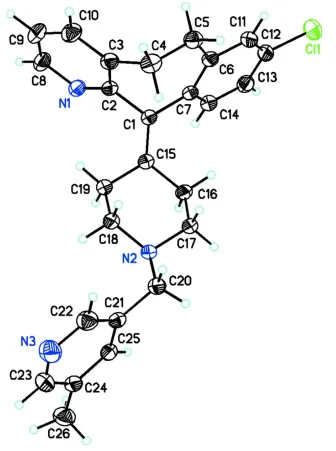
In (I), the dihedral angle between the mean planes of the chlorophenyl and cyclohepta[1,2-b]pyridinyl rings fused to the
cycloheptane ring is 56.6 (1)° (Fig. 1). The mean planes of the cyclohepta[1,2-b]pyridinyl and 5-methyl-3-pyridinyl rings
are twisted by 64.9 (4)°. The central 6-membered piperizene group adopts a slightly distorted chair configuration with
puckering parameters Q, θ and φ of 0.5613 (16)Å, 3.31 (16)°, and 348 (3)°, respectively. A weak C—H···O
intramolecular interaction is observed between the cyclohepta[1,2-b]pyridinyl and 4-piperidinylidene moieties. In the
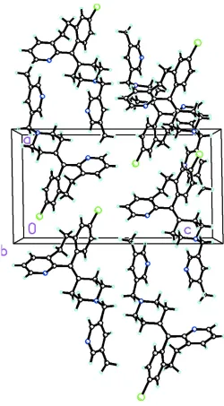
crystal, the molecules pack in a normal head-to tail dimer-like arrangement (Fig. 2).
S2. Experimental
4-methyl-3-chloromethyl pyridine hydrochloride (3.5 g, 0.02 mol), desloratadine (6.2 g, 0.02 mol), potassium carbonate
(6.9 g, 0.05 mol) was charged into acetonitrile (30 ml) Fig. 3). The reaction mass was heated to 313–318 K and stirred for
10-12 h (Agarwal et al., 2008). The reaction mass was cooled to 298–303 K and the inorganic material filtered. The
solvent was removed under reduced pressure. Toluene (40 ml) was added to residue and heated to 328-333 K to get a
clear solution. The toluene layer was washed with a saturated sodium chloride solution (40 ml) and water (25 ml). Half
the quantity of toluene was distilled out under vacuum and single crystals were grown from toluene using the slow
evaporation technique (m. p.: 409–410 K).
S3. Refinement
All of the H atoms were placed in their calculated positions and then refined using the riding model with Atom—H
lengths of 0.95Å (CH), 0.99Å (CH2) or 0.98Å (CH3). Idealised Me was refined as a rotating group:
C26(H26A,H26B,H26C). Isotropic displacement parameters for these atoms were set to 1.2 (CH, CH2) or 1.5 (CH3 times
Figure 1
Molecular structure of the title compound showing the atom labeling scheme and 30% probability displacement
supporting information
Figure 2
Figure 3
Reaction scheme for the synthesis of rupatadine free base.
8-Chloro-11-{1-[(5-methylpyridin-3-yl)methyl]piperidin-4-ylidene}-6,11-dihydro-5H -benzo[5,6]cyclohepta[1,2-b]pyridine
Crystal data
C26H26ClN3
Mr = 415.95 Monoclinic, P21/n
a = 10.2655 (3) Å
b = 11.3341 (4) Å
c = 18.8111 (6) Å
β = 90.874 (3)°
V = 2188.43 (11) Å3
Z = 4
F(000) = 880
Dx = 1.262 Mg m−3
Cu Kα radiation, λ = 1.5418 Å Cell parameters from 4864 reflections
θ = 3.9–72.2°
µ = 1.67 mm−1
T = 173 K
Irregular, clear orangish orange 0.42 × 0.38 × 0.22 mm
Data collection
Agilent Xcalibur (Eos, Gemini) diffractometer
Radiation source: Enhance (Cu) X-ray Source Graphite monochromator
Detector resolution: 16.0416 pixels mm-1
ω scans
Absorption correction: multi-scan
(CrysAlis PRO and CrysAlis RED; Agilent, 2012)
Tmin = 0.673, Tmax = 1.000 13849 measured reflections 4281 independent reflections 3565 reflections with I > 2σ(I)
Rint = 0.026
θmax = 72.4°, θmin = 4.6°
h = −11→12
k = −8→13
l = −21→23
Refinement
Refinement on F2 Least-squares matrix: full
R[F2 > 2σ(F2)] = 0.041
wR(F2) = 0.122
S = 1.05 4281 reflections 273 parameters 0 restraints
Primary atom site location: structure-invariant direct methods
Hydrogen site location: inferred from neighbouring sites
H-atom parameters constrained
w = 1/[σ2(F
o2) + (0.0616P)2 + 0.3876P] where P = (Fo2 + 2Fc2)/3
supporting information
Δρmin = −0.29 e Å−3 Extinction correction: SHELXL2012 (Sheldrick, 2008), Fc*=kFc[1+0.001xFc2λ3/sin(2θ)]-1/4 Extinction coefficient: 0.0043 (3)
Special details
Experimental. (HPLC purity 99.75 %) FT IR (KBr) : 1350.2, 1475.6, 1583.6; 1H NMR (300 MHz, DMSO d6) δ 2.072
(s, 1H), 2.127-2.164 (m, 3H), 2.247 (s, 3H), 2.264-2.320 (m, 2H), 2.545-2.580 (m, 2H), 2.725-2.827 (m, 2H),
3.217-3.324 (m, 2H), 3.406 (s, 2H), 7.011-7.038 (d, 1H), 7.124-7.193 (m, 2H), 7.242-7.248 (d, 1H), 7.474-7.537 (m, 2H), 8.250-8.313 (dd, 3H); MS m/z (EI): 416 (M + 1).
Geometry. All esds (except the esd in the dihedral angle between two l.s. planes) are estimated using the full covariance
matrix. The cell esds are taken into account individually in the estimation of esds in distances, angles and torsion angles; correlations between esds in cell parameters are only used when they are defined by crystal symmetry. An approximate (isotropic) treatment of cell esds is used for estimating esds involving l.s. planes.
Fractional atomic coordinates and isotropic or equivalent isotropic displacement parameters (Å2)
x y z Uiso*/Ueq
H17A 0.8738 0.5333 0.0257 0.053* H17B 0.7984 0.6116 0.0826 0.053* C18 0.97594 (15) 0.61314 (15) 0.18277 (8) 0.0445 (4) H18A 0.9108 0.6764 0.1897 0.053* H18B 1.0604 0.6412 0.2024 0.053* C19 0.93417 (14) 0.50352 (15) 0.22298 (8) 0.0440 (4) H19A 1.0030 0.4426 0.2199 0.053* H19B 0.9227 0.5233 0.2738 0.053* C20 1.02633 (15) 0.69742 (15) 0.06862 (8) 0.0462 (4) H20A 0.9631 0.7606 0.0795 0.055* H20B 1.0215 0.6823 0.0168 0.055* C21 1.16119 (15) 0.73962 (13) 0.08791 (8) 0.0412 (3) C22 1.17979 (18) 0.84145 (16) 0.12707 (10) 0.0566 (4) H22 1.1046 0.8829 0.1421 0.068* C23 1.39933 (18) 0.82602 (18) 0.12289 (10) 0.0604 (5) H23 1.4831 0.8563 0.1350 0.072* C24 1.39372 (16) 0.72266 (16) 0.08313 (9) 0.0485 (4) C25 1.27098 (15) 0.67932 (15) 0.06625 (8) 0.0432 (3) H25 1.2621 0.6083 0.0398 0.052* C26 1.51532 (18) 0.6591 (2) 0.06151 (12) 0.0698 (6) H26A 1.5056 0.5745 0.0709 0.105* H26B 1.5899 0.6897 0.0889 0.105* H26C 1.5297 0.6715 0.0107 0.105*
Atomic displacement parameters (Å2)
U11 U22 U33 U12 U13 U23
supporting information
C17 0.0387 (8) 0.0608 (10) 0.0343 (7) −0.0050 (7) 0.0030 (6) −0.0020 (7) C18 0.0418 (8) 0.0552 (9) 0.0364 (7) −0.0072 (7) 0.0024 (6) −0.0051 (7) C19 0.0394 (8) 0.0564 (9) 0.0364 (7) −0.0036 (7) 0.0023 (6) 0.0012 (7) C20 0.0411 (8) 0.0540 (9) 0.0436 (8) −0.0003 (7) 0.0021 (6) 0.0062 (7) C21 0.0443 (8) 0.0447 (8) 0.0348 (7) −0.0038 (6) 0.0048 (6) 0.0052 (6) C22 0.0586 (10) 0.0550 (10) 0.0563 (10) −0.0035 (8) 0.0110 (8) −0.0088 (8) C23 0.0531 (10) 0.0690 (12) 0.0589 (10) −0.0227 (9) −0.0024 (8) 0.0007 (9) C24 0.0437 (8) 0.0598 (10) 0.0420 (8) −0.0050 (7) 0.0037 (6) 0.0096 (7) C25 0.0470 (8) 0.0472 (8) 0.0356 (7) −0.0027 (7) 0.0021 (6) 0.0007 (6) C26 0.0456 (10) 0.0882 (15) 0.0758 (13) 0.0045 (10) 0.0078 (9) 0.0121 (11)
Geometric parameters (Å, º)
Cl1—C12 1.7465 (17) C12—C13 1.378 (3) N1—C2 1.343 (2) C13—H13 0.9500 N1—C8 1.340 (2) C13—C14 1.388 (2) N2—C17 1.4597 (18) C14—H14 0.9500 N2—C18 1.4688 (18) C15—C16 1.510 (2) N2—C20 1.465 (2) C15—C19 1.508 (2) N3—C22 1.334 (2) C16—H16A 0.9900 N3—C23 1.330 (3) C16—H16B 0.9900 C1—C2 1.4973 (19) C16—C17 1.520 (2) C1—C7 1.499 (2) C17—H17A 0.9900 C1—C15 1.341 (2) C17—H17B 0.9900 C2—C3 1.397 (2) C18—H18A 0.9900 C3—C4 1.505 (2) C18—H18B 0.9900 C3—C10 1.382 (2) C18—C19 1.520 (2) C4—H4A 0.9900 C19—H19A 0.9900 C4—H4B 0.9900 C19—H19B 0.9900 C4—C5 1.527 (2) C20—H20A 0.9900 C5—H5A 0.9900 C20—H20B 0.9900 C5—H5B 0.9900 C20—C21 1.504 (2) C5—C6 1.514 (2) C21—C22 1.381 (2) C6—C7 1.411 (2) C21—C25 1.385 (2) C6—C11 1.398 (2) C22—H22 0.9500 C7—C14 1.399 (2) C23—H23 0.9500 C8—H8 0.9500 C23—C24 1.391 (3) C8—C9 1.375 (3) C24—C25 1.385 (2) C9—H9 0.9500 C24—C26 1.503 (2) C9—C10 1.380 (3) C25—H25 0.9500 C10—H10 0.9500 C26—H26A 0.9800 C11—H11 0.9500 C26—H26B 0.9800 C11—C12 1.376 (3) C26—H26C 0.9800
supporting information
C13—C14—H14 118.8 H26B—C26—H26C 109.5 C1—C15—C16 124.75 (14)
Cl1—C12—C13—C14 177.98 (13) C7—C1—C15—C16 5.0 (2) N1—C2—C3—C4 −179.86 (14) C7—C1—C15—C19 179.20 (14) N1—C2—C3—C10 −0.6 (2) C7—C6—C11—C12 0.7 (2) N1—C8—C9—C10 −0.4 (3) C8—N1—C2—C1 −179.61 (14) N2—C18—C19—C15 −56.69 (17) C8—N1—C2—C3 0.0 (2) N2—C20—C21—C22 −109.94 (17) C8—C9—C10—C3 −0.2 (3) N2—C20—C21—C25 71.12 (18) C10—C3—C4—C5 −106.83 (19) N3—C23—C24—C25 −0.3 (3) C11—C6—C7—C1 179.20 (13) N3—C23—C24—C26 −178.44 (19) C11—C6—C7—C14 −2.9 (2) C1—C2—C3—C4 −0.3 (2) C11—C12—C13—C14 −1.7 (3) C1—C2—C3—C10 179.00 (14) C12—C13—C14—C7 −0.6 (2) C1—C7—C14—C13 −179.02 (14) C15—C1—C2—N1 −68.7 (2) C1—C15—C16—C17 122.92 (17) C15—C1—C2—C3 111.71 (17) C1—C15—C19—C18 −122.30 (16) C15—C1—C7—C6 −126.93 (17) C2—N1—C8—C9 0.5 (3) C15—C1—C7—C14 55.1 (2) C2—C1—C7—C6 52.68 (19) C15—C16—C17—N2 55.82 (17) C2—C1—C7—C14 −125.27 (15) C16—C15—C19—C18 52.61 (17) C2—C1—C15—C16 −174.60 (14) C17—N2—C18—C19 59.11 (16) C2—C1—C15—C19 −0.4 (2) C17—N2—C20—C21 −172.79 (13) C2—C3—C4—C5 72.40 (19) C18—N2—C17—C16 −58.83 (17) C2—C3—C10—C9 0.7 (3) C18—N2—C20—C21 66.99 (16) C3—C4—C5—C6 −61.48 (19) C19—C15—C16—C17 −51.94 (18) C4—C3—C10—C9 179.93 (17) C20—N2—C17—C16 −179.79 (13) C4—C5—C6—C7 5.9 (2) C20—N2—C18—C19 178.72 (12) C4—C5—C6—C11 −173.99 (14) C20—C21—C22—N3 −178.84 (17) C5—C6—C7—C1 −0.7 (2) C20—C21—C25—C24 178.02 (14) C5—C6—C7—C14 177.28 (14) C22—N3—C23—C24 −0.5 (3) C5—C6—C11—C12 −179.44 (14) C22—C21—C25—C24 −1.0 (2) C6—C7—C14—C13 2.9 (2) C23—N3—C22—C21 0.6 (3) C6—C11—C12—Cl1 −178.00 (12) C23—C24—C25—C21 1.0 (2) C6—C11—C12—C13 1.6 (3) C25—C21—C22—N3 0.1 (3) C7—C1—C2—N1 111.69 (16) C26—C24—C25—C21 179.15 (15) C7—C1—C2—C3 −67.92 (18)
Hydrogen-bond geometry (Å, º)
D—H···A D—H H···A D···A D—H···A