4-(4-Nitrobenzyl)pyridine
Deeb Taher, Firas F. Awwadi and Mohammed H. Kailani*
Department of Chemistry, The University of Jordan, Amman 11942, Jordan Correspondence e-mail: [email protected]
Received 27 May 2013; accepted 20 June 2013
Key indicators: single-crystal X-ray study;T= 293 K; mean(C–C) = 0.002 A˚; Rfactor = 0.047;wRfactor = 0.106; data-to-parameter ratio = 14.7.
The title compound, C12H10N2O2, has a twisted conformation, with a dihedral angle between the planes of the pyridine and benzene rings of 78.4 (2). The nitro group is coplanar with the attached benzene ring within experimental error. The mol-ecules form centrosymmetric dimers via Car—H O inter-actions (H O = 2.49 A˚ ) and the dimers are-stacked along the b axis [the separation between ring centroids is 3.788 (2) A˚ ].
Related literature
For adducts of the title compound with different organic acids, see: Smithet al.(1997); Smith & Wermuth (2010, 2013). For a zinc complex of the title compound, see: Smith et al.(2011). For the analysis of-stacking interactions, see: Dolomanovet al.(2009).
Experimental
Crystal data
C12H10N2O2
Mr= 214.22
Monoclinic,P21=c
a= 11.4138 (9) A˚
b= 6.1241 (5) A˚
c= 15.5812 (13) A˚
V= 1054.13 (15) A˚
Z= 4
MoKradiation
T= 293 K
0.40.20.15 mm
Data collection
Agilent Xcalibur Eos diffractometer Absorption correction: multi-scan
(CrysAlis PRO; Agilent, 2011)
Tmin= 0.770,Tmax= 1.000
4351 measured reflections 2136 independent reflections 1514 reflections withI> 2(I)
Rint= 0.018
Refinement
R[F2> 2(F2)] = 0.047
wR(F2) = 0.106
S= 1.03 2136 reflections
145 parameters
H-atom parameters constrained
max= 0.12 e A˚3 min=0.15 e A˚
3
Table 1
Hydrogen-bond geometry (A˚ ,).
D—H A D—H H A D A D—H A
C9—H9A O2i
0.93 2.49 3.302 (2) 146
Symmetry code: (i)x;yþ1;z.
Data collection: CrysAlis PRO(Agilent, 2011); cell refinement: CrysAlis PRO; data reduction: CrysAlis PRO; program(s) used to solve structure: SHELXS97(Sheldrick, 2008); program(s) used to refine structure:SHELXL97(Sheldrick, 2008); molecular graphics: XP in SHELXTL (Sheldrick, 2008); software used to prepare material for publication:SHELXTL(Sheldrick, 2008).
The authors thank the University of Jordan and Hamdi Mango Center for Scientific Research for providing support and time to collect the single-crystal X-ray diffraction data set.
Supplementary data and figures for this paper are available from the IUCr electronic archives (Reference: LD2106).
References
Agilent (2011).CrysAlis PRO. Agilent Technologies, Yarnton, Oxfordshire, England.
Dolomanov, O. V., Bourhis, L. J., Gildea, R. J., Howard, J. A. K. & Puschmann, H. (2009).J. Appl. Cryst.42, 339–341.
Sheldrick, G. M. (2008).Acta Cryst.A64, 112–122.
Smith, G., Lynch, D. E., Byriel, K. A. & Kennard, C. H. L. (1997).J. Chem. Crystallogr.27, 307–317.
Smith, G. & Wermuth, U. D. (2010).Acta Cryst.E66, o1173. Smith, G. & Wermuth, U. D. (2013).Acta Cryst.E69, o206.
Smith, G., Wermuth, U. D. & Williams, M. L. (2011).Acta Cryst.E67, m359.
Structure Reports
Online
supporting information
Acta Cryst. (2013). E69, o1164 [https://doi.org/10.1107/S1600536813017145]
4-(4-Nitrobenzyl)pyridine
Deeb Taher, Firas F. Awwadi and Mohammed H. Kailani
S1. Comment
X-Ray structure of the title compound was never reported before in its non-coordinated form, even though several works
have been published on it's pyridiunium salts/adducts. The adducts with carboxilic acids were reported for
4-amino-benzoic (Smith et al., 1997) and 5-nitrosalicylic acid (Smith & Wermuth, 2010). Recently, a structure of an adduct with
3-carboxy-4-hydroxybenzenesulfonic acid was also determined (Smith & Wermuth, 2013). The structures of the adducts
are dominated by N(pyridine)—H···O hydrogen bonding interactions. In addition, X-ray structure of a zinc complex of
the title compound (Diiodidobis[4-(4-nitrobenzyl)pyridine-κN1]zinc) has also been determined (Smith et al., 2011).
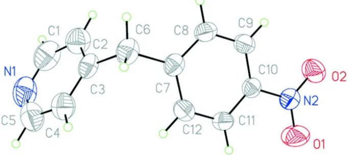
The title compound (Fig. 1) gives colorless crystals. The angle between the planes of benzene and pyridine rings is
78.43° and the nitro group is coplanar with the benzene ring. The two aromatic planes are twisted relative to each other,
which result in reduction of molecular symmetry from Cs to C1: the dihedral angle C2—C3···C7—C8 is 30.5 (2)°. Two
molecular units of the title compound inter-associate through duplex C9—H···O2 hydrogen bonds to form a cyclic dimer
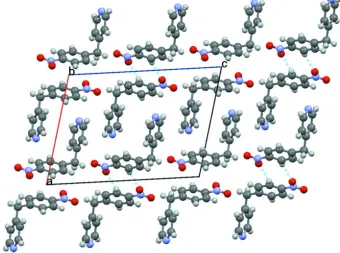
(Fig. 2 and Table 1). Then, these dimers are stacked viaπ···π interactions between benzene rings to form ribbon structure
extending parallel to b-axis (Fig. 3); the angle between the two planes, centroid-centroid distance and shift distance are
0°, 3.788 Å and 1.613 Å, respectively, as determined by Olex2 program package (Dolomanov et al., 2009). Subsequently,
these ribbons are interdigitated to form the final three-dimensional structure (Fig. 4).
The nitro group of the title compound, was found to be a major factor in determining the interactions in the crystal
form, unlike in the previously published structures where the pyridinic nitrogen was the main driving force for
amolecular association.
S2. Experimental
Crystals of the title compound were obtained by dissolving 1 mmol of 4-(4-nitrobenzyl) pyridine in 30 ml of hot 96%
ethanol. Partial evaporation of the hot-filtered solution at room temperature yielded colourless crystals from which a
block section was cleaved for the X-ray analysis.
S3. Refinement
The structure was solved by direct methods and refined by least squares method on F2 using the SHELXTL program
package. All atoms were refined anisotropically. Hydrogen atoms were placed at the calculated positions using a riding
model with C(aromatic)— H = 0.95 Å and Uiso(H) = 1.2Ueq(C), and with C(aliphatic)—H = 0.98 Å and Uiso(H) =
Figure 1
[image:3.610.63.477.275.535.2]Molecular unit of the title compound. Thermal ellipsoids are shown at 50% probability.
Figure 2
Structure of the cyclic dimer.
Figure 3
Figure 4
Illustration of the three dimensional structure of the title compound viewed along the b-axis.
4-(4-Nitrobenzyl)pyridine
Crystal data
C12H10N2O2
Mr = 214.22 Monoclinic, P21/c
Hall symbol: -P 2ybc a = 11.4138 (9) Å b = 6.1241 (5) Å c = 15.5812 (13) Å β = 104.561 (9)° V = 1054.13 (15) Å3
Z = 4
F(000) = 448 Dx = 1.350 Mg m−3
Mo Kα radiation, λ = 0.71073 Å Cell parameters from 949 reflections θ = 3.3–29.0°
µ = 0.09 mm−1
T = 293 K Needle, white 0.4 × 0.2 × 0.15 mm
Data collection
Agilent Xcalibur Eos diffractometer
Radiation source: Enhance (Mo) X-ray Source Graphite monochromator
Detector resolution: 16.0534 pixels mm-1
ω scans
Absorption correction: multi-scan (CrysAlis PRO; Agilent, 2011) Tmin = 0.770, Tmax = 1.000
4351 measured reflections 2136 independent reflections 1514 reflections with I > 2σ(I) Rint = 0.018
θmax = 26.3°, θmin = 3.6°
Refinement on F2
Least-squares matrix: full R[F2 > 2σ(F2)] = 0.047
wR(F2) = 0.106
S = 1.03 2136 reflections 145 parameters 0 restraints
Primary atom site location: structure-invariant direct methods
Secondary atom site location: difference Fourier map
Hydrogen site location: inferred from neighbouring sites
H-atom parameters constrained w = 1/[σ2(F
o2) + (0.0401P)2 + 0.1216P]
where P = (Fo2 + 2Fc2)/3
(Δ/σ)max < 0.001
Δρmax = 0.12 e Å−3
Δρmin = −0.15 e Å−3
Special details
Experimental. Absorption correction CrysAlis PRO (Agilent, 2011). Empirical absorption correction using spherical
harmonics, implemented in SCALE3 ABSPACK scaling algorithm.
Geometry. All e.s.d.'s (except the e.s.d. in the dihedral angle between two l.s. planes) are estimated using the full
covariance matrix. The cell e.s.d.'s are taken into account individually in the estimation of e.s.d.'s in distances, angles and torsion angles; correlations between e.s.d.'s in cell parameters are only used when they are defined by crystal symmetry. An approximate (isotropic) treatment of cell e.s.d.'s is used for estimating e.s.d.'s involving l.s. planes.
Refinement. Refinement of F2 against ALL reflections. The weighted R-factor wR and goodness of fit S are based on F2,
conventional R-factors R are based on F, with F set to zero for negative F2. The threshold expression of F2 > σ(F2) is used
only for calculating R-factors(gt) etc. and is not relevant to the choice of reflections for refinement. R-factors based on F2
are statistically about twice as large as those based on F, and R- factors based on ALL data will be even larger.
Fractional atomic coordinates and isotropic or equivalent isotropic displacement parameters (Å2)
x y z Uiso*/Ueq
H5A 0.5263 −0.6770 −0.0881 0.080* C1 0.51831 (19) −0.2128 (4) −0.16440 (15) 0.0731 (6) H1B 0.5676 −0.1088 −0.1812 0.088*
Atomic displacement parameters (Å2)
U11 U22 U33 U12 U13 U23
N2 0.0485 (9) 0.0470 (9) 0.0436 (9) 0.0015 (7) 0.0142 (7) −0.0001 (7) C10 0.0327 (8) 0.0392 (9) 0.0376 (9) −0.0024 (7) 0.0108 (7) 0.0001 (7) C9 0.0387 (9) 0.0401 (9) 0.0448 (10) 0.0031 (7) 0.0098 (8) 0.0074 (8) C7 0.0308 (8) 0.0477 (10) 0.0398 (9) −0.0043 (7) 0.0104 (7) −0.0016 (8) O2 0.0808 (10) 0.0477 (8) 0.0618 (9) 0.0179 (7) 0.0189 (7) −0.0018 (7) C11 0.0430 (9) 0.0428 (9) 0.0363 (9) 0.0032 (8) 0.0086 (8) 0.0070 (8) C8 0.0424 (9) 0.0498 (10) 0.0350 (9) −0.0003 (8) 0.0082 (8) 0.0064 (8) C12 0.0396 (9) 0.0385 (9) 0.0494 (10) 0.0037 (7) 0.0109 (8) 0.0026 (8) C6 0.0413 (10) 0.0621 (11) 0.0455 (10) −0.0024 (8) 0.0113 (8) −0.0088 (9) O1 0.1380 (14) 0.0749 (10) 0.0378 (8) 0.0326 (10) 0.0224 (8) 0.0078 (7) C3 0.0436 (9) 0.0551 (11) 0.0361 (9) −0.0042 (9) 0.0126 (8) −0.0120 (8) C4 0.0519 (11) 0.0561 (11) 0.0601 (12) −0.0023 (9) 0.0172 (10) −0.0061 (10) N1 0.0481 (10) 0.0950 (14) 0.0719 (12) 0.0042 (10) 0.0176 (9) −0.0145 (11) C2 0.0540 (12) 0.0668 (13) 0.0653 (13) −0.0035 (10) 0.0211 (10) 0.0019 (11) C5 0.0585 (13) 0.0708 (14) 0.0692 (14) 0.0117 (12) 0.0114 (11) −0.0119 (11) C1 0.0535 (13) 0.0956 (17) 0.0759 (15) −0.0169 (13) 0.0271 (12) −0.0053 (14)
Geometric parameters (Å, º)
N2—O1 1.2101 (17) C6—C3 1.506 (2) N2—O2 1.2194 (17) C6—H6A 0.9700 N2—C10 1.464 (2) C6—H6B 0.9700 C10—C9 1.374 (2) C3—C4 1.377 (2) C10—C11 1.377 (2) C3—C2 1.378 (2) C9—C8 1.379 (2) C4—C5 1.370 (3) C9—H9A 0.9300 C4—H4A 0.9300 C7—C8 1.383 (2) N1—C5 1.320 (3) C7—C12 1.391 (2) N1—C1 1.325 (3) C7—C6 1.513 (2) C2—C1 1.382 (3) C11—C12 1.372 (2) C2—H2A 0.9300 C11—H11A 0.9300 C5—H5A 0.9300 C8—H8A 0.9300 C1—H1B 0.9300 C12—H12A 0.9300
C8—C9—H9A 120.7 C5—C4—C3 119.94 (18) C8—C7—C12 118.32 (15) C5—C4—H4A 120.0 C8—C7—C6 121.02 (15) C3—C4—H4A 120.0 C12—C7—C6 120.66 (15) C5—N1—C1 115.11 (18) C12—C11—C10 118.48 (15) C3—C2—C1 119.2 (2) C12—C11—H11A 120.8 C3—C2—H2A 120.4 C10—C11—H11A 120.8 C1—C2—H2A 120.4 C9—C8—C7 121.25 (15) N1—C5—C4 124.7 (2) C9—C8—H8A 119.4 N1—C5—H5A 117.6 C7—C8—H8A 119.4 C4—C5—H5A 117.6 C11—C12—C7 121.44 (15) N1—C1—C2 124.7 (2) C11—C12—H12A 119.3 N1—C1—H1B 117.7 C7—C12—H12A 119.3 C2—C1—H1B 117.7 C3—C6—C7 111.59 (13)
O1—N2—C10—C9 178.75 (15) C6—C7—C12—C11 179.59 (14) O2—N2—C10—C9 −0.5 (2) C8—C7—C6—C3 119.47 (17) O1—N2—C10—C11 −0.3 (2) C12—C7—C6—C3 −60.7 (2) O2—N2—C10—C11 −179.55 (15) C7—C6—C3—C4 98.12 (19) C11—C10—C9—C8 −1.2 (2) C7—C6—C3—C2 −80.4 (2) N2—C10—C9—C8 179.84 (13) C2—C3—C4—C5 1.5 (3) C9—C10—C11—C12 1.2 (2) C6—C3—C4—C5 −177.18 (16) N2—C10—C11—C12 −179.81 (13) C4—C3—C2—C1 −1.4 (3) C10—C9—C8—C7 0.2 (2) C6—C3—C2—C1 177.24 (17) C12—C7—C8—C9 0.6 (2) C1—N1—C5—C4 −1.2 (3) C6—C7—C8—C9 −179.57 (14) C3—C4—C5—N1 −0.1 (3) C10—C11—C12—C7 −0.3 (2) C5—N1—C1—C2 1.3 (3) C8—C7—C12—C11 −0.6 (2) C3—C2—C1—N1 0.0 (3)
Hydrogen-bond geometry (Å, º)
D—H···A D—H H···A D···A D—H···A
C9—H9A···O2i 0.93 2.49 3.302 (2) 146