research communications
Acta Cryst.(2018). E74, 1093–1096 https://doi.org/10.1107/S205698901800912X
1093
Received 29 May 2018Accepted 22 June 2018
Edited by M. Zeller, Purdue University, USA
Keywords:crystal structure; 5-substituted dime-thyl isophthalates; I O C interaction; C— H O and C—H I hydrogen bonding;– stacking.
CCDC references:780476; 780475
Supporting information:this article has supporting information at journals.iucr.org/e
Crystal structures of dimethyl 5-iodoisophthalate
and dimethyl 5-ethynylisophthalate
Ines Hauptvogel, Wilhelm Seichter and Edwin Weber*
TU Bergakademie Freiberg, Leipziger Str. 29, D-09596 Freiberg/Sachsen, Germany. *Correspondence e-mail: [email protected]
In dimethyl 5-iodoisophthalate, C10H9IO4, (I), the planes through the methyl
carboxylate moieties are tilted with respect to the benzene ring, whereas the molecular framework of dimethyl 5-ethynylisophthalate, C12H10O4, (II), is
perfectly planar. The crystal structure of (I) is stabilized by a three-dimensional supramolecular network comprising C—H O C hydrogen bonds, as well as I O C interactions. In the crystal of (II), the molecules are connectedviaC— Hethynyl O C hydrogen bonds to infinite strands. Moreover, – arene
stacking interactions connect the molecular chains into two-dimensional supramolecular aggregates.
1. Chemical context
In recent years, the design of solid porous framework mate-rials (MacGillivray, 2010; Furukawaet al., 2013; Eddaoudiet al., 2015) has become a very important topic in the field of supramolecular crystal engineering (Desiraju et al., 2011). Associated with it, so-called linker molecules featuring a geometrically rigid structure frequently being of linear, trigonal or tetrahedral shape and having carboxylic acid functions as terminal groups play a key role in building such systems (Linet al., 2006; Hausdorf et al., 2009; Zheng et al., 2010). In the course of the synthesis of the respective linkers, the title compounds (I) and (II), both being 5-substituted
dimethyl isophthalates, are much used intermediates.
However, these compounds are not only synthetically signifi-cant but also show interesting structures in the crystalline state, as demonstrated herein.
2. Structural commentary
The molecular structures of the title compounds, (I) and (II), are illustrated in Fig. 1a and 1b, respectively. Taking into account experimental error, the bond distances within the isophthalate framework agree well with those found in the crystal structure of dimethyl isophthalate (Gallagher, 2012). Compound (I) crystallizes in the orthorhombic space group
Pna21with one molecule in the asymmetric unit. The molecule
by the methyl carboxylate moieties inclined at angles of 12.6 (2) and 6.0 (2)with respect to the plane of the benzene ring. Compound (II) crystallizes in the orthorhombic space groupPnmawith the molecule located on a symmetry plane,
i.e. the molecule is perfectly planar. However, the molecule adopts approximateC2vsymmetry with the atoms C2, C5, C11 and C12 lying on a non-crystallographic bisecting symmetry plane.
3. Supramolecular features
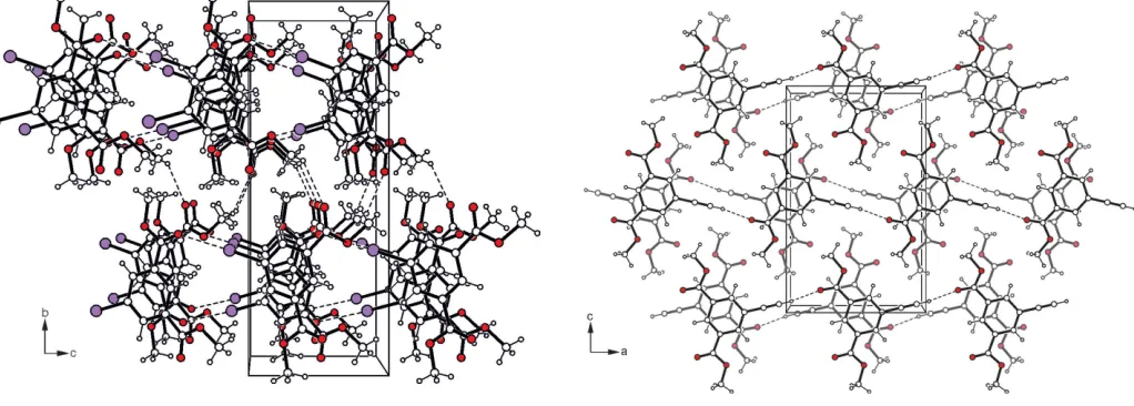
Infinite strands with the molecules connected via I O C interactions [I1 O3—C9(x1
2,y+ 3
2,z1;D A= 3.129 (2)
(Desiraju & Steiner, 1999) (Politzeret al.2007; Desirajuet al., 2013), represent the basic supramolecular aggregates of the crystal structure of (I). Association of the molecular strands by C—H O C type hydrogen bonds (Table 1) (Desiraju & Steiner, 1999) and – stacking interactions [centroid– centroid distance = 4.149 (2) A˚ ] (Tiekink &
Zukerman-Schpector, 2012) generate a three-dimensional
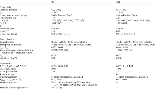
supra-molecular network (Fig. 2). In the crystal structure of (II), the molecules are connected via Cethynyl—H O C bonds
(Table 2) into infinite strands, which are further arranged into molecular sheets that extend parallel to theacplane (Fig. 3).
Furthermore,–arene interactions with a centroid–centroid distance of 3.566 (1) A˚ and a slippage of 1.325 A˚ between the interacting aromatic rings stabilize the crystal structure along the stacking axis of the molecular sheets.
4. Database survey
The search in the Cambridge Structural Database (CSD, Version 5.38, update May 2017; Groomet al., 2016) formeta -substituted derivatives of dimethyl isophthalate excluding their metal complexes, solvates and salts gave 18 hits. None of these compounds represents a 5-halogen- and 5-alkynyl-substituted dimethyl isophalate. The parent compound, dimethyl isophthalate (CSD refcode GOHRUS; Gallagher & Mocilac, 2012) crystallizes in space group Pna21 with two
conformationally similar molecules in the asymmetric unit. The independent molecules participate in different ways in non-covalent bonding. One of them is involved in the formation of linear strands with the molecules connected by C—Haryl O C bonds. Interstrand association is
accom-plished by – arene stacking. Molecules related by the twofold screw axis are also linked via C—Haryl O C
1094
Hauptvogelet al. C [image:2.610.43.298.72.154.2]10H9IO4and C12H10O4 Acta Cryst.(2018). E74, 1093–1096
research communications
Table 1
Hydrogen-bond geometry (A˚ ,) for (I).
D—H A D—H H A D A D—H A
C8—H8A O1i 0.98 2.55 3.257 (4) 129
[image:2.610.312.566.178.211.2]Symmetry code: (i)xþ1;yþ1;z1 2.
Figure 2
Packing diagram of compound (I) viewed down theaaxis. Dashed lines represent hydrogen-bonding interactions.
Table 2
Hydrogen-bond geometry (A˚ ,) for (II).
D—H A D—H H A D A D—H A
C12—H12 O1i 0.94 2.29 3.223 (1) 172
[image:2.610.47.312.526.711.2] [image:2.610.52.563.535.718.2]Symmetry code: (i)x1;y;z.
Figure 3
Packing excerpt of compound (II) viewed down thebaxis. Dashed lines represent hydrogen-bonding interactions.
Figure 1
[image:2.610.305.563.539.716.2]bonding to form helical strands. In addition, these strands are stabilized by–stacking forces.
5. Synthesis and crystallization
Compounds (I) and (II) were synthesized following literature procedures. This involves a diazotization/iodination reaction of dimethyl 5-aminoisophthalate (Mazik & Ko¨nig, 2006) to give compound (I). Subsequent reaction of (I) with 2-methylbut-3-yne-2-ol (MEBYNOL) using a Pd-catalysed Sonogashira coupling procedure (Doucet & Hierso, 2007; Rafael & Carmen, 2007) yielded the corresponding blocked acetylenic diester as an intermediate (Hauptvogelet al., 2011). Removal of the 2-hydroxypropyl blocking group was under-taken using sodium hydride in toluene and quenching with water to result in the title compound (II) (Havens & Hergenrother, 1985; Hauptvogelet al., 2011).
6. Refinement
Crystal data, data collection and structure refinement details are summarized in Table 3. Hydrogen atoms were positioned geometrically and refined using a riding model with C—H distances of 0.94–0.98 A˚ and Uiso(H) = 1.5Ueq(C-methyl) or
Uiso(H) = 1.2Ueq(C) for other H atoms.
Funding information
We acknowledge the financial support by the Deutsche Forschungsgemeinschaft (DFG Priority Program 1362 ‘Porous Metal-Organic Frameworks’).
References
Bruker (2014). APEX2 and SAINT. Bruker AXS Inc., Madison, Wisconsin, USA.
Desiraju, G. R., Ho, P. S., Kloo, L., Legon, A. C., Marquardt, R., Metrangolo, P., Politzer, P., Resnati, G. & Rissanen, K. (2013).Pure Appl. Chem.85, 1711–1713.
Desiraju, G. R. & Steiner, T. (1999). InThe Weak Hydrogen Bond. Oxford University Press.
Desiraju, G. R., Vittal, J. J. & Ramanan, A. (2011). Crystal Engineering. Singapore: World Scientific Publications.
Doucet, H. & Hierso, J. C. (2007).Angew. Chem. Int. Ed.46, 834– 871.
Eddaoudi, M., Sava, D. F., Eubank, J. F., Adil, K. & Guillerm, V. (2015).Chem. Soc. Rev.44, 228–249.
Farrugia, L. J. (2012).J. Appl. Cryst.45, 849–854.
Furukawa, H., Cordova, K. E., O’Keeffe, M. & Yaghi, O. M. (2013).
Science,341, 1230444.
Gallagher, C. F. & Mocilac, P. (2012).CSD Communication (Refcode GOHRUS). CCDC, Cambridge, England.
Groom, C. R., Bruno, I. J., Lightfoot, M. P. & Ward, S. C. (2016).Acta Cryst.B72, 171–179.
research communications
Acta Cryst.(2018). E74, 1093–1096 Hauptvogelet al. C
[image:3.610.48.562.94.391.2]10H9IO4and C12H10O4
1095
Table 3
Experimental details.
(I) (II)
Crystal data
Chemical formula C10H9IO4 C12H10O4
Mr 320.07 218.20
Crystal system, space group Orthorhombic,Pna21 Orthorhombic,Pnma
Temperature (K) 143 223
a,b,c(A˚ ) 7.7483 (2), 19.3451 (6), 7.2338 (2) 10.1206 (5), 6.6219 (4), 16.3658 (8)
V(A˚3) 1084.29 (5) 1096.80 (10)
Z 4 4
Radiation type MoK MoK
(mm1) 2.94 0.10
Crystal size (mm) 0.300.220.15 0.540.120.10
Data collection
Diffractometer Bruker APEXII CCD area detector Bruker APEXII CCD area detector
Absorption correction Multi-scan (SADABS; Sheldrick, 2008a) Multi-scan (SADABS; Sheldrick, 2008a)
Tmin,Tmax 0.472, 0.666 0.948, 0.990
No. of measured, independent and observed [I> 2(I)] reflections
22794, 2909, 2806 12397, 1292, 932
Rint 0.026 0.033
(sin/)max(A˚
1) 0.684 0.638
Refinement
R[F2> 2(F2)],wR(F2),S 0.015, 0.038, 1.05 0.039, 0.110, 1.03
No. of reflections 2909 1292
No. of parameters 139 87
No. of restraints 1 0
H-atom treatment H-atom parameters constrained H-atom parameters constrained
max,min(e A˚
3) 0.47,0.44 0.17,0.18
Absolute structure Flackxdetermined using 1255 quotients
[(I+)(I
)]/[(I+)+(I
)] (Parsonset al., 2013)
–
Absolute structure parameter 0.004 (8) –
Computer programs:APEX2andSAINT(Bruker, 2014),SHELXT(Sheldrick, 2015a),SHELXL2018(Sheldrick, 2015b),ORTEP-3 for Windows(Farrugia, 2012) andSHELXTL
Hauptvogel, I. M., Seichter, W. & Weber, E. (2011).Supramol. Chem.
23, 398–406.
Hausdorf, S., Seichter, W., Weber, E. & Mertens, F. O. R. L. (2009).
Dalton Trans.pp. 1107–1113.
Havens, S. J. & Hergenrother, P. M. (1985).J. Org. Chem.50, 1863– 1865.
Lin, X., Jia, J., Zhao, X., Thomas, K. M., Blake, A. J., Walker, G. S., Champness, N. R., Hubberstey, P. & Schro¨der, M. (2006).Angew. Chem. Int. Ed.45, 7358–7364.
MacGillivray, L. R. (2010). Metal-Organic Frameworks. Hoboken: Wiley.
Mazik, M. & Ko¨nig, A. (2006).J. Org. Chem.71, 7854–7857. Parsons, S., Flack, H. D. & Wagner, T. (2013).Acta Cryst.B69, 249–
259.
Politzer, P., Lane, P., Concha, M. C., Ma, Y. & Murray, J. S. (2007).J. Mol. Model.13, 305–311.
Rafael, C. & Carmen, N. (2007).Chem. Rev.B107, 874–922. Sheldrick, G. M. (2008a). SADABS. University of Go¨ttingen,
Germany.
Sheldrick, G. M. (2008b).Acta Cryst.A64, 112–122. Sheldrick, G. M. (2015a).Acta Cryst.A71, 3–8. Sheldrick, G. M. (2015b).Acta Cryst.C71, 3–8.
Tiekink, E. R. T. & Zukerman-Schpector, J. (2012). In The Importance of Pi-Interactions in Crystal Engineering. Frontiers in Crystal Engineering. Chichester: Wiley.
Zheng, B., Liang, Z., Li, G., Huo, Q. & Liu, Y. (2010).Cryst. Growth Des.10, 3405–3409.
1096
Hauptvogelet al. Csupporting information
sup-1 Acta Cryst. (2018). E74, 1093-1096
supporting information
Acta Cryst. (2018). E74, 1093-1096 [https://doi.org/10.1107/S205698901800912X]
Crystal structures of dimethyl iodoisophthalate and dimethyl
5-ethynylisophthalate
Ines Hauptvogel, Wilhelm Seichter and Edwin Weber
Computing details
For both structures, data collection: APEX2 (Bruker, 2014); cell refinement: SAINT (Bruker, 2014); data reduction:
SAINT (Bruker, 2014); program(s) used to solve structure: SHELXT (Sheldrick, 2015a). Program(s) used to refine
structure: SHELXL2018 (Sheldrick, 2015b) for (I); SHELXL2014 (Sheldrick, 2015b) for (II). For both structures,
molecular graphics: ORTEP-3 for Windows (Farrugia, 2012); software used to prepare material for publication:
SHELXTL (Sheldrick, 2008b).
1,3-Dimethyl 1-iodocyclohexa-3,5-diene-1,3-dicarboxylate (I)
Crystal data
C10H9IO4
Mr = 320.07
Orthorhombic, Pna21
a = 7.7483 (2) Å
b = 19.3451 (6) Å
c = 7.2338 (2) Å
V = 1084.29 (5) Å3
Z = 4
F(000) = 616
Dx = 1.961 Mg m−3
Mo Kα radiation, λ = 0.71073 Å Cell parameters from 5755 reflections
θ = 3.0–33.7°
µ = 2.94 mm−1
T = 143 K
Irregular, colourless 0.30 × 0.22 × 0.15 mm
Data collection
Bruker APEXII CCD area detector diffractometer
φ and ω scans
Absorption correction: multi-scan (SADABS; Sheldrick, 2008a)
Tmin = 0.472, Tmax = 0.666
22794 measured reflections
2909 independent reflections 2806 reflections with I > 2σ(I)
Rint = 0.026
θmax = 29.1°, θmin = 1.1°
h = −10→10
k = −26→26
l = −9→9
Refinement
Refinement on F2
Least-squares matrix: full
R[F2 > 2σ(F2)] = 0.015
wR(F2) = 0.038
S = 1.05 2909 reflections 139 parameters 1 restraint
Hydrogen site location: inferred from neighbouring sites
H-atom parameters constrained
w = 1/[σ2(F
o2) + (0.019P)2 + 0.3689P]
where P = (Fo2 + 2Fc2)/3
(Δ/σ)max = 0.002
Δρmax = 0.47 e Å−3
Δρmin = −0.44 e Å−3
Absolute structure: Flack x determined using 1255 quotients [(I+)-(I-)]/[(I+)+(I-)] (Parsons et
al., 2013)
supporting information
sup-2 Acta Cryst. (2018). E74, 1093-1096
Special details
Geometry. All esds (except the esd in the dihedral angle between two l.s. planes) are estimated using the full covariance matrix. The cell esds are taken into account individually in the estimation of esds in distances, angles and torsion angles; correlations between esds in cell parameters are only used when they are defined by crystal symmetry. An approximate (isotropic) treatment of cell esds is used for estimating esds involving l.s. planes.
Refinement. Refined as a 2-component twin.
Fractional atomic coordinates and isotropic or equivalent isotropic displacement parameters (Å2)
x y z Uiso*/Ueq
I1 0.81504 (2) 0.66115 (2) 0.83115 (6) 0.02386 (5)
O1 0.5202 (3) 0.54751 (11) 0.1864 (3) 0.0327 (5)
O2 0.3831 (3) 0.63417 (11) 0.0411 (3) 0.0260 (4)
O3 0.4971 (3) 0.87789 (10) 0.2029 (3) 0.0281 (5)
O4 0.6557 (3) 0.89701 (11) 0.4576 (3) 0.0282 (5)
C1 0.5514 (4) 0.66267 (12) 0.2993 (4) 0.0190 (8)
C2 0.5347 (3) 0.73272 (14) 0.2585 (4) 0.0198 (5)
H2 0.479369 0.746965 0.147720 0.024*
C3 0.5994 (3) 0.78152 (14) 0.3805 (3) 0.0194 (5)
C4 0.6796 (3) 0.76086 (15) 0.5445 (4) 0.0206 (5)
H4 0.723931 0.794439 0.627770 0.025*
C5 0.6940 (3) 0.69088 (15) 0.5850 (4) 0.0207 (5)
C6 0.6295 (4) 0.64128 (15) 0.4637 (4) 0.0209 (5)
H6 0.638485 0.593502 0.492478 0.025*
C7 0.4856 (4) 0.60819 (14) 0.1721 (4) 0.0223 (5)
C8 0.3221 (5) 0.58545 (18) −0.0951 (5) 0.0330 (7)
H8A 0.418544 0.571014 −0.173823 0.049*
H8B 0.232932 0.607288 −0.171341 0.049*
H8C 0.273509 0.544937 −0.032593 0.049*
C9 0.5768 (3) 0.85636 (11) 0.3323 (8) 0.0214 (4)
C10 0.6363 (5) 0.97101 (16) 0.4293 (5) 0.0338 (7)
H10A 0.688803 0.984036 0.311034 0.051*
H10B 0.693716 0.995947 0.529896 0.051*
H10C 0.513396 0.982919 0.427800 0.051*
Atomic displacement parameters (Å2)
U11 U22 U33 U12 U13 U23
I1 0.02804 (8) 0.02336 (8) 0.02019 (8) −0.00006 (6) −0.00121 (12) 0.00381 (11)
O1 0.0500 (14) 0.0174 (10) 0.0308 (12) 0.0032 (9) −0.0060 (10) −0.0006 (9)
O2 0.0300 (11) 0.0187 (10) 0.0293 (11) 0.0017 (8) −0.0066 (9) −0.0048 (8)
O3 0.0347 (12) 0.0194 (10) 0.0301 (11) 0.0027 (9) −0.0082 (9) 0.0019 (8)
O4 0.0382 (12) 0.0165 (9) 0.0299 (11) −0.0011 (8) −0.0073 (9) −0.0004 (9)
C1 0.0216 (11) 0.0183 (10) 0.017 (2) 0.0009 (9) 0.0020 (10) −0.0004 (9)
C2 0.0196 (12) 0.0190 (12) 0.0209 (11) 0.0022 (10) 0.0042 (10) 0.0019 (10)
C3 0.0191 (11) 0.0183 (11) 0.0208 (13) 0.0013 (9) 0.0025 (9) 0.0012 (8)
C4 0.0217 (13) 0.0191 (13) 0.0211 (12) −0.0014 (10) 0.0017 (10) −0.0012 (10)
supporting information
sup-3 Acta Cryst. (2018). E74, 1093-1096
C6 0.0242 (13) 0.0183 (12) 0.0201 (12) 0.0016 (10) 0.0024 (11) 0.0013 (10)
C7 0.0256 (13) 0.0197 (12) 0.0217 (13) −0.0011 (10) 0.0035 (11) −0.0011 (10)
C8 0.0409 (19) 0.0257 (15) 0.0323 (15) 0.0005 (13) −0.0088 (13) −0.0075 (13)
C9 0.0218 (10) 0.0173 (9) 0.0250 (10) −0.0001 (8) 0.0096 (18) −0.002 (2)
C10 0.0453 (19) 0.0174 (14) 0.0387 (19) 0.0014 (13) −0.0064 (15) −0.0021 (12)
Geometric parameters (Å, º)
I1—C5 2.093 (3) C3—C4 1.398 (4)
O1—C7 1.209 (3) C3—C9 1.499 (4)
O2—C7 1.335 (4) C4—C5 1.390 (4)
O2—C8 1.443 (4) C4—H4 0.9500
O3—C9 1.196 (5) C5—C6 1.393 (4)
O4—C9 1.347 (5) C6—H6 0.9500
O4—C10 1.454 (4) C8—H8A 0.9800
C1—C2 1.393 (3) C8—H8B 0.9800
C1—C6 1.398 (4) C8—H8C 0.9800
C1—C7 1.489 (4) C10—H10A 0.9800
C2—C3 1.386 (4) C10—H10B 0.9800
C2—H2 0.9500 C10—H10C 0.9800
C7—O2—C8 115.7 (2) C1—C6—H6 120.4
C9—O4—C10 115.7 (3) O1—C7—O2 124.0 (3)
C2—C1—C6 120.5 (3) O1—C7—C1 124.0 (3)
C2—C1—C7 121.8 (3) O2—C7—C1 112.1 (2)
C6—C1—C7 117.7 (2) O2—C8—H8A 109.5
C3—C2—C1 119.7 (3) O2—C8—H8B 109.5
C3—C2—H2 120.2 H8A—C8—H8B 109.5
C1—C2—H2 120.2 O2—C8—H8C 109.5
C2—C3—C4 120.4 (3) H8A—C8—H8C 109.5
C2—C3—C9 117.9 (3) H8B—C8—H8C 109.5
C4—C3—C9 121.7 (3) O3—C9—O4 123.9 (2)
C5—C4—C3 119.5 (3) O3—C9—C3 125.3 (3)
C5—C4—H4 120.2 O4—C9—C3 110.7 (3)
C3—C4—H4 120.2 O4—C10—H10A 109.5
C4—C5—C6 120.6 (3) O4—C10—H10B 109.5
C4—C5—I1 118.9 (2) H10A—C10—H10B 109.5
C6—C5—I1 120.5 (2) O4—C10—H10C 109.5
C5—C6—C1 119.2 (3) H10A—C10—H10C 109.5
C5—C6—H6 120.4 H10B—C10—H10C 109.5
C6—C1—C2—C3 1.3 (4) C8—O2—C7—O1 −4.1 (4)
C7—C1—C2—C3 −179.4 (2) C8—O2—C7—C1 176.3 (3)
C1—C2—C3—C4 −0.6 (4) C2—C1—C7—O1 168.0 (3)
C1—C2—C3—C9 −179.3 (3) C6—C1—C7—O1 −12.7 (4)
C2—C3—C4—C5 −0.1 (4) C2—C1—C7—O2 −12.5 (4)
C9—C3—C4—C5 178.5 (3) C6—C1—C7—O2 166.8 (3)
supporting information
sup-4 Acta Cryst. (2018). E74, 1093-1096
C3—C4—C5—I1 179.86 (19) C10—O4—C9—C3 −177.5 (3)
C4—C5—C6—C1 0.7 (4) C2—C3—C9—O3 5.3 (5)
I1—C5—C6—C1 −179.1 (2) C4—C3—C9—O3 −173.3 (3)
C2—C1—C6—C5 −1.3 (4) C2—C3—C9—O4 −176.1 (3)
C7—C1—C6—C5 179.4 (2) C4—C3—C9—O4 5.3 (4)
Hydrogen-bond geometry (Å, º)
D—H···A D—H H···A D···A D—H···A
C8—H8A···O1i 0.98 2.55 3.257 (4) 129
Symmetry code: (i) −x+1, −y+1, z−1/2.
1,3-Dimethyl 1-ethynylcyclohexa-3,5-diene-1,3-dicarboxylate (II)
Crystal data
C12H10O4
Mr = 218.20
Orthorhombic, Pnma a = 10.1206 (5) Å
b = 6.6219 (4) Å
c = 16.3658 (8) Å
V = 1096.80 (10) Å3
Z = 4
F(000) = 456
Dx = 1.321 Mg m−3
Mo Kα radiation, λ = 0.71073 Å Cell parameters from 2950 reflections
θ = 2.4–23.1°
µ = 0.10 mm−1
T = 223 K
Column, colourless 0.54 × 0.12 × 0.10 mm
Data collection
Bruker APEXII CCD area detector diffractometer
φ and ω scans
Absorption correction: multi-scan (SADABS; Sheldrick, 2008a)
Tmin = 0.948, Tmax = 0.990
12397 measured reflections
1292 independent reflections 932 reflections with I > 2σ(I)
Rint = 0.033
θmax = 27.0°, θmin = 2.5°
h = −12→12
k = −8→5
l = −20→19
Refinement
Refinement on F2
Least-squares matrix: full
R[F2 > 2σ(F2)] = 0.039
wR(F2) = 0.110
S = 1.03 1292 reflections 87 parameters 0 restraints
Hydrogen site location: inferred from neighbouring sites
H-atom parameters constrained
w = 1/[σ2(F
o2) + (0.0486P)2 + 0.2932P]
where P = (Fo2 + 2Fc2)/3
(Δ/σ)max < 0.001
Δρmax = 0.17 e Å−3
Δρmin = −0.18 e Å−3
Special details
supporting information
sup-5 Acta Cryst. (2018). E74, 1093-1096
Fractional atomic coordinates and isotropic or equivalent isotropic displacement parameters (Å2)
x y z Uiso*/Ueq Occ. (<1)
O1 1.25864 (13) 0.2500 0.40733 (10) 0.0552 (4)
O2 1.08848 (14) 0.2500 0.32081 (9) 0.0519 (4)
O3 1.15356 (16) 0.2500 0.70802 (10) 0.0759 (6)
O4 0.94247 (15) 0.2500 0.74402 (9) 0.0617 (5)
C1 0.98919 (18) 0.2500 0.60451 (11) 0.0339 (4)
C2 1.08100 (18) 0.2500 0.54143 (12) 0.0350 (4)
H2 1.1718 0.2500 0.5536 0.042*
C3 1.03958 (18) 0.2500 0.46063 (12) 0.0339 (4)
C4 0.90505 (19) 0.2500 0.44325 (12) 0.0361 (4)
H4 0.8767 0.2500 0.3886 0.043*
C5 0.81192 (17) 0.2500 0.50582 (12) 0.0348 (4)
C6 0.85496 (18) 0.2500 0.58682 (12) 0.0340 (4)
H6 0.7929 0.2500 0.6296 0.041*
C7 1.0392 (2) 0.2500 0.68995 (13) 0.0428 (5)
C8 0.9818 (3) 0.2500 0.82935 (14) 0.0791 (9)
H8A 1.0203 0.1202 0.8431 0.119* 0.5
H8B 0.9050 0.2740 0.8634 0.119* 0.5
H8C 1.0465 0.3558 0.8385 0.119* 0.5
C9 1.1418 0.2500 0.3949 0.039
C10 1.1813 0.2500 0.2530 0.067
H10A 1.1353 0.2149 0.2030 0.101* 0.5
H10B 1.2505 0.1519 0.2633 0.101* 0.5
H10C 1.2202 0.3832 0.2474 0.101* 0.5
C11 0.67260 (19) 0.2500 0.48649 (12) 0.0406 (5)
C12 0.5604 (2) 0.2500 0.47022 (14) 0.0532 (6)
H12 0.4700 0.2500 0.4571 0.064*
Atomic displacement parameters (Å2)
U11 U22 U33 U12 U13 U23
O1 0.0293 (8) 0.0828 (11) 0.0536 (10) 0.000 0.0049 (7) 0.000
O2 0.0409 (9) 0.0783 (11) 0.0364 (8) 0.000 0.0066 (7) 0.000
O3 0.0329 (9) 0.1475 (18) 0.0473 (10) 0.000 −0.0079 (8) 0.000
O4 0.0362 (9) 0.1153 (14) 0.0336 (8) 0.000 −0.0026 (7) 0.000
C1 0.0294 (10) 0.0371 (10) 0.0352 (11) 0.000 −0.0006 (8) 0.000
C2 0.0257 (9) 0.0385 (10) 0.0408 (11) 0.000 −0.0029 (8) 0.000
C3 0.0295 (10) 0.0338 (9) 0.0385 (11) 0.000 0.0023 (8) 0.000
C4 0.0343 (11) 0.0404 (10) 0.0334 (10) 0.000 −0.0020 (9) 0.000
C5 0.0275 (9) 0.0379 (10) 0.0389 (11) 0.000 −0.0002 (8) 0.000
C6 0.0276 (9) 0.0398 (10) 0.0347 (11) 0.000 0.0011 (8) 0.000
C7 0.0299 (11) 0.0568 (12) 0.0416 (12) 0.000 −0.0013 (9) 0.000
C8 0.0550 (16) 0.151 (3) 0.0315 (13) 0.000 −0.0049 (12) 0.000
C9 0.034 0.043 0.039 0.000 0.003 0.000
C10 0.063 0.098 0.041 0.000 0.018 0.000
supporting information
sup-6 Acta Cryst. (2018). E74, 1093-1096
C12 0.0340 (12) 0.0849 (17) 0.0409 (13) 0.000 −0.0041 (10) 0.000
Geometric parameters (Å, º)
O1—C9 1.1998 (14) C4—C5 1.392 (3)
O2—C9 1.3272 (15) C4—H4 0.9400
O2—C10 1.4544 (14) C5—C6 1.395 (3)
O3—C7 1.195 (3) C5—C11 1.445 (3)
O4—C7 1.319 (3) C6—H6 0.9400
O4—C8 1.452 (3) C8—H8A 0.9700
C1—C2 1.389 (3) C8—H8B 0.9700
C1—C6 1.389 (3) C8—H8C 0.9700
C1—C7 1.487 (3) C10—H10A 0.9700
C2—C3 1.387 (3) C10—H10B 0.9700
C2—H2 0.9400 C10—H10C 0.9700
C3—C4 1.391 (3) C11—C12 1.166 (3)
C3—C9 1.4925 (18) C12—H12 0.9400
C9—O2—C10 115.75 (10) O3—C7—O4 123.6 (2)
C7—O4—C8 116.19 (18) O3—C7—C1 124.21 (19)
C2—C1—C6 119.96 (18) O4—C7—C1 112.23 (17)
C2—C1—C7 118.13 (17) O4—C8—H8A 109.5
C6—C1—C7 121.91 (17) O4—C8—H8B 109.5
C3—C2—C1 120.42 (17) H8A—C8—H8B 109.5
C3—C2—H2 119.8 O4—C8—H8C 109.5
C1—C2—H2 119.8 H8A—C8—H8C 109.5
C2—C3—C4 119.39 (18) H8B—C8—H8C 109.5
C2—C3—C9 118.53 (15) O1—C9—O2 123.76 (10)
C4—C3—C9 122.09 (16) O1—C9—C3 124.12 (11)
C3—C4—C5 120.83 (18) O2—C9—C3 112.12 (9)
C3—C4—H4 119.6 O2—C10—H10A 109.5
C5—C4—H4 119.6 O2—C10—H10B 109.5
C4—C5—C6 119.18 (17) H10A—C10—H10B 109.5
C4—C5—C11 119.98 (18) O2—C10—H10C 109.5
C6—C5—C11 120.84 (17) H10A—C10—H10C 109.5
C1—C6—C5 120.21 (17) H10B—C10—H10C 109.5
C1—C6—H6 119.9 C12—C11—C5 179.5 (2)
C5—C6—H6 119.9 C11—C12—H12 180.0
C6—C1—C2—C3 0.000 (1) C8—O4—C7—O3 0.000 (1)
C7—C1—C2—C3 180.000 (1) C8—O4—C7—C1 180.000 (1)
C1—C2—C3—C4 0.000 (1) C2—C1—C7—O3 0.000 (1)
C1—C2—C3—C9 180.000 (1) C6—C1—C7—O3 180.000 (1)
C2—C3—C4—C5 0.000 (1) C2—C1—C7—O4 180.000 (1)
C9—C3—C4—C5 180.000 (1) C6—C1—C7—O4 0.000 (1)
C3—C4—C5—C6 0.000 (1) C10—O2—C9—O1 0.000 (1)
C3—C4—C5—C11 180.000 (1) C10—O2—C9—C3 180.000 (1)
supporting information
sup-7 Acta Cryst. (2018). E74, 1093-1096
C7—C1—C6—C5 180.000 (1) C4—C3—C9—O1 180.000 (1)
C4—C5—C6—C1 0.000 (1) C2—C3—C9—O2 180.000 (1)
C11—C5—C6—C1 180.000 (1) C4—C3—C9—O2 0.000 (1)
Hydrogen-bond geometry (Å, º)
D—H···A D—H H···A D···A D—H···A
C12—H12···O1i 0.94 2.29 3.223 (1) 172