The use of biologics for immune modulation in
allergic disease
Willem van de Veen, Mübeccel Akdis
J Clin Invest. 2019;
129(4)
:1452-1462.
https://doi.org/10.1172/JCI124607
.
The rising prevalence of allergies represents an increasing socioeconomic burden. A
detailed understanding of the immunological mechanisms that underlie the development of
allergic disease, as well as the processes that drive immune tolerance to allergens, will be
instrumental in designing therapeutic strategies to treat and prevent allergic disease.
Improved characterization of individual patients through the use of specific biomarkers and
improved definitions of disease endotypes are paving the way for the use of targeted
therapeutic approaches for personalized treatment. Allergen-specific immunotherapy and
biologic therapies that target key molecules driving the Th2 response are already used in
the clinic, and a wave of novel drug candidates are under development. In-depth analysis of
the cells and tissues of patients treated with such targeted interventions provides a wealth of
information on the mechanisms that drive allergies and tolerance to allergens. Here, we aim
to deliver an overview of the current state of specific inhibitors used in the treatment of
allergy, with a particular focus on asthma and atopic dermatitis, and provide insights into the
roles of these molecules in immunological mechanisms of allergic disease.
Review Series
Find the latest version:
Immunological mechanisms of allergy
and tolerance
Allergic sensitization and type I and IV hypersensitivity reactions.
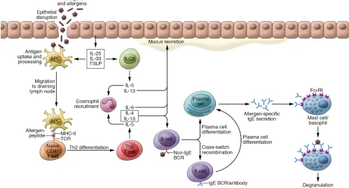
Allergies develop as a result of an immune response to distinct environmental protein antigens, called allergens. In recent years, substantial progress has been made in elucidating mechanisms that drive the initiation and persistence of allergic reactions. Development of allergic diseases starts with a sensitization phase (Figure 1). Key processes driving allergic sensitization include activation and maturation of dendritic cells (DCs) upon contact with allergens, epithelial alarmins, and infectious agents, fol-lowed by clonal expansion of allergen-specific Th2 cells that are polarized toward producing type 2 cytokines, including IL-4, IL-5, IL-9, IL-13, IL-25, IL-31, IL-33, and thymic stromal lymphopoietin (TSLP) (1). Th2 cells induce class switch recombination of B cells to the IgE isotype and differentiation to IgE-producing plasma cells. IgE can bind to the high-affinity IgE receptor FcεRI on basophils and mast cells. Moreover, IgE can also enhance allergen uptake and presentation through so-called facilitated antigen presenta-tion (2, 3). These processes form the basis of allergic sensitizapresenta-tion. Subsequent exposure to the culprit allergen can trigger IgE- mediated FcεRI cross-linking on mast cells and basophils, lead-ing to a type I hypersensitivity reaction. Durlead-ing the early phase of this reaction, which starts within minutes after allergen expo-sure, mast cells and/or basophils degranulate and release a range of preformed and newly synthesized mediators including his-tamine, leukotrienes, heparin, and several proteases, as well as
cytokines and growth factors such as TNF-α and VEGFA (4). After 2 to 6 hours of exposure, the late phase of the reaction manifests, characterized by edema, erythema in skin, and airway narrowing and mucus secretion in the airways. These symptoms are caused by the recruitment and activation of Th2 cells, eosinophils, baso-phils, and tissue- resident mast cells. Persistent allergen exposure can produce a chronic phase of inflammation that represents a distinct Th2 type of type IV hypersensitivity (5). Classical type IV hypersensitivity denotes tuberculin-type hypersensitivity with the involvement of Th1 cells. In allergic inflammation a cellular late-phase response develops, characterized by tissue inflamma-tion with eosinophils, Th2 cells, and type 2 innate lymphoid cells (ILC2s); remodeling of vasculature and smooth muscle cells; and extracellular matrix formation and fibrosis (4, 5). In the clinical setting, this chronic inflammation prevails and forms the basis for the main tissue inflammation mechanisms referred to as Th2 or type 2 inflammation in asthma, atopic dermatitis (AD), chronic rhi-nosinusitis (CRS) with nasal polyps, and allergic rhinitis. Notably, approximately 20% of pediatric and adult AD patients and 10% to 20% of pediatric and 50% of adult asthma patients show a non-allergic and non- eosinophilic phenotype. Most of the CRS without nasal polyps patients also fall into this non–type 2 category. So far, these non–type 2 diseases have not been efficiently targeted with biologics, and their pathogenetic mechanisms are associated with a broad range of environmental and/or host factors, such as smok-ing, exposure to pollutants, work-related agents, chronic infec-tions, and obesity (6).
Mechanisms of immune tolerance to allergens. In recent years,
substantial efforts have been made to improve the classification of patients suffering from allergic disease in order to facilitate the development of personalized medicine. A key concept in this regard comes from the determination of so-called disease
The rising prevalence of allergies represents an increasing socioeconomic burden. A detailed understanding of the immunological mechanisms that underlie the development of allergic disease, as well as the processes that drive immune tolerance to allergens, will be instrumental in designing therapeutic strategies to treat and prevent allergic disease. Improved characterization of individual patients through the use of specific biomarkers and improved definitions of disease endotypes are paving the way for the use of targeted therapeutic approaches for personalized treatment. Allergen-specific immunotherapy and biologic therapies that target key molecules driving the Th2 response are already used in the clinic, and a wave of novel drug candidates are under development. In-depth analysis of the cells and tissues of patients treated with such targeted interventions provides a wealth of information on the mechanisms that drive allergies and tolerance to allergens. Here, we aim to deliver an overview of the current state of specific inhibitors used in the treatment of allergy, with a particular focus on asthma and atopic dermatitis, and provide insights into the roles of these molecules in immunological mechanisms of allergic disease.
The use of biologics for immune modulation
in allergic disease
Willem van de Veen1,2 and Mübeccel Akdis1
1Swiss Institute of Allergy and Asthma Research (SIAF), University of Zurich, Davos, Switzerland. 2Christine Kühne – Center for Allergy Research and Education (CK-CARE), Davos, Switzerland.
Conflict of interest: The authors have declared that no conflict of interest exists.
Copyright: © 2019 American Society for Clinical Investigation
injection induced the cat allergen– blocking antibody effect within a week to the same extent as did AIT (28). Knowledge obtained from these studies paved the way for the development of diagnos-tic tools and innovative drugs, pardiagnos-ticularly biologics that specifical-ly target select mediators of allergic immune responses (7, 27, 29, 30). To date there is no direct evidence that any of the biologics discussed in this Review induce allergen tolerance; however, their usage together with immune tolerance–inducing AIT is open for further studies.
Targeting the mediators of the allergic response
Improved understanding of the immunological mechanisms that regulate allergy development and allergen tolerance allows a systematic strategy for drug design aimed at targeting specific molecules with a known or suspected role in these processes. The feedback from preclinical work and clinical trial results provide an invaluable data resource to further elucidate these mechanisms. Here, we describe the major strategies aimed at prevention and treatment of allergic disease and discuss the insights they provide into the immunological mechanisms of allergy and tolerance.
Recent advances in the field of therapeutic antibodies have led to the development of a wide range of biologic drug candi-dates (mostly mAbs) for the treatment of allergic diseases (listed in Table 1). These can be broadly grouped into two distinct groups: (a) biologics targeting cytokines and cytokine receptors, and (b) biologics targeting soluble and membrane-bound IgE.
endotypes, which describe a subtype of a disease condition defined by distinct pathophysiological mechanisms, in contrast to disease phenotypes, which define disease characteristics without implying a mechanism. Identification of biomarkers that accurately and objectively examine pathogenic process-es and rprocess-esponsprocess-es to therapeutic interventions is critical for the development of individual therapies. The term “theratype” has been coined to describe subsets of patients that respond well to a certain therapeutic intervention (7–12).
[image:3.585.41.539.64.332.2]The induction of immune tolerance involves molecular mech-anisms of anergy, deletion, suppression, immune privilege, and ignorance. This Review mainly focuses on immunological mech-anisms of peripheral tolerance to allergens. Various models have been used to study the mechanisms that drive immune tolerance to allergens. These include the in-depth analysis of immunologi-cal parameters in allergic patients treated with allergen-specific immunotherapy (AIT) and in healthy individuals who are chron-ically exposed to high-doses of allergens, such as cat owners and nonallergic beekeepers (13, 14). Early desensitization of mast cells and basophils (15), induction of regulatory T (13, 16–18) and B cells (19–22), anergy in Th2 cells, and probably apoptosis of highly activated Th2 cells (23), suppression of eosinophil activation and migration (24), and production of allergen- specific IgG4 antibod-ies (25, 26) are key processes in the development of immune toler-ance in response to AIT (27). In humans most of these findings are correlative, but a recent study reported that anti–Fel d 1–IgG4 mAb
Figure 1. Key cellular and molecular players in allergic sensitization. Allergens and pathogens that have passed the skin or mucosal epithelium are phagocytosed by antigen-presenting cells (APCs). These APCs mature as a result of cytokines produced by epithelial cells, then migrate to draining lymph nodes, where they present allergen-derived peptides to CD4+ T cells and induce differentiation to Th2 cells. ILC2s are also activated by epithelial cell–
unsuccessful studies cannot rule out any relevant molecular mechanisms, because these drugs depend on many pharmacody-namic factors, such as dose, affinity, half-life, etc. A clinical study in 2000 showed that a single dose of anti–IL-5 mAb decreased blood eosinophils for up to 16 weeks and sputum eosinophils at 4 weeks, which has considerable therapeutic potential for asthma and allergy. However, the data did not support the treatment effect of anti–IL-5 mAb for late asthmatic response and airway hyperre-sponsiveness (38).
Dupilumab’s use for other applications is currently being explored. Promising results have been obtained for patients with chronic sinusitis and nasal polyposis (39, 40). A recent case report described the successful treatment of a recalcitrant bullous pem-phigoid patient with dupilumab (41). Several other biologics have been developed to target IL-4Rα (42, 43). Among these is pitrak-inra (trade name Aerovant), a biologic drug that, in contrast to other biologics targeting the IL-4/IL-13 pathway, is not an mAb. Pitrakinra is a recombinant IL-4 that contains two targeted point mutations and functions as an antagonist of IL-4Rα (44, 45). Thus, its expected mode of action is analogous to that of dupi-lumab. Inhalation treatment of asthma patients with pitrakinra did not show an overall significant improvement of asthma exac-erbations (45). Interestingly, a subgroup analysis revealed that patients with a specific SNP (rs8832GG) in the 3′-untranslated region of the IL4RA gene did significantly improve exacerbations
Targeting classical Th2 effectors using IL-4/IL-13
interference
IL-4 and IL-13 are structurally and functionally related cytokines that display approximately 25% sequence homology. Both are composed of four α-helix bundles. These cytokines are pivotal for both IgE sensitization and late-phase allergic responses. IL-4 and IL-13 share many functional characteristics, including their capacity to induce IgE class switch recombination (albeit only in humans, as IL-13 cannot induce IgE in mice), airway hyperreac-tivity, mucus production, goblet cell hyperplasia, smooth muscle cell contraction, and airway remodeling (1, 31).
[image:4.585.39.542.77.404.2]Both IL-4 and IL-13 are produced by Th2 cells, basophils, mast cells, and NKT cells. ILC2s can also produce small amounts of IL-4, but their capacity to produce IL-13 is much higher (1, 32). IL-4 signals through receptors consisting of heterodimers of the IL-4Rα and the common Υ chain and through receptors compris-ing a heterodimer of IL-4Rα chain and IL-13Rα1 or IL-13Rα2, a sig-naling pathway it shares with IL-13 (Figure 2 and ref. 1). The fact that IL-4 and IL-13 share a receptor explains their large function-al overlap. Severfunction-al drugs targeting IL-4, IL-13, and their common receptor have been developed. However, so far only dupilumab, an mAb targeting IL-4Rα, has proven efficacious, and it has been licensed for the treatment of AD (33) and asthma (34–37). These findings suggest that inhibiting both IL-4 and IL-13 is necessary for clinically successful results, but it should be noted here that
Table 1. Biologic drug candidates for the treatment of allergic diseases
Target Biologic Type of drug Effect on disease
IL-4Rα Dupilumab mAb Efficacious for atopic dermatitis and asthma
IL-4Rα Pitrakinra Mutated IL-4 molecule No significant improvement of asthma exacerbations but efficacious in a subgroup of asthma patients with a specific SNP in the IL4RA gene
IL-13 Lebrikizumab mAb No significant improvement of asthma exacerbations but efficacious in a subgroup of patients with high serum periostin levels
IL-4/IL-13 QBX258 Combination of mAbs
targeting IL-4 and IL-13 No results published yet
IL-5 Mepolizumab mAb Efficacious for treatment of severe eosinophilic asthma; reduced need for surgery in severe nasal polyposis patients; reduction in blood and esophageal eosinophilia in EoE patients
IL-5 Reslizumab mAb Efficacious for treatment of severe eosinophilic asthma IL-5Rα Benralizumab mAb Efficacious for treatment of severe eosinophilic asthma IL-9 Enokizumab mAb No significant improvement of asthma symptoms IL-31 BMS-981164 mAb No results published yet
IL-31 Nemolizumab mAb Significantly reduced the level of pruritus in patients with moderate to severe AD
TSLP Tezepelumab mAb Efficacious for treatment of allergic asthma; no significant improvement in atopic dermatitis patients IL-33 Etokimab mAb Trials ongoing for treatment of peanut allergy, atopic dermatitis, allergic asthma, chronic
rhinosinusitis with nasal polyps
IL-33 MEDI3506 mAb Trial ongoing for treatment of chronic obstructive pulmonary disease
IL-33R (ST2) RG 6149/AMG 282 mAb Trial in chronic rhinosinusitis with nasal polyps completed, no results published; trial ongoing for treatment of chronic obstructive pulmonary disease
IgE Omalizumab mAb Efficacious for treatment of allergic asthma and spontaneous urticaria IgE Ligelizumab mAb Efficacious for treatment of allergic asthma
IL-4 (dectrekumab, also known as VAK 694) and anti–IL-13 (QAX576, listed under the name QBX258) agents. This approach has the potential to tackle the redundancy prob-lem, but results have not yet been published.
Interfering with eosinophil
responses by targeting IL-5
IL-5 is produced by Th2 cells, mast cells, NKT cells, activated eosinophils, and ILC2s (50–53). It binds to IL-5R, a heterod-imeric receptor composed of an IL-5Rα
subunit responsible for binding IL-5 and the common β chain responsible for sig-naling. IL-5R is expressed on eosinophils, basophils, mast cells, and B cells (Figure 2). The common β chain is also responsible for IL-3 and GM-CSF signaling (1). Eosin-ophils inflict tissue damage by degranu-lating and secreting a range of mediators including histamine, arylsulfatase, eosino-phil peroxidase, and others (54). Because of its potent induction of eosinophil activa-tion, survival, and proliferaactiva-tion, IL-5 con-tributes substantially to the development of allergic airway inflammation in asthma (1, 55). This also makes it an attractive tar-get for therapeutic intervention.
Several biologics directly target IL-5 itself (mepolizumab and reslizumab) or IL-5Rα (benralizumab) (55). All three drugs were found to reduce the rate of asthma exacerbations in patients with severe eosino-philic asthma by approximately half and are now approved for the treatment of eosino-philic asthma (56–61). Long-term intrave-nous application of reslizumab produced sustained improvements in lung function and asthma exacerbations for up to 2 years in patients with moderate to severe eosinophilic asthma (62).
Identification of accurate predictive biomarkers to identify which patients will respond to therapy will help to further improve the success of treatment with such IL-5–targeting biolog-ics. Patients with a count of ≥150 eosinophils per microliter dis-played the strongest reduction in asthma exacerbation upon treat-ment with mepolizumab (63). Anti–IL-5 or anti–IL-5Rα therapy was also investigated in other patient groups that suffer from eosinophil- associated diseases. Patients with eosinophilic nasal polyposis who were treated with mepolizumab every 4 weeks for 25 weeks were less likely to require surgery as a treatment for their condition (64). Another condition that may benefit from anti–IL-5 therapy is eosinophilic esophagitis (EoE). Anti–IL-5 therapy led to marked decreases in peripheral blood and esoph-ageal eosinophilia in EoE patients and improved clinical out-comes in both adults and children (65–67) in some studies, but (45). Therefore, this genetic variant could be considered a
bio-marker for patients with a high likelihood of responding to this treatment. Contemporaneous with this trial, a successful phase II trial with lebrikizumab (an anti–IL-13 mAb) for treatment of asthma indicated the secreted protein periostin as a biomarker for patient selection (46); however, lebrikizumab did not show efficacy in a multicenter phase III trial (47).
[image:5.585.39.378.61.481.2]Several biologics that directly target IL-4 or IL-13, rather than their receptors, have failed to meet their clinical endpoints in asth-ma (43, 48, 49). The asth-main reason for this most likely stems from the large degree of redundancy in biological function between IL-4 and IL-13. A recent study (NCT01479595; ClinicalTrials.gov) assessed the efficacy of treating asthma patients with a combination of anti–
82). It has been shown to promote Th2-driven inflammation (83). The main property of IL-31 that gives it a unique role is its ability to induce pruritus in conditions such as AD (82). This is most likely related to the fact that the IL-31 receptor complex is expressed at high levels on dorsal root ganglia, where the cell bodies of cutaneous neurons are located.
Currently two mAbs that target IL-31 signaling are under development. BMS-981164 binds IL-31, while nemolizumab is directed against IL-31Rα (82). A phase I trial of BMS-981164 in AD patients was completed in 2015 (NCT01614756), but no results have been published yet. A phase II trial demonstrated that sub-cutaneous monthly treatment with nemolizumab significantly reduced the level of pruritus after 3 months in patients with mod-erate to severe AD (84). A later study showed that this beneficial effect was maintained for up to 64 weeks (85). Interestingly, an experiment in which healthy controls and AD patients were chal-lenged with topical IL-31 by skin prick testing showed no direct induction of itch at the provocation site in most individuals, while a late-onset itch response, with a delay of over 2 hours, was reported by 9 of 30 subjects. This suggests that IL-31 may exert its pruritic effects indirectly through keratinocyte activation rather than directly through interaction with cutaneous nerves (86).
Targeting IL-25–, IL-33–, and TSLP-mediated
epithelium–immune cell interactions
Epithelial cells are instrumental in orchestrating allergic immune responses. They are an important source of the Th2- inducing cytokines IL-25, IL-33, and TSLP (87). Considerable efforts have been made to elucidate the distinct roles of IL-25, IL-33, and TSLP in both early and late phases of allergic responses. Exposure to allergens, infections, or tissue damage can pro-mote the release of these cytokines from the epithelium. Early priming of the allergen- specific immune responses with these cytokines may play an important role in the development of Th2 priming by DCs and type 2 ILCs. Decreased production of these cytokines represents an important target for the priming of type 2 immune responses as discussed below. Currently, IL-33 and TSLP are promising targets, but IL-25–related studies are not ongoing on a large scale. This may be due to more exhaustive effects of IL-33 and TSLP in in vitro and in vivo studies com-pared with IL-25, although further studies are needed (88).
IL-25. IL-25 (also referred to as IL-17E) is produced by epithelial
cells in the gut, lung, and sinus, as well as by Th2 cells, basophils, mast cells, and eosinophils (1, 89). Recent studies demonstrated that intestinal tuft cells constitutively produce IL-25 to sustain ILC2 homeostasis in the murine lamina propria (90). IL-25 binds to a heterodimeric receptor consisting of IL-17RA and IL-17RB (Figure 2). IL-25 induces secretion of IL-4, IL-5, and IL-13 from not recent ones (68). Identification of biologic-responsive EoE
endotypes and investigation of biologics combined with other therapies may help elucidate a role for biologics in EoE (69).
Interfering in IL-9–mediated Th2 and mast cell
responses
IL-9 is produced by Th2 cells, ILC2s, Th9 cells, and granulocytes (including basophils, eosinophils, and possibly neutrophils), as well as mast cells (70–76). It signals through the IL-9 receptor complex, comprising an IL-9Rα chain and the common γ chain (γc), which is expressed on B and T cells, epithelial cells, and mast cells (Figure 2 and ref. 1). IL-9 plays a role in allergic disease by promoting IgE production by B cells, chemokine production and mucus secretion by epithelial cells, and mast cell proliferation (77, 78). The anti–IL-9 mAb enokizumab (MEDI528) has been evaluated in moderate to severe asthma but failed to demon-strate an improvement in any of the trials (79). The impressive impact of IL-9 in mouse models was not replicated in human asthma. Further studies could possibly identify subgroups of patients with distinct asthma endotypes that may benefit from anti–IL-9 therapy. Anti–IL-9 could also be considered in the con-text of a combination therapy together with AIT: OVA-sensitized mice treated with an oral immunotherapy regimen and anti– IL-9 showed significant reductions in allergic symptoms, OVA- specific IgE levels, and eosinophil counts, as well as reduced Th2 responses and increased FoxP3 and IL-10 expression (80).
Fighting the itch response by targeting IL-31
IL-31 is produced mainly by Th2 cells and to a lesser extent by CD8+ T cells. It signals through a heterodimeric receptor
[image:6.585.40.281.55.311.2]consisting of IL-31Rα and oncostatin M receptor β, which is expressed by keratinocytes, epithelial cells, eosinophils, baso-phils, monocytes, and dorsal root ganglia (Figure 2 and refs. 81,
Th2 cells and induces IL-5 and IL-13 secretion from ILC2s (89). Therefore, IL-25 is a promising drug target for allergic disease. Several studies have demonstrated the potential of IL-25 neutral-ization in the context of allergic airway inflammation and CRS with nasal polyps, but human therapeutics targeting IL-25 are not yet in clinical development (91, 92).
Neutralization of IL-25 during the sensitization phase in a murine model of allergic airway inflammation significantly reduced IL-5 and IL-13 production, reduced eosinophil inflam-mation and IgE production, and prevented allergic airway hyper-responsiveness (AHR). When administered during the challenge phase, anti–IL-25 could also prevent AHR, indicating that IL-25 may also play a role during the effector phase of the allergic response (93).
TSLP. TSLP is a member of the IL-2 cytokine family that is
produced by epithelial cells of the skin, gut, and lung (94, 95). Basophils, mast cells, and DCs have also been shown to produce TSLP (96–98). TSLP has two isoforms, of which the short isoform is constitutively expressed, while the long isoform is released in response to pathogen exposure, TLR engagement, and stimu-lation with cytokines such as IL-4, IL-13, IL-1β, and TNF-α. The heterodimeric receptor for TSLP, which is composed of the TSLP receptor (TSLPR) and IL-7Rα, is expressed by DCs, monocytes, B cells, mast cells, and ILC2s (Figure 2 and ref. 99). DCs and ILCs are considered prime target cells for TSLP in the context of allergic responses (100–102). TSLP-stimulated DCs upregulate OX40L, CD80, and CD86 and induce Th2 differentiation from naive CD4+ T cells (103). Since TSLP acts on DCs that prime Th2 cells,
blocking TSLP is expected to reduce the development or enhance-ment of Th2 responses. TSLP is upregulated in airways of asthma patients, and it was found that an SNP in the TSLP locus was asso-ciated with protection from asthma (104, 105).
The TSLP-targeting therapeutic antibody tezepelumab (AMG 157) is currently under investigation for the treatment of asthma and AD. A phase I trial in 31 patients with mild allergic asthma showed that three monthly doses of tezepelumab attenuated most measures of allergen-induced early and late asthmatic responses. This was associated with decreases in blood and sputum eosino-phils and exhaled nitric oxide (106). In a phase II study of tezepe-lumab for the treatment of AD, improvements over placebo were not statistically significant (107). A phase II trial with a duration of 1 year assessing the efficacy and safety of monthly treatment with tezepelumab in patients with uncontrolled asthma showed a signif-icant reduction in asthma exacerbations as well as improved lung function. This clinical improvement was independent of eosinophil counts at baseline (108). Tezepelumab is also currently being tested in combination with AIT with the aim of enhancing the efficacy of immunotherapy in cat-allergic patients (NCT02237196). These results indicate that anti-TSLP therapy may be effective for the treatment of asthma.
IL-33. IL-33 is a member of the IL-1 cytokine family that is
produced by epithelial cells (109). Its activity is significantly increased upon proteolytic cleavage by elastase or cathepsin G (1, 110). IL-33 signals through a heterodimeric receptor comprising ST2 and IL-1 receptor accessory protein (IL-1RAP), the latter of which is expressed on mast cells, macrophages, hematopoietic stem cells, eosinophils, ILC2s, a subset of Th2 cells, and NKT cells
(Figure 2 and refs. 1, 110). IL-33 plays a pivotal role in allergic sen-sitization through its induction of Th2 responses (111), and is an important player in DC responses to house dust mites (112). IL-33 and ST2 have been identified as susceptibility loci for asthma. A recently identified subset of Th2 cells, called Th2A cells, included all allergen-specific Th2 cells in a cohort of peanut-allergic indi-viduals. These Th2A cells displayed upregulated ST2 expression and produced IL-4 and IL-5 upon IL-33 stimulation (113). Soluble ST2 functions as a decoy receptor that suppresses IL-33–mediated responses. Recent data showed that IL-33–mediated allergic sen-sitization in a murine model for gastrointestinal allergy occurred independently of TSLP, while TSLP-driven allergic disease could not be induced in mice deficient in the IL-33 receptor, indicating that IL-33 functions independently or downstream of TSLP. Inter-estingly, loss of IL-33 expression ameliorated allergic symptoms even after sensitization, indicating that IL-33 may be an important target in both prevention and treatment of allergies (114).
Therapeutic strategies aimed at targeting IL-33 are being explored for their potential in treatment of allergies. Neutral-izing IL-33 using subcutaneous injection of an anti–IL-33 mAb significantly improved AD-like symptoms in a murine model of 2,4-dinitrochlorobenzene–induced AD, as demonstrated by reduced tissue eosinophil and mast cell counts and reduced serum IgE levels (115). Therapeutic antibodies targeting IL-33 (etokimab/ANB020 and MEDI350) or the IL-33 receptor ST2 (RG 6149/AMG 282) are currently under development. Clinical trials are ongoing for the use of etokimab in AD, peanut allergy, and allergic asthma. A phase IIa proof-of-concept study on the use of etokimab in peanut allergy showed that a single dose of 300 mg etokimab improved tolerance to an oral challenge in 6 of 13 patients who exhibited moderate to severe symptoms at baseline (NCT02920021). It is difficult to conclusively inter-pret the induction of immune tolerance with this number of patients. A recent phase IIa proof-of-concept trial for the treat-ment of moderate to severe AD demonstrated that a single dose of etokimab led to decreased clinical severity (demonstrated by 50% reduction of the eczema area and severity index [EASI] score 57 days after treatment). This was associated with a decrease in circulating eosinophils and IL-33–mediated IFN-γ
release (NCT03533751) (116). Etokimab is currently also being investigated in phase II clinical trials for its effect on CRS with nasal polyps (NCT03614923) and severe eosinophilic asth-ma (NCT03469934). MEDI3506 is currently being tested in a phase I trial for chronic obstructive pulmonary disease (COPD) (NCT03096795). The anti-ST2 drug candidate RG 6149/AMG 282 has been tested in a phase I trial in CRS patients with nasal polyps, but no results have been published (NCT02170337). Moreover, recruitment for a phase II trial for treatment of COPD is ongoing (NCT03615040).
led to reductions in eosinophils, fibrosis, and ILC2s. Similar results were found in a model of house dust mite–induced allergic airway hypersensitivity (117). These data indicate that monotherapy tar-geting individual cytokines may not be sufficient in certain cases of progressive type 2–driven disease, and combination therapy could be considered. While these preclinical studies targeting TSLP, IL-33, and IL-25 simultaneously showed a superior effect on the inhibition of Th2 responses, it should noted here that combination therapy with three new biologics in humans is currently not pos-sible. The increased complexity associated with the use of combi-nations of novel biologics may lead to unexpected side effects and increased toxicity. One innovative approach that is being pursued is the design of novel highly potent bispecific anti–TSLP/IL-13 anti-bodies called Zweimabs (monovalent bispecific) and Doppelmabs (bivalent bispecific) that concurrently inhibit signaling by these two cytokines (118).
Targeting IgE and IgE-expressing B cells
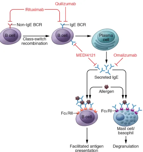
Because of its key role in type I hypersensitivity reactions, IgE has always been a prime target for intervention in allergic disease. This has been approached from two different angles: (a) target-ing soluble IgE and (b) targettarget-ing B cells that express surface IgE (119). Another less specific approach for targeting IgE production has been the depletion of all B cells using the anti-CD20 antibody rituximab (Figure 3).
Despite its critical role in allergies, surprisingly little is known regarding the regulation of IgE production in humans. This fact relates primarily to the extremely low frequency of IgE-switched memory B cells. It remains challenging to accurately detect and purify these cells from humans. A large body of research has con-vincingly demonstrated that IgE production is highly dependent on Th2 responses, in particular IL-4 and IL-13 signaling (120). The main question that still remains incompletely answered per-tains to the location of IgE memory. Which cells form the basis of IgE memory? This is a critical issue, because these cells should be targeted to efficiently eliminate allergen-specific IgE. There are three possible major cell types that could form a reservoir for the IgE memory response: (a) IgE-switched memory B cells, (b) long- living plasma cells that primarily reside in the bone marrow (121), and (c) memory B cells that switched to any isotype within the IgH (immunoglobulin heavy chain) locus located between IgHD and IgHE (in humans these are IgG3, IgG1, IgA1, IgG2, and IgG4, and in mice, IgG3, IgG1, IgG2b, and IgG2a), because these cells can develop into IgE-switched B cells through a process called sequential class switch recombination (122, 123). Our ability to design the optimal strategy for targeting IgE memory responses will strongly improve if we can define which IgH isotype is the major contributor to IgE memory.
Currently, omalizumab is the only efficacious IgE- targeting antibody licensed for the treatment of allergic asthma and chronic spontaneous urticaria (124–130). Preliminary data indi-cate that patients suffering from systemic mastocytosis may also benefit from omalizumab treatment (131). Omalizumab binds soluble IgE, thereby preventing its binding to IgE receptors on effector cells (Figure 3). Interestingly, omalizumab treatment also leads to a reduction of FcεRI on mast cells, basophils, and DCs (132). Omalizumab is currently being tested for a wide
range of allergic conditions and other pathologies (133). An additional anti-IgE antibody, ligelizumab (QGE031), that binds the Cε3 domain of IgE with higher affinity than omalizumab is currently in clinical testing (Figure 3). Ligelizumab was found to have a larger suppressive effect than omalizumab on circulating IgE, basophil FcεRI expression, and skin prick test responses to allergens (134). A controlled trial in 37 mild allergic asthma patients demonstrated that ligelizumab has greater efficacy than omalizumab against inhaled and skin allergic responses (135). However, both antibodies neutralize serum IgE without affecting IgE production by plasma cells. While this type of approach neu-tralizes IgE-mediated type I hypersensitivity reactions, it has no impact on the IgE production, necessitating treatment at regular intervals of 2–4 weeks (125). Clinical studies are ongoing for urticaria (NCT03437278, NCT02649218).
An alternative strategy to interfere with IgE production involves the use of antibodies that bind to the membrane-bound form of IgE, thereby targeting B cells expressing an IgE B cell receptor (BCR). Quilizumab (MEMP1972A/RG7449) is a humanized mAb that binds to the M1′ segment of IgE (136). This segment is only present on membrane IgE and not on free, soluble IgE (137). There-fore, quilizumab targets IgE+ memory B cells and plasmablasts,
while long-lived IgE-switched plasma cells, which do not express surface IgE, are not targeted (Figure 3). Preclinical studies demon-strated that targeting the M1′ domain of IgE efficiently reduced serum IgE without affecting other isotypes. Anti–M1′ domain was an effective prophylactic and therapeutic therapy in murine models of allergic asthma and helminth infection (136). Despite yielding a significant reduction of total and allergen- specific IgE, quilizumab treatment did not impact asthma exacerbations, lung function, and symptom scores in a phase II trial that included 578 patients with inadequately controlled allergic asthma (138). Quili-zumab reduced circulating IgE antibodies between 30% and 40%, while omalizumab treatment resulted in an 89% to 98% reduction (129). This indicates that targeting membrane IgE is not sufficient to remove the allergen-specific IgE to a degree that confers clini-cal improvement. Therefore, other potential compartments of IgE memory appear to play a key role in the production of clinically rel-evant, allergen-specific IgE. These may include long-living plasma cells or subpopulations of memory B cells of other isotypes that can undergo a secondary switch to IgE upon reactivation.
MEDI4212, another IgE-targeting biologic, binds to soluble and membrane IgE. The main potential advantage of this approach is that it may achieve an immediate clinical benefit by directly tar-geting free IgE, while limiting the generation of new IgE-switched B cells and plasma cells (ref. 139 and Figure 3). In a phase I study, atopic individuals were treated with a single dose of MEDI4212, omalizumab, or placebo. MEDI4212 treatment decreased serum IgE more rapidly than omalizumab treatment, but IgE recovery was also much more rapid than in the omalizumab- treated group (140). The short-lived suppression of serum IgE in MEDI4212-treated patients corresponded with a rapid decrease in serum levels of the therapeutic antibody, a feature that limits the potential for dosing- schedule advantages over omalizumab.
1. Akdis M, et al. Interleukins (from IL-1 to IL-38), interferons, transforming growth factor β, and TNF-α: receptors, functions, and roles in diseas-es. J Allergy Clin Immunol. 2016;138(4):984–1010. 2. van Neerven RJ, Knol EF, Ejrnaes A,
Würtz-en PA. IgE-mediated allergWürtz-en presWürtz-entation and blocking antibodies: regulation of T-cell activation in allergy. Int Arch Allergy Immunol. 2006;141(2):119–129.
3. Mudde GC, Bheekha R, Bruijnzeel-Koomen CA. IgE-mediated antigen presentation. Allergy. 1995;50(3):193–199.
4. Galli SJ, Tsai M, Piliponsky AM. The devel-opment of allergic inflammation. Nature. 2008;454(7203):445–454.
5. Gerber BO, Pichler WJ. Cellular mechanisms of T cell mediated drug hypersensitivity. Curr Opin Immunol. 2004;16(6):732–737.
6. Esteban-Gorgojo I, Antolín-Amérigo D, Domín-guez-Ortega J, Quirce S. Non-eosinophilic asthma: current perspectives. J Asthma Allergy. 2018;11:267–281.
7. Muraro A, et al. Precision medicine in allergic disease—food allergy, drug allergy, and anaphy-laxis—PRACTALL document of the European Academy of Allergy and Clinical Immunology and the American Academy of Allergy, Asthma
and Immunology. Allergy. 2017;72(7):1006–1021. 8. Muraro A, et al. Precision medicine in patients
with allergic diseases: airway diseases and atopic dermatitis—PRACTALL document of the Euro-pean Academy of Allergy and Clinical Immu-nology and the American Academy of Allergy, Asthma & Immunology. J Allergy Clin Immunol. 2016;137(5):1347–1358.
9. Bachert C, Akdis CA. Phenotypes and emerging endotypes of chronic rhinosinusitis. J Allergy Clin Immunol Pract. 2016;4(4):621–628.
10. Akdis CA, et al. Endotypes and phenotypes of chronic rhinosinusitis: a PRACTALL document of the European Academy of Allergy and Clinical Immunology and the American Academy of Allergy, Asthma & Immunology. J Allergy Clin Immunol. 2013;131(6):1479–1490.
11. Agache I, Akdis C, Jutel M, Virchow JC. Untangling asthma phenotypes and endotypes. Allergy. 2012;67(7):835–846.
12. Lötvall J, et al. Asthma endotypes: a new approach to classification of disease entities within the asthma syndrome. J Allergy Clin Immu-nol. 2011;127(2):355–360.
13. van de Veen W, Wirz OF, Globinska A, Akdis M. Novel mechanisms in immune tolerance to allergens during natural allergen exposure and
allergen-specific immunotherapy. Curr Opin Immunol. 2017;48:74–81.
14. Jutel M, et al. International Consensus on Aller-gen Immunotherapy II: mechanisms, standard-ization, and pharmacoeconomics. J Allergy Clin Immunol. 2016;137(2):358–368.
15. Novak N, et al. Early suppression of basophil acti-vation during allergen-specific immunotherapy by histamine receptor 2. J Allergy Clin Immunol. 2012;130(5):1153–1158.e2.
16. Akdis M, et al. Immune responses in healthy and allergic individuals are characterized by a fine balance between allergen-specific T regulatory 1 and T helper 2 cells. J Exp Med. 2004;199(11):1567–1575.
17. Jutel M, Van de Veen W, Agache I, Azkur KA, Akdis M, Akdis CA. Mechanisms of allergen-spe-cific immunotherapy and novel ways for vaccine development. Allergol Int. 2013;62(4):425–433. 18. Meiler F, Zumkehr J, Klunker S, Rückert B, Akdis
CA, Akdis M. In vivo switch to IL-10-secreting T regulatory cells in high dose allergen exposure. J Exp Med. 2008;205(12):2887–2898. 19. van de Veen W. The role of regulatory B cells in
allergen immunotherapy. Curr Opin Allergy Clin Immunol. 2017;17(6):447–452.
20. Boonpiyathad T, et al. High-dose bee venom
fighting the itch response by targeting IL-31; and interfering with epithelium–immune cell interaction by targeting IL-25, IL-33, and TSLP, or by targeting soluble or cell surface–bound IgE. It should be noted here that downregulation of a general type 2 response may have certain consequences for immune responses in which Th2 cells, eosinophils, and type 2 effector cytokines such as IL-4, IL-5, and IL-13 play a role. Parasitic infections and downregulation and control of extensive type 1 immune responses that are domi-nant in autoimmunity represent two main conditions that must be sorted out in multicenter studies. All of these effector molecules target upstream or downstream events in type 2 immune response, omitting only a fraction of patients who lack a predominant type 2 immune response. Patients with non–type 2 response, such as intrinsic type of AD, non-eosinophilic asthma, CRS without nasal polyps, neutrophilic asthma, and obesity-induced asthma, have not so far been efficiently targeted with biologics. While there are obvi-ous similarities, there are also evident clinical, immunological, and pathological divergences between asthma, allergic rhinitis, nasal polyps, food allergy, and EoE. Current approaches targeting type 2 immunity seem to affect overlapping patient groups and produce similar final physiopathological outcomes. Extensive research in the area is needed, and given the rapid developments in such a short time, a brighter future is ahead for a variety of allergic diseases.
Acknowledgments
Funding is provided by the Center for Allergy Research and Educa-tion (CARE) of Switzerland (to CK) and the Swiss NaEduca-tional Science Foundation (grant 320030–159870, 310030_179428).
Address correspondence to: Mübeccel Akdis, Swiss Institute of Allergy and Asthma Research (SIAF), University of Zurich, Oberestrasse 22, CH-7270 Davos Platz, Switzerland. Phone: 41.0.814100848; Email: [email protected].
B cells that replenish the IgE-switched plasma cell compartment (141). It remains to be determined whether the same applies to other models and how this finding translates to the human system. Notably, treating AD patients with the anti-CD20 antibody ritux-imab, which depletes all circulating B cells (but only a fraction of B cells in various tissues), did not result in a reduction of allergen- specific IgE 4 to 8 weeks after treatment. This suggests that plasma cells can maintain IgE production for weeks to months without replenishment from circulating memory cells, or, alternatively, that tissue- resident memory B cells can replace circulating cells following depletion (142). Strategies that target these populations could be considered but may cause undesired side effects, because broadly targeting plasma cells or major memory B cell populations will impair protective humoral immunity against pathogens.
Conclusion
exposure induces similar tolerogenic B-cell responses in allergic patients and healthy bee-keepers. Allergy. 2017;72(3):407–415. 21. van de Veen W, Stanic B, Wirz OF, Jansen K,
Globinska A, Akdis M. Role of regulatory B cells in immune tolerance to allergens and beyond. J Allergy Clin Immunol. 2016;138(3):654–665. 22. van de Veen W, et al. IgG4 production is confined
to human IL-10-producing regulatory B cells that suppress antigen-specific immune responses. J Allergy Clin Immunol. 2013;131(4):1204–1212. 23. Ryan JF, et al. Successful immunotherapy
induc-es previously unidentified allergen-specific CD4+ T-cell subsets. Proc Natl Acad Sci U S A.
2016;113(9):E1286–E1295.
24. Wilson DR, et al. Grass pollen immunotherapy inhibits seasonal increases in basophils and eosinophils in the nasal epithelium. Clin Exp Allergy. 2001;31(11):1705–1713.
25. Aalberse RC, Platts-Mills TA, Rispens T. The developmental history of IgE and IgG4 antibod-ies in relation to atopy, eosinophilic esophagitis, and the modified TH2 response. Curr Allergy Asthma Rep. 2016;16(6):45.
26. van de Veen W, Akdis M. Role of IgG4 in IgE-me-diated allergic responses. J Allergy Clin Immunol. 2016;138(5):1434–1435.
27. Shamji MH, et al. Biomarkers for monitoring clini-cal efficacy of allergen immunotherapy for allergic rhinoconjunctivitis and allergic asthma: an EAACI Position Paper. Allergy. 2017;72(8):1156–1173. 28. Orengo JM, et al. Treating cat allergy with
monoclonal IgG antibodies that bind allergen and prevent IgE engagement. Nat Commun. 2018;9(1):1421.
29. Larsen LF, et al. A comparative study on baso-phil activation test, histamine release assay, and passive sensitization histamine release assay in the diagnosis of peanut allergy. Allergy. 2018;73(1):137–144.
30. Boyman O, et al. EAACI IG Biologicals task force paper on the use of biologic agents in allergic dis-orders. Allergy. 2015;70(7):727–754.
31. Lim HF, Nair P. Airway inflammation and inflam-matory biomarkers. Semin Respir Crit Care Med. 2018;39(1):56–63.
32. Mjösberg J, et al. The transcription factor GATA3 is essential for the function of human type 2 innate lymphoid cells. Immunity. 2012;37(4):649–659.
33. Hamilton JD, Ungar B, Guttman-Yassky E. Drug evaluation review: dupilumab in atopic dermati-tis. Immunotherapy. 2015;7(10):1043–1058. 34. Simpson EL, et al. Two phase 3 trials of
dupilum-ab versus placebo in atopic dermatitis. N Engl J Med. 2016;375(24):2335–2348.
35. Beck LA, et al. Dupilumab treatment in adults with moderate-to-severe atopic dermatitis. N Engl J Med. 2014;371(2):130–139.
36. Rabe KF, et al. Efficacy and safety of dupilumab in glucocorticoid-dependent severe asthma. N Engl J Med. 2018;378(26):2475–2485. 37. Castro M, et al. Dupilumab efficacy and safety in
moderate-to-severe uncontrolled asthma. N Engl J Med. 2018;378(26):2486–2496.
38. Leckie MJ, et al. Effects of an interleukin-5 block-ing monoclonal antibody on eosinophils, airway hyper-responsiveness, and the late asthmatic
response. Lancet. 2000;356(9248):2144–2148. 39. Bachert C, et al. Effect of subcutaneous
dupilum-ab on nasal polyp burden in patients with chronic sinusitis and nasal polyposis: a randomized clini-cal trial. JAMA. 2016;315(5):469–479. 40. Jonstam K, et al. Dupilumab reduces local type 2
pro-inflammatory biomarkers in chronic rhinosi-nusitis with nasal polyposis [published online ahead of print November 28, 2018]. Allergy. https://doi.org/10.1111/all.13685. 41. Kaye A, Gordon SC, Deverapalli SC, Her MJ,
Rosmarin D. Dupilumab for the treatment of recalcitrant bullous pemphigoid. JAMA Dermatol. 2018;154(10):1225–1226.
42. Corren J, et al. A randomized, controlled, phase 2 study of AMG 317, an IL-4Ralpha antagonist, in patients with asthma. Am J Respir Crit Care Med. 2010;181(8):788–796.
43. Pelaia G, Vatrella A, Maselli R. The potential of biologics for the treatment of asthma. Nat Rev Drug Discov. 2012;11(12):958–972.
44. Wenzel S, Wilbraham D, Fuller R, Getz EB, Long-phre M. Effect of an interleukin-4 variant on late phase asthmatic response to allergen challenge in asthmatic patients: results of two phase 2a studies. Lancet. 2007;370(9596):1422–1431. 45. Slager RE, et al. IL-4 receptor polymorphisms
predict reduction in asthma exacerbations during response to an anti-IL-4 receptor α antagonist. J Allergy Clin Immunol. 2012;130(2):516–522.e4. 46. Corren J, et al. Lebrikizumab treatment in adults
with asthma. N Engl J Med. 2011;365(12):1088–1098. 47. Korenblat P, et al. Efficacy and safety of
lebriki-zumab in adult patients with mild-to-moderate asthma not receiving inhaled corticosteroids. Respir Med. 2018;134:143–149.
48. Borish LC, et al. Interleukin-4 receptor in mod-erate atopic asthma. A phase I/II randomized, placebo-controlled trial. Am J Respir Crit Care Med. 1999;160(6):1816–1823.
49. Borish LC, et al. Efficacy of soluble IL-4 receptor for the treatment of adults with asthma. J Allergy Clin Immunol. 2001;107(6):963–970. 50. Nussbaum JC, et al. Type 2 innate lymphoid
cells control eosinophil homeostasis. Nature. 2013;502(7470):245–248.
51. Warren HS, Kinnear BF, Phillips JH, Lanier LL. Production of IL-5 by human NK cells and regu-lation of IL-5 secretion by IL-4, IL-10, and IL-12. J Immunol. 1995;154(10):5144–5152.
52. Desreumaux P, et al. Interleukin 5 messenger RNA expression by eosinophils in the intestinal mucosa of patients with coeliac disease. J Exp Med. 1992;175(1):293–296.
53. Yanagibashi T, Satoh M, Nagai Y, Koike M, Takatsu K. Allergic diseases: from bench to clinic — contribution of the discovery of interleukin-5. Cytokine. 2017;98:59–70.
54. Robida PA, Puzzovio PG, Pahima H, Levi-Schaf-fer F, Bochner BS. Human eosinophils and mast cells: birds of a feather flock together. Immunol Rev. 2018;282(1):151–167.
55. Bagnasco D, Ferrando M, Varricchi G, Puggioni F, Passalacqua G, Canonica GW. Anti-interleukin 5 (IL-5) and IL-5Ra biological drugs: efficacy, safe-ty, and future perspectives in severe eosinophilic asthma. Front Med (Lausanne). 2017;4:135. 56. Farne HA, Wilson A, Powell C, Bax L, Milan SJ.
Anti-IL5 therapies for asthma. Cochrane Database Syst Rev. 2017;9:CD010834.
57. Goldman M, Hirsch I, Zangrilli JG, Newbold P, Xu X. The association between blood eosinophil count and benralizumab efficacy for patients with severe, uncontrolled asthma: subanalyses of the Phase III SIROCCO and CALIMA studies. Curr Med Res Opin. 2017;33(9):1605–1613. 58. Bleecker ER, et al. Efficacy and safety of
benral-izumab for patients with severe asthma uncon-trolled with high-dosage inhaled corticosteroids and long-acting β2-agonists (SIROCCO): a ran-domised, multicentre, placebo-controlled phase 3 trial. Lancet. 2016;388(10056):2115–2127. 59. Chupp GL, et al. Efficacy of mepolizumab add-on
therapy on health-related quality of life and mark-ers of asthma control in severe eosinophilic asth-ma (MUSCA): a randomised, double-blind, place-bo-controlled, parallel-group, multicentre, phase 3b trial. Lancet Respir Med. 2017;5(5):390–400. 60. Ortega HG, et al. Mepolizumab treatment in
patients with severe eosinophilic asthma. N Engl J Med. 2014;371(13):1198–1207.
61. Castro M, et al. Reslizumab for inadequately controlled asthma with elevated blood eosinophil counts: results from two multicentre, parallel, dou-ble-blind, randomised, placebo-controlled, phase 3 trials. Lancet Respir Med. 2015;3(5):355–366. 62. Murphy K, et al. Long-term safety and efficacy of
reslizumab in patients with eosinophilic asthma. J Allergy Clin Immunol Pract. 2017;5(6):1572–1581.e3. 63. Ortega HG, et al. Severe eosinophilic asthma
treated with mepolizumab stratified by baseline eosinophil thresholds: a secondary analysis of the DREAM and MENSA studies. Lancet Respir Med. 2016;4(7):549–556.
64. Bachert C, et al. Reduced need for surgery in severe nasal polyposis with mepolizum-ab: randomized trial. J Allergy Clin Immunol. 2017;140(4):1024–1031.e14.
65. Stein ML, et al. Anti-IL-5 (mepolizumab) therapy for eosinophilic esophagitis. J Allergy Clin Immu-nol. 2006;118(6):1312–1319.
66. Straumann A, et al. Anti-interleukin-5 antibody treatment (mepolizumab) in active eosinophilic oesophagitis: a randomised, placebo-controlled, double-blind trial. Gut. 2010;59(1):21–30. 67. Assa’ad AH, et al. An antibody against IL-5
reduc-es numbers of reduc-esophageal intraepithelial eosin-ophils in children with eosinophilic esophagitis. Gastroenterology. 2011;141(5):1593–1604. 68. Hassani M, Koenderman L. Immunological and
hematological effects of IL-5(Rα)-targeted thera-py: an overview. Allergy. 2018;73(10):1979–1988. 69. Otani IM, Nadeau KC. Biologic therapies for
immunoglobulin E-mediated food allergy and eosinophilic esophagitis. Immunol Allergy Clin North Am. 2017;37(2):369–396.
70. Schmitt E, et al. IL-9 production of naive CD4+
T cells depends on IL-2, is synergistically enhanced by a combination of TGF-beta and IL-4, and is inhibited by IFN-γ. J Immunol. 1994;153(9):3989–3996.
71. Gounni AS, et al. IL-9 expression by human eosinophils: regulation by IL-1β and TNF-α. J Allergy Clin Immunol. 2000;106(3):460–466. 72. Stassen M, et al. Murine bone marrow-derived
costimulato-ry function of IL-10 and kit ligand in the presence of IL-1. J Immunol. 2000;164(11):5549–5555. 73. Abdelilah S, et al. Functional expression of
IL-9 receptor by human neutrophils from asth-matic donors: role in IL-8 release. J Immunol. 2001;166(4):2768–2774.
74. Nagato T, et al. Expression of interleukin-9 in nasal natural killer/T-cell lymphoma cell lines and patients. Clin Cancer Res. 2005;11(23):8250–8257. 75. Veldhoen M, et al. Transforming growth factor-β
‘reprograms’ the differentiation of T helper 2 cells and promotes an interleukin 9-producing subset. Nat Immunol. 2008;9(12):1341–1346. 76. Blom L, Poulsen BC, Jensen BM, Hansen A,
Poulsen LK. IL-33 induces IL-9 production in human CD4+ T cells and basophils. PLoS One.
2011;6(7):e21695.
77. Sehra S, et al. TH9 cells are required for tissue mast cell accumulation during allergic inflammation. J Allergy Clin Immunol. 2015;136(2):433–440.e1. 78. Brough HA, et al. IL-9 is a key component of
memory TH cell peanut-specific responses from children with peanut allergy. J Allergy Clin Immu-nol. 2014;134(6):1329–1338.e10.
79. Oh CK, Leigh R, McLaurin KK, Kim K, Hultquist M, Molfino NA. A randomized, controlled trial to evaluate the effect of an anti-interleukin-9 monoclonal antibody in adults with uncontrolled asthma. Respir Res. 2013;14:93.
80. Shin JH, et al. Anti-interleukin-9 antibody increases the effect of allergen-specific immuno-therapy in murine allergic rhinitis. Allergy Asthma Immunol Res. 2017;9(3):237–246.
81. Bağci IS, Ruzicka T. IL-31: a new key player in dermatology and beyond. J Allergy Clin Immunol. 2018;141(3):858–866.
82. Furue M, Yamamura K, Kido-Nakahara M, Naka-hara T, Fukui Y. Emerging role of interleukin-31 and interleukin-31 receptor in pruritus in atopic dermatitis. Allergy. 2018;73(1):29–36. 83. Stott B, Lavender P, Lehmann S, Pennino D,
Durham S, Schmidt-Weber CB. Human IL-31 is induced by IL-4 and promotes TH2-driv-en inflammation. J Allergy Clin Immunol. 2013;132(2):446–454.e5.
84. Ruzicka T, et al. Anti-interleukin-31 receptor a antibody for atopic dermatitis. N Engl J Med. 2017;376(9):826–835.
85. Kabashima K, et al. Nemolizumab in patients with moderate-to-severe atopic dermatitis: ran-domized, phase II, long-term extension study. J Allergy Clin Immunol. 2018;142(4):1121–1130.e7. 86. Hawro T, Saluja R, Weller K, Altrichter S, Metz
M, Maurer M. Interleukin-31 does not induce immediate itch in atopic dermatitis patients and healthy controls after skin challenge. Allergy. 2014;69(1):113–117.
87. Divekar R, Kita H. Recent advances in epi-thelium-derived cytokines (IL-33, IL-25, and thymic stromal lymphopoietin) and allergic inflammation. Curr Opin Allergy Clin Immunol. 2015;15(1):98–103.
88. Ogasawara N, et al. Epithelial activators of type 2 inflammation: elevation of thymic stromal lymphopoietin, but not IL-25 or IL-33, in chronic rhinosinusitis with nasal polyps in Chicago, Illi-nois. Allergy. 2018;73(11):2251–2254. 89. Xu M, Dong C. IL-25 in allergic inflammation.
Immunol Rev. 2017;278(1):185–191. 90. von Moltke J, Ji M, Liang HE, Locksley RM.
Tuft-cell-derived IL-25 regulates an intesti-nal ILC2-epithelial response circuit. Nature. 2016;529(7585):221–225.
91. Lee M, Kim DW, Shin HW. Targeting IL-25 as a novel therapy in chronic rhinosinusitis with nasal polyps. Curr Opin Allergy Clin Immunol. 2017;17(1):17–22.
92. Shin HW, et al. IL-25 as a novel therapeutic target in nasal polyps of patients with chronic rhinosinusitis. J Allergy Clin Immunol. 2015;135(6):1476–1485.e7. 93. Ballantyne SJ, et al. Blocking IL-25 prevents air-way hyperresponsiveness in allergic asthma. J Allergy Clin Immunol. 2007;120(6):1324–1331. 94. Rusinova GG, Sharova EG, Libinzon RE. [The
fate of homologous and heterologous DNA in animals]. Biokhimiia. 1969;34(3):464–471. 95. Soumelis V, et al. Human epithelial cells trigger
dendritic cell mediated allergic inflammation by producing TSLP. Nat Immunol. 2002;3(7):673–680. 96. Sokol CL, Barton GM, Farr AG, Medzhitov R.
A mechanism for the initiation of allergen- induced T helper type 2 responses. Nat Immunol. 2008;9(3):310–318.
97. Moon PD, Kim HM. Thymic stromal lymphopoi-etin is expressed and produced by caspase-1/ NF-κB pathway in mast cells. Cytokine. 2011;54(3):239–243.
98. Spadoni I, Iliev ID, Rossi G, Rescigno M. Den-dritic cells produce TSLP that limits the differen-tiation of Th17 cells, fosters Treg development, and protects against colitis. Mucosal Immunol. 2012;5(2):184–193.
99. Ziegler SF, Roan F, Bell BD, Stoklasek TA, Kitajima M, Han H. The biology of thymic stromal lymphopoietin (TSLP). Adv Pharmacol. 2013;66:129–155.
100. Reche PA, et al. Human thymic stromal lympho-poietin preferentially stimulates myeloid cells. J Immunol. 2001;167(1):336–343.
101. Zhou B, et al. Thymic stromal lymphopoietin as a key initiator of allergic airway inflammation in mice. Nat Immunol. 2005;6(10):1047–1053. 102. Kabata H, Moro K, Koyasu S. The group 2 innate
lymphoid cell (ILC2) regulatory network and its underlying mechanisms. Immunol Rev. 2018;286(1):37–52.
103. Ito T, et al. TSLP-activated dendritic cells induce an inflammatory T helper type 2 cell response through OX40 ligand. J Exp Med. 2005;202(9):1213–1223.
104. Ying S, et al. Thymic stromal lymphopoietin expression is increased in asthmatic airways and correlates with expression of Th2-attracting chemokines and disease severity. J Immunol. 2005;174(12):8183–8190.
105. He JQ, et al. A thymic stromal lymphopoietin gene variant is associated with asthma and air-way hyperresponsiveness. J Allergy Clin Immunol. 2009;124(2):222–229.
106. Gauvreau GM, et al. Effects of an TSLP anti-body on allergen-induced asthmatic responses. N Engl J Med. 2014;370(22):2102–2110. 107. Simpson EL, et al. An anti-TSLP monoclonal
antibody, in the treatment of moderate to severe atopic dermatitis: a randomized phase 2a clinical trial. J Am Acad Dermatol. 2019;80(4):1013–1021.
108. Corren J, et al. Tezepelumab in adults with uncontrolled asthma. N Engl J Med. 2017;377(10):936–946.
109. Schmitz J, et al. IL-33, an interleukin-1-like cyto-kine that signals via the IL-1 receptor-related pro-tein ST2 and induces T helper type 2-associated cytokines. Immunity. 2005;23(5):479–490. 110. Molofsky AB, Savage AK, Locksley RM.
Interleu-kin-33 in tissue homeostasis, injury, and inflam-mation. Immunity. 2015;42(6):1005–1019. 111. Cayrol C, Girard JP. Interleukin-33 (IL-33): a
nuclear cytokine from the IL-1 family. Immunol Rev. 2018;281(1):154–168.
112. Chu DK, et al. IL-33, but not thymic stromal lymphopoietin or IL-25, is central to mite and peanut allergic sensitization. J Allergy Clin Immu-nol. 2013;131(1):187–200.e1.
113. Wambre E, et al. A phenotypically and function-ally distinct human TH2 cell subpopulation is associated with allergic disorders. Sci Transl Med. 2017;9(401):eaam9171.
114. Han H, Roan F, Johnston LK, Smith DE, Bryce PJ, Ziegler SF. IL-33 promotes gastrointestinal allergy in a TSLP-independent manner. Mucosal Immunol. 2018;11(2):394–403.
115. Peng G, et al. Anti-IL-33 antibody has a therapeu-tic effect in an atopic dermatitis murine model induced by 2, 4-dinitrochlorobenzene. Inflam-mation. 2018;41(1):154–163.
116. Chen Y, et al. First-in-class phase 2a study of anb020 (anti-il-33) in the treatment of moder-ate-to-severe atopic dermatitis. Oral abstract presented at: European Academy of Allergy and Clinical Immunology, Congress 2018. 2018 May 26–30. https://onlinelibrary.wiley.com/ doi/epdf/10.1111/all.13535 (abstract no. 0137). Accessed February 28, 2019.
117. Vannella KM, et al. Combinatorial targeting of TSLP, IL-25, and IL-33 in type 2 cytokine-driv-en inflammation and fibrosis. Sci Transl Med. 2016;8(337):337ra65.
118. Venkataramani S, et al. Design and charac-terization of Zweimab and Doppelmab, high affinity dual antagonistic anti-TSLP/IL13 bispe-cific antibodies. Biochem Biophys Res Commun. 2018;504(1):19–24.
119. Hu J, Chen J, Ye L, Cai Z, Sun J, Ji K. Anti-IgE therapy for IgE-mediated allergic diseases: from neutralizing IgE antibodies to eliminating IgE+ B cells. Clin Transl Allergy. 2018;8:27.
120. He JS, Narayanan S, Subramaniam S, Ho WQ, Lafaille JJ, Curotto de Lafaille MA. Biology of IgE production: IgE cell differentiation and the memory of IgE responses. Curr Top Microbiol Immunol. 2015;388:1–19.
121. Luger EO, Wegmann M, Achatz G, Worm M, Renz H, Radbruch A. Allergy for a lifetime? Allergol Int. 2010;59(1):1–8.
122. Looney TJ, et al. Human B-cell isotype switch-ing origins of IgE. J Allergy Clin Immunol. 2016;137(2):579–586.e7.
123. Xiong H, Dolpady J, Wabl M, Curotto de Lafaille MA, Lafaille JJ. Sequential class switching is required for the generation of high affinity IgE antibodies. J Exp Med. 2012;209(2):353–364.
N Engl J Med. 2013;368(10):924–935.
125. Strunk RC, Bloomberg GR. Omalizumab for asth-ma. N Engl J Med. 2006;354(25):2689–2695. 126. Holgate ST, et al. Efficacy and safety of a
recombinant anti-immunoglobulin E antibody (omalizumab) in severe allergic asthma. Clin Exp Allergy. 2004;34(4):632–638.
127. Milgrom H, et al. Treatment of childhood asthma with anti-immunoglobulin E antibody (omali-zumab). Pediatrics. 2001;108(2):E36. 128. Solèr M, et al. The anti-IgE antibody
omal-izumab reduces exacerbations and steroid requirement in allergic asthmatics. Eur Respir J. 2001;18(2):254–261.
129. Busse W, et al. Omalizumab, anti-IgE recombi-nant humanized monoclonal antibody, for the treatment of severe allergic asthma. J Allergy Clin Immunol. 2001;108(2):184–190.
130. Trischler J, et al. Omalizumab effectively protects against early and late allergic responses in asth-ma after 4 weeks. Allergy. 2017;72(12):1912–1915. 131. Broesby-Olsen S, et al. Omalizumab prevents
anaphylaxis and improves symptoms in systemic mastocytosis: efficacy and safety observations.
Allergy. 2018;73(1):230–238.
132. Beck LA, Marcotte GV, MacGlashan D, Togias A, Saini S. Omalizumab-induced reductions in mast cell FcεRI expression and function. J Allergy Clin Immunol. 2004;114(3):527–530.
133. Incorvaia C, Mauro M, Makri E, Leo G, Ridolo E. Two decades with omalizumab: what we still have to learn. Biologics. 2018;12:135–142. 134. Arm JP, et al. Pharmacokinetics,
pharmacody-namics and safety of QGE031 (ligelizumab), a novel high-affinity anti-IgE antibody, in atopic subjects. Clin Exp Allergy. 2014;44(11):1371–1385. 135. Gauvreau GM, et al. Efficacy and safety of
multiple doses of QGE031 (ligelizumab) versus omalizumab and placebo in inhibiting aller-gen-induced early asthmatic responses. J Allergy Clin Immunol. 2016;138(4):1051–1059. 136. Brightbill HD, et al. Antibodies specific for a
seg-ment of human membrane IgE deplete IgE-pro-ducing B cells in humanized mice. J Clin Invest. 2010;120(6):2218–2229.
137. Zhang K, Saxon A, Max EE. Two unusual forms of human immunoglobulin E encoded by alter-native RNA splicing of epsilon heavy chain
mem-brane exons. J Exp Med. 1992;176(1):233–243. 138. Harris JM, et al. A randomized trial of the efficacy
and safety of quilizumab in adults with inade-quately controlled allergic asthma. Respir Res. 2016;17:29.
139. Nyborg AC, et al. Development of an antibody that neutralizes soluble IgE and eliminates IgE express-ing B cells. Cell Mol Immunol. 2016;13(3):391–400. 140. Sheldon E, et al. Pharmacokinetics, pharmaco-dynamics, and aafety of MEDI4212, an anti-ige monoclonal antibody, in subjects with atopy: a phase I study. Adv Ther. 2016;33(2):225–251. 141. Jiménez-Saiz R, et al. Lifelong memory responses
perpetuate humoral TH2 immunity and ana-phylaxis in food allergy. J Allergy Clin Immunol. 2017;140(6):1604–1615.e5.
142. Simon D, Hösli S, Kostylina G, Yawalkar N, Simon HU. Anti-CD20 (rituximab) treatment improves atopic eczema. J Allergy Clin Immunol. 2008;121(1):122–128.