Pathology and Surgery
Jae Hyoung Kim, Robert D. Tien, Gary J. Felsberg, Alan K. Osumi, Namsoo Lee, and Allan H. Friedman
PURPOSE: To identify the extent of hippocampal sclerosis in temporal lobe epilepsy with fast
spin-echo MR and correlate it with histopathologic findings and surgical outcome. METHODS: MR images of 30 patients with temporal lobe epilepsy and pathologically proved hippocampal scle-rosis and 30 control subjects were obtained using a fast spin-echo technique with 4000/100/4 (repetition time/echo time/excitations), 16 echo train, 2- to 3-mm section thickness with inter-leave, 2563 256 matrix, and 18-cm field of view. Criteria for MR diagnosis of hippocampal sclerosis included hippocampal atrophy diagnosed with MR volumetry and/or T2-weighted signal change. Hippocampal sectional areas were plotted, and T2 signal changes were topographically evaluated to identify the extent of hippocampal sclerosis, which was subsequently correlated with histopathologic findings and surgical outcome. RESULTS: Hippocampal sclerosis was diffuse, involving both hippocampal head and body, in 96.7% of patients (29 of 30 patients). One patient had normal MR findings. Focal hippocampal sclerosis was not seen. Histopathologic findings of hippocampal sclerosis were present in all 29 patients who had abnormal MR findings. Eighty-six percent of patients (18 of 21 patients), who were followed for at least 1 year after temporal lobectomy, were seizure free (81%, 17 of 21 patients) or significantly improved (5%, 1 of 21 patients). CONCLUSION: Fast spin-echo MR enables accurate definition of the extent of hip-pocampal sclerosis in patients with temporal lobe epilepsy. All cases of hiphip-pocampal sclerosis identified in this study involved the hippocampus diffusely. However, leaving the posterior portion of the hippocampus during surgery does not seem to be a major factor influencing surgical outcome.
Index terms: Brain, magnetic resonance; Brain, temporal lobe; Sclerosis, hippocampal; Seizures
AJNR Am J Neuroradiol16:627–636, April 1995
Hippocampal sclerosisrefers to neuronal loss and gliosis of the hippocampus. Hippocampal sclerosis is the most common pathologic abnor-mality associated with medically intractable temporal lobe epilepsy (1, 2). The radiologic diagnosis of hippocampal sclerosis is based on the magnetic resonance (MR) findings of atro-phy and/or T2 signal change of the hippocam-pus (3–10). Neuropathologically, the
distribu-tion and extent of neuronal loss and gliosis within the hippocampus (topographical pattern of hippocampal sclerosis) may be related to different surgical outcomes in patients with temporal lobe epilepsy treated by surgery (2, 11, 12). Several distinct topographical types of hippocampal atrophy in patients with temporal lobe epilepsy have been described with MR im-aging (13).
Recently, fast spin-echo MR imaging has been used to obtain high-resolution anatomic information and T2 signal character of the hip-pocampus with one pulse sequence (3, 14). In this study, we identified the extent or topo-graphical distribution of hippocampal sclerosis in patients with temporal lobe epilepsy by fast spin-echo MR examination and correlated the extent of hippocampal sclerosis with his-topathologic examination and with patient out-come after surgery.
Received June 16, 1994; accepted after revision October 24. This study was supported in part by a Merritt-Putnam Fellowship from the Epilepsy Foundation of America (Dr Lee).
From the Department of Radiology (J.H.K., R.D.T., G.J.F., A.K.O.) and the Divisions of Neurology (N.L.) and Neurosurgery (A.H.F.), Duke Uni-versity Medical Center, Durham, NC.
Address reprint requests to Robert D. Tien, MD, Department of Radiol-ogy, Box 3808, Duke University Medical Center, Durham, NC 27710.
AJNR 16:627–636, Apr 1995 0195-6108/95/1604 –0627
qAmerican Society of Neuroradiology
Subjects and Methods
During the period of 2.6 years, 33 patients, who had no focal epileptogenic extrahippocampal lesions, underwent anterior temporal lobectomy for the treatment of intracta-ble temporal lobe epilepsy caused by presumed hip-pocampal sclerosis. Of these 33 patients, 30 had diag-noses of hippocampal sclerosis, and 3 patients had normal findings on histopathologic examinations. The pathologic diagnosis of hippocampal sclerosis was made qualitatively without hippocampal neuronal counting by an experi-enced neuropathologist. Thirty patients with pathologi-cally proved hippocampal sclerosis were included in this study. During the surgical procedure, the hippocampal head and anterior body were resected en bloc, and the posterior body was then removed by subpial aspiration. This technique leaves a small portion of the posterior body and tail of the hippocampus remaining. There were 16 male and 14 female patients with a mean age of 31.7 years (range, 13 to 57 years). All patients underwent a standard-ized preoperative protocol for location of seizure focus (15). Intracranial depth electroencephalography was used in several patients. A control group consisted of both volunteers and patients without clinical history of seizures. They were 15 male and 15 female patients with a mean age of 33.2 years (range, 14 to 56 years). All patients and control subjects underwent MR examina-tions on a 1.5-T system. The imaging protocol included conventional spin-echo T1- and T2-weighted axial im-ages and T1-weighted coronal imim-ages. Additionally, high-resolution coronal T2-weighted fast spin-echo im-aging of the temporal lobe was performed as previously described (3, 14): 4000/100/4 (repetition time/echo time/excitations), 16 echo train length, 2- to 3-mm sec-tion thickness with interleave, 256 3 256 matrix, and 18-cm field of view. The imaging plane was perpendicu-lar to the long axis of the hippocampus.
Visual analysis of all fast spin-echo MR images was performed to assess the T2 signal change of the hip-pocampus. The presence or absence of increased T2 sig-nal at each image was visually determined by the consen-sus of three neuroradiologists. The location of fast spin-echo images demonstrating T2 signal change was recorded for correlation with the topographical distribution of hippocampal atrophy.
All fast spin-echo MR images were loaded on the Signa console (General Electric, Milwaukee, Wis) for volumetry. In a blinded fashion, a neuroradiologist with experience in MR volumetry measured the cross-sectional areas of each hippocampus manually, tracing the hippocampus from the head to tail on each successive section as previously reported (3). Hippocampal volumes were then calculated by summing areas and multiplying by section thickness. To determine unilateral hippocampal atrophy, a mean vol-ume difference (right hippocampal volvol-ume minus left hip-pocampal volume) and standard deviation were used. Val-ues beyond 2 SD of mean volume difference of control subjects were determined to be abnormal.
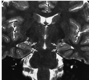
To identify the topographical distribution type of hip-pocampal sclerosis, diagrams of hiphip-pocampal successive cross-sectional areas measured on MR images were plot-ted and combined with the T2 signal information of the hippocampus. Successive cross-sectional areas of both hippocampi were plotted against the long axis of the hip-pocampi to compare easily both hippocampal volumes section to section as previously performed (13). Because visual comparison of cross-sectional areas of both hip-pocampi was sensitive to the patient’s head rotation in the coronal plane, the degree of head rotation was estimated by comparing the bilateral symmetric structures of the gyrus intralimbicus, which is an internal marker separating the head and body of the hippocampus. When the degree of head rotation in the coronal plane was more than one section thickness, the area curve plots were shifted ac-cordingly to correct for rotation. The gyrus intralimbicus is a small structure composed of the cornu Ammonis sub-fields 3 and 4 and constitutes the medial portion of the most posterior segment of the hippocampal head. When tracing the coronal MR images from the hippocampal head toward the tail, the level of the gyrus intralimbicus can be identified by recognizing the absent digitations and the bulbous medial mass of the hippocampal head (Fig 1). The extent of hippocampal sclerosis was determined, by the consensus of three neuroradiologists, by visually com-paring area-curve plots of the patients with those of the 30 control subjects. The topographical distribution of hip-pocampal atrophy was classified into diffuse (involving the hippocampal head and the body), anterior focal (involving the head to the level of the gyrus intralimbicus), and pos-terior focal types (only involving the body and/or tail). The level of the gyrus intralimbicus and that of the quadrigem-inal plate were considered the most caudal sections of the hippocampal head and body, respectively. The distribu-tion of T2 signal change was also classified similarly. These two parameters (hippocampal atrophy and T2
[image:2.612.341.524.523.688.2]nal change) were then incorporated into a final determi-nation of the topographical distribution of hippocampal sclerosis. On any section of an individual hippocampus, if either hippocampal atrophy or T2 signal change existed, that individual section was considered positive for hip-pocampal sclerosis.
The fast spin-echo MR-derived topographical types of hippocampal sclerosis were compared with the his-topathologic extents of hippocampal neuronal loss. The presence or absence of hippocampal neuronal loss was qualitatively determined by a neurologist experienced in neuropathologic examination of hippocampi, without quantitative neuronal counting, by reviewing specimens of both the hippocampal head and body. The hippocampal head and body were differentiated by histologic character-istics; the hippocampal head has the folding of the gran-ular cell layer of the dentate gyrus and the pyramidal cell layer of the cornu Ammonis, which are absent in the hip-pocampal body. Because of the method of surgical re-moval, an exact section-to-section comparison between
the fast spin-echo MR images and the histopathologic specimen was not possible.
Surgical outcomes were assessed in all patients who were evaluated postsurgically for at least 1 year and were compared with the fast spin-echo MR-derived extent of hippocampal sclerosis. Surgical outcomes were classified into seizure free, significantly improved (defined as 10 or less seizures per year and more than a 75% decrease in seizure rate), and not significantly improved (defined as more than 10 seizures per year or less than a 75% decrease in seizure rate) as previously defined (15).
Results
The data of MR-based topographical types, T2 signal change, histopathologic examina-tions, and surgical outcomes of hippocampal sclerosis are summarized in the Table.
Summary of MR volumetry, topographical type, and surgical outcome
Patient MR Volumetric Lateralization*
MR-Based Topographical Type Pathologic
Correlation?
Surgical Outcome
Atrophy T2 signal Combined
1 R (20.666) P D D yes SF
2 R (21.086) D D D yes
3 R (21.281) D D D yes SF
4 R (20.854) D D D yes SF
5 R (20.774) D D D yes SF
6 R (20.570) D D D yes
7 R (21.161) D D D yes
8 R (20.680) D D D yes SF
9 R (20.843) D D D yes
10 R (20.372) D D D yes SF
11 R (21.236) D D D yes
12 R (20.528) D NS D yes SF
13 R (20.759) D NS D yes SI
14 R (21.022) D D D yes
15 L (0.981) D D D yes SF
16 L (0.720) D D D yes
17 L (0.584) D D D yes SF
18 L (0.891) D D D yes
19 L (0.986) D D D yes SF
20 L (1.215) D D D yes SF
21 L (0.886) D D D yes SF
22 L (1.090) D D D yes SF
23 L (1.060) D D D yes SF
24 L (0.764) D D D yes NSI
25 L (1.224) D D D yes
26 NL (0.222) NA NS N no† NSI
27 R (20.420) P Db Db yes NSI
28 NL (0.156) Db Db Db yes SF
29 NL (0.240) Db Db Db yes SF
30 NL (0.172) Db D‡ Db yes SF
Note.—D indicates diffuse; Db, bilaterally diffuse; N, normal; NA, no atrophy detected; NL, no lateralization detected; NS, no T2 signal change detected; NSI, not significantly improved; P, posterior; SF, seizure-free; and SI, significantly improved.
* The number in parentheses is the hippocampal volume difference (right minus left) in cubic centimeters.
Fast Spin-Echo MR Volumetric Lateralization
The mean difference of hippocampal vol-umes of controls was 0.079 cm3 (SD, 0.177 cm3). Lateralization of the abnormal hippocam-pus with fast spin-echo MR volumetry was per-formed in 26 of the 30 patients with hippocam-pal sclerosis. There was no false lateralization. Four patients were not lateralized. Three of these four patients (patients 28, 29, and 30) had evidence of bilateral hippocampal atrophy based on MR volumetry. All three had corrobo-rative evidence of bilateral hippocampal sclero-sis by depth electroencephalographic findings, and the dominant seizure-producing hippocam-pus was resected. One patient (patient 26) with pathologically proved hippocampal sclerosis showed no lateralization and bilaterally reduced hippocampal volumes, although the right hip-pocampus was resected based on the depth electroencephalographic finding.
Topographical Distribution of Signal Abnormality
Of the 26 patients who were lateralized by fast spin-echo MR volumetry, 23 also had visu-ally identified T2 signal change ipsilatervisu-ally; the signal change was diffuse in all 23. Another patient (patient 27) had bilaterally increased T2 signal suggesting bilateral hippocampal sclero-sis; the right hippocampus was resected based on MR evidence of unilateral atrophy and the consistent depth electroencephalographic find-ing of right-sided seizure activity. Two other patients (patients 12 and 13) who were lateral-ized by volumetry showed no signal abnormal-ity. Of the 3 patients with definite bilateral hip-pocampal atrophy, 2 patients (patients 28 and 29) showed bilateral diffuse T2 signal change, and 1 patient (patient 30) showed unilateral diffuse signal change in the dominant seizure-producing hippocampus. One patient (patient 26), who was not lateralized and did not show bilaterally reduced hippocampal volumes, did not show signal abnormality.
Topographical Distribution of Atrophy
From the area curves of the 30 control sub-jects, a close approximation between the right and left hippocampal area curves was demon-strated against the entire hippocampal axis (Fig 2), although a minimal discrepancy between both curves was often found at the level of the
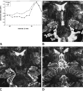
hippocampal head (Fig 3). Twenty-six patients with fast spin-echo MR-volumetric lateralization showed definite discrepancies between both area curves, suggesting hippocampal atrophy; the atrophy was of the diffuse type in 24 (Figs 4 and 5) and the posterior focal type in 2 (patients 1 and 27) (Fig 6). Three patients (patients 28, 29, and 30) showed bilateral diffuse atrophy, there were no definite discrepancies between both area curves but definitely reduced absolute volumes bilaterally when compared with con-trols (Fig 7). One patient (patient 26) did not show any discrepancy suggesting atrophy.
Combination of Topographical Distribution of Atrophy and Signal (Topographical
Distribution of Hippocampal Sclerosis)
[image:4.612.340.529.100.228.2]Of the 30 patients, 22 showed both unilateral diffuse hippocampal atrophy and T2 signal change. Two patients (patients 12 and 13)
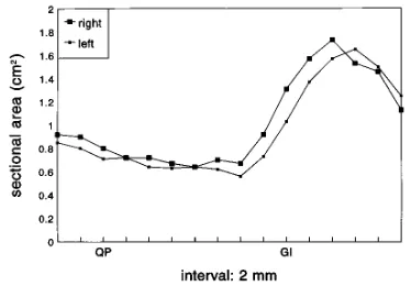
Fig 2. Hippocampal area curves of the control subjects. Close approximation between the right and left hippocampal curves is noted along the entire hippocampal axis. The hippocampal head is on right side and the tail is on left side. GI andQP on the hippocampal long axis indicate the level of the gyrus intralimbicus and that of the quadrigeminal plate, respectively.
[image:4.612.341.528.580.710.2]showed unilateral diffuse hippocampal atrophy but no T2 signal change. Two patients had pos-terior focal atrophy; 1 (patient 1) had ipsilateral diffuse T2 signal change, and the other (patient 27) had bilaterally diffuse T2 signal change sug-gesting bilateral hippocampal sclerosis.
Three patients had bilateral diffuse hip-pocampal atrophy; 2 (patients 28 and 29) also had bilateral diffuse T2 signal abnormality, and 1 (patient 30) had diffuse signal only in the dominant seizure-producing hippocampus. One patient had normal fast spin-echo MR find-ings. Overall, considering both hippocampal at-rophy and T2 signal change, 29 of the 30 pa-tients were determined to have the diffuse type of hippocampal sclerosis (25 unilateral diffuse type and 4 bilateral diffuse type).
Histopathologic Correlation
Sixteen patients with intact histopathologic specimens of both the hippocampal head and
body showed good correlations between the MR-determined and histopathologic extent of hippocampal sclerosis. Thirteen patients with intact histopathologic specimens of either the hippocampal head or body also demonstrated positive correlations between the MR-deter-mined extent of hippocampal sclerosis and his-topathologies; all showed neuronal loss on each available specimen (of the hippocampal head in five patients and the body in eight patients). One patient (patient 26) with normal MR find-ings showed mild neuronal loss on the his-topathologic specimen of only the hippocampal body.
Surgical Outcomes
Clinical follow-up after temporal lobectomy for the 30 patients ranged from 6 months to 2.6 years. Surgical outcomes were assessed in 21 patients who were postsurgically evaluated for
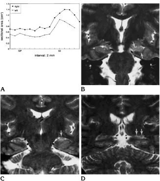
Fig 4. A, Hippocampal area curves of the diffuse type of hippocampal sclerosis (patient 18). Discrepancy between the right and left curves is noted diffusely from the hippocampal head to the tail.
[image:5.612.59.385.99.467.2]at least 1 year (mean, 1.8 years) with 17 pa-tients seizure free, 1 patient significantly im-proved, and 3 patients (patients 24, 26, and 27) not significantly improved. Of these three, 1 patient (patient 24) had definite MR evidence of unilateral hippocampal sclerosis; another pa-tient (papa-tient 27) had bilateral diffuse T2 signal change; and the other patient (patient 26) had normal fast spin-echo MR findings.
Discussion
Hippocampal sclerosis refers to an entity of neuronal loss and atrophy with associated glio-sis involving the hippocampus and is the most common cause of medically intractable tempo-ral lobe epilepsy (1, 2). Tempotempo-ral lobectomy with hippocampectomy is a widely accepted surgical treatment for patients with hippocam-pal sclerosis (16 –18). Accurate decision as to which hippocampus to resect is essential before surgical treatment of hippocampal sclerosis.
MR imaging has been used to assess the hip-pocampus for the evaluation of patients with temporal lobe epilepsy and demonstrated its valuable role in depicting the abnormalities of the hippocampus (atrophy and/or increased T2 signal of the hippocampus) and lateralizing the seizure focus (3–10). Significant correlations between the hippocampal size measured on MR imaging and hippocampal neuronal density in surgical specimens have been found in patients with hippocampal sclerosis (9, 19, 20) (Lee N, Lewis D, Tien R, et al, “Hippocampal Sclerosis: Fast Spin Echo MRI Volumetry and Neuronal Loss” [abstract], Epilepsia 1993;34 [suppl 6]:129 –130). Increased T2 signal of the hip-pocampus, reflecting gliosis, is another impor-tant indicator of hippocampal sclerosis (3, 7, 10, 21, 22). Previous MR imaging studies have described a variable frequency of T2 signal change of the hippocampus, occurring in 12% to 65% of patients with hippocampal sclerosis (4, 10, 23). However, with the improvement of
[image:6.612.227.551.101.455.2]MR imaging techniques, several recent MR studies have verified T2 signal change of the hippocampus to be a highly sensitive MR find-ing suggestfind-ing hippocampal sclerosis, occur-ring in 84% to 100% (3, 22) (Ojemann LM, Tsuruda JS, Holmes MD, Alvord EC, Ojemann GA, Hayes CF, “Comparison of Clinical Fea-tures, Histology and High Resolution Fast Spin MRI Using a Phased Array Coil in Patients Un-dergoing Surgery for Temporal Lobe Epilepsy” [abstract], Epilepsia1993;34 [suppl 6]:136).
Several topographical types of hippocampal atrophy in patients with temporal lobe epilepsy were described by the MR volumetric and mor-phometric method (13). In this method, right and left hippocampal volumes could be easily compared with each other along the hippocam-pal long axis. We combined the method of MR volumetry and T2 signal change of the hip-pocampi to identify a more accurate topograph-ical distribution of hippocampal sclerosis. This information could be obtained by fast spin-echo
coronal hippocampal MR images, and with this technique, all patients but one showed evidence of diffuse hippocampal sclerosis. Posterior focal atrophy of the hippocampus with diffuse T2 sig-nal change was found in two patients. Addition-ally, because hippocampal sclerosis was found in our study to be a diffuse process, commonly observed minor variations in hippocampal anat-omy from side to side on MR should not indicate hippocampal sclerosis.
MR volumetry for the classification of topo-graphical distribution of hippocampal sclerosis has some technical drawbacks. First, mild dis-crepancy between area curves bilaterally was often found at the level of the hippocampal head in the control group. This finding reflects the complex morphology of the hippocampal head as opposed to the uniform morphology of the hippocampal body. A discrepancy in the hip-pocampal head area curves was also noted even with slight rotation of the patient’s head.
[image:7.612.58.387.103.461.2]Therefore, when the degree of patient head ro-tation was more than one section thickness (2 to 3 mm) by comparing the gyrus intralimbicus bilaterally, an appropriate correction was made on the corresponding area curve. Second, the discrepancy in the anterior-to-posterior dimen-sions of both hippocampi occurred normally, which may result in difficulty in matching the hippocampal area curves. Therefore, we used the gyrus intralimbicus as an anatomic land-mark for matching both hippocampal area curves. However, mild focal atrophy of the hip-pocampal head may not be detected easily in this manner, so the consideration of T2 signal becomes a significant parameter in the diagno-sis of hippocampal sclerodiagno-sis.
Combining the information obtained from the area-curve plots and T2 signal change, diffuse hippocampal sclerosis was the only topograph-ical type seen in our study. Similar findings have been reported (Ojemann et al, “Comparison of Clinical Features”; Kuzniecky R, Faught E, Morawetz R, Black L, “MRI Patterns of Mesio-temporal Atrophy in Intractable Temporal Lobe Epilepsy” [abstract], Epilepsia 1993;34 [suppl 6]:141); however, a recent investigation, al-though described in patients who did not un-dergo surgery, reported that the anterior focal type was more frequent (60%) than the diffuse type (35%) (13). This discrepancy may result from differences in patient selection, the defini-tion of hippocampal head and body, and the scan technique (eg, the lack of T2-weighted scans). Our results are supported by correlative
histopathologic examination in 16 patients, al-though our technique was a qualitative method without exact section-to-section comparison between the MR sections and histopathologic sections. The validity of our technique is also supported by previous studies describing that the hippocampal size or volume correlates well with hippocampal neuronal density (9, 19, 20) (Lee et al, “Hippocampal Sclerosis”).
Most patients with hippocampal sclerosis be-come seizure free or significantly improved after surgical treatment; however, some patients do not improve. Two patterns of hippocampal scle-rosis have been classified by means of his-topathologic examination with implications concerning surgical outcomes: the diffuse pat-tern (neuronal loss in the hippocampal head and body equally) with a worse surgical out-come than that of the focal pattern (neuronal loss in the hippocampal head predominantly) (2, 11, 12). Better surgical outcomes have also been reported in patients whose resected hip-pocampi showed definite neuronal loss and at-rophy than in the patients without those findings (24 –26). The persistence of the most posterior portion of the hippocampus (presumably con-taining similar neuronal loss and gliosis) in pa-tients after temporal lobectomy has been pos-tulated to have an influence on surgical outcomes (2, 12). However, most of the pa-tients with the diffuse type of hippocampal scle-rosis in our study were seizure free or signifi-cantly improved during at least a 1-year follow-up period despite the remainder of the presumably atrophied and gliotic posterior hip-pocampus. Further follow-up is needed, be-cause seizures may recur in a minority of pa-tients after a longer postoperative period (15). Our results are supported by several reports in which similar surgical outcomes of hippocam-pal sclerosis have been documented regardless of the extent of the hippocampal resection (16, 25, 27–29). It has been postulated that suffi-cient removal of the entorhinal cortex is a major factor to control seizure activity, because it is a functional area (Brodmann’s area 28) conduct-ing the propagation of seizures arisconduct-ing from the hippocampus (30). This area is located in the anterior portion of the parahippocampal gyrus, but its posterior extension along the parahip-pocampal gyrus is uncertain (31). Therefore, it may be that the posterior extent of hippocampal sclerosis will not greatly influence surgical
[image:8.612.88.267.102.282.2]comes if the surgical procedure includes re-moval of the anterior hippocampus and a suf-ficient amount of the entorhinal cortex. In-sufficient resection of the entorhinal cortex may have been the cause of poor surgical outcome in one patient (patient 24). Another possible cause is propagation of the seizure activity along the fornix to the frontal cortex or the cin-gulate gyrus instead of propagating via the en-torhinal cortex to other cortical areas (30). Pre-liminary investigation suggests surgical failure may occur if the seizure focus is bilateral or widespread in the temporal lobe (2), and this may be the cause of the poor postoperative outcome of another patient (patient 27). The third patient (patient 26) with poor outcome had a normal fast spin-echo MR finding, and histopathological examination of this case re-vealed only very mild hippocampal neuronal loss. Prior reports have shown that surgical out-come is not optimal in patients whose resected hippocampi do not show significant neuronal loss and atrophy (24 –26).
Our study has identified the relationship among the extent of hippocampal sclerosis de-termined by fast spin-echo MR, pathologic find-ings, and surgical outcome. All cases of hip-pocampal sclerosis identified on fast spin-echo MR involved the hippocampus diffusely from the head to the posterior body or tail, and focal involvement was not seen. The surgical out-come of patients with the diffuse type of hip-pocampal sclerosis was excellent in our study, and therefore it can be assumed that persis-tence of the posterior portion of the pathologic hippocampus is not a factor influencing surgical outcome. Awareness of the topographical dis-tribution of hippocampal sclerosis within the hippocampus aids in the interpretation of hip-pocampal sclerosis of hiphip-pocampal MR images in patients with seizure and in the presurgical evaluation of these patients.
References
1. Falconer MA. Mesial temporal (Ammon’s horn) sclerosis as a common cause of epilepsy: etiology, treatment, and prevention.
Lancet1974;2:767–770
2. Babb TL, Brown WJ. Pathological findings in epilepsy. In: Engel J Jr, ed. Surgical Treatment of the Epilepsies. New York: Raven Press; 1987:511–540
3. Tien RD, Felsberg GJ, de Castro CC, et al. Complex partial sei-zures and mesial temporal sclerosis: evaluation with fast spin-echo MR imaging.Radiology1993;189:835– 842
4. Jack CR Jr, Sharbrough FW, Twomey CK, et al. Temporal lobe seizures: lateralization with MR volume measurements of the hip-pocampal formation.Radiology1990;175:423– 429
5. Ashtari M, Barr WB, Schaul N, Bogerts B. Three-dimensional fast low-angle shot imaging and computerized volume measurement of the hippocampus in patients with chronic epilepsy of the tem-poral lobe.AJNR Am J Neuroradiol1991;12:941–947
6. Jackson GD, Berkovic SF, Tress BM, Kalnins RM, Fabinyi GCA, Bladin PF. Hippocampal sclerosis can be reliably detected by magnetic resonance imaging.Neurology1990;40:1869 –1875 7. Jackson GD, Berkovic SF, Duncan JS, Connelly A. Optimizing the
diagnosis of hippocampal sclerosis using MR imaging.AJNR Am J Neuroradiol1993;14:753–762
8. Spencer SS, McCarthy G, Spencer DD. Diagnosis of medial tem-poral lobe seizure onset: relative specificity and sensitivity of quantitative MRI.Neurology1993;43:2117–2124
9. Casino GD, Jack CR Jr, Parisi JE, et al. Magnetic resonance imaging-based volume studies in temporal lobe epilepsy: patho-logical correlations.Ann Neurol1991;30:31–36
10. Kuzniecky R, de la Sayette V, Ethier R, et al. Magnetic resonance imaging in temporal lobe epilepsy: pathological correlation.Ann Neurol1987;22:341–347
11. Babb TL, Lieb JP, Brown WJ, Pretorius J, Crandall PH. Distribu-tion of pyramidal cell density and hyperexcitability in the epileptic human hippocampal formation.Epilepsia1984;25:721–728 12. Babb TL, Brown WJ, Pretorius J, Davenport C, Lieb JP, Crandall
PH. Temporal lobe volumetric cell densities in temporal lobe epilepsy.Epilepsia1984;25:729 –740
13. Cook MJ, Fish DR, Shorvon SD, Straughan K, Stevens JM. Hip-pocampal volumetric and morphometric studies in frontal and temporal lobe epilepsy.Brain1992;115:1001–1015
14. Tien RD, Felsberg GJ, Crain B. Normal anatomy of the hippocam-pus and adjacent temporal lobe: high-resolution fast spin-echo MR images in volunteers correlated with cadaveric histologic sec-tions.AJR Am J Roentgenol1992;159:1309 –1313
15. Walczak TS, Radtke RA, McNamara JO, et al. Anterior temporal lobectomy for complex partial seizure: evaluation, results, and long-term follow-up in 100 cases.Neurology1990;40:413– 418 16. Nayel MH, Awad IA, Lu¨ders H. Extent of mesiobasal resection
determines outcome after temporal lobectomy for intractable complex partial seizures.Neurosurgery1991;29:55– 61 17. Awad IA, Katz A, Hahn JF, Kong AK, Ahl J, Lu¨ders H. Extent of
resection in temporal lobectomy for epilepsy, I: interobserver analysis and correlation with seizure outcome.Epilepsia1989;30: 756 –762
18. Jack CR Jr, Sharbrough FW, Marsh WR. Use of MR imaging for quantitative evaluation of resection for temporal lobe epilepsy.
Radiology1988;169:463– 468
19. Bronen RA, Cheung G, Charles JT, et al. Imaging findings in hippocampal sclerosis: correlation with pathology.AJNR Am J Neuroradiol1991;12:933–940
20. Lencz T, McCarthy G, Bronen RA, et al. Quantitative magnetic resonance imaging in temporal lobe epilepsy: relationship to neu-ropathology and neuropsychological function.Ann Neurol1992; 31:629 – 637
21. Jackson GD, Connelly A, Duncan JS, Gru¨newald RA, Gadian DG. Detection of hippocampal pathology in intractable partial epi-lepsy: increased sensitivity with quantitative magnetic resonance T2 relaxometry.Neurology1993;43:1793–1799
23. Casino GD, Jack CR Jr, Sharbrough FW, Kelly PJ, Marsh WR. Magnetic resonance-imaging-based volume studies in patients with bitemporal epileptiform abnormalities. J Epilepsy1992;5: 210 –213
24. Jack CR Jr, Sharbrough FW, Casino GD, Hirschorn KA, O’Brien PC, Marsh WR. Magnetic resonance image-based hippocampal volumetry: correlation with outcome after temporal lobectomy.
Ann Neurol1992;31:138 –146
25. Davidson S, Falconer MA. Outcome of surgery of 40 children with temporal-lobe epilepsy.Lancet1975;1:1260 –1263
26. Duncan JS, Sagar HJ. Seizure characteristics, pathology, and outcome after temporal lobectomy.Neurology1987;37:405– 409 27. Spencer DD, Spencer SS, Mattson RH, et al. Access to the pos-terior medial temporal lobe structures in the surgical treatment of temporal lobe epilepsy.Neurosurgery1984;15:667– 671
28. Yasargil MG, Weiser HG. Selective amygdalohippocampectomy at the University Hospital, Zurich. In: Engel J Jr, ed. Surgical Treatment of the Epilepsies. New York: Raven Press; 1987:653– 658
29. Oliver A. Surgical management of complex partial seizures.Prog Clin Biol Res1983;124:309 –324
30. Goldring S, Edwards I, Harding GW, Bernardo KL. Results of anterior temporal lobectomy that spares the amygdala in patients with complex partial seizures.J Neurosurg1992;77:185–193 31. Duvernoy HM.The Human Hippocampus: An Atlas of Applied