Arterial Occlusion Revealed by CT Angiography
Predicts NIH Stroke Score and Acute Outcomes
after IV tPA Treatment
John R. Sims, Guy Rordorf, Eric E. Smith, Walter J. Koroshetz, Michael H. Lev, Ferdinando Buonanno, and Lee H. Schwamm
BACKGROUND AND PURPOSE:The relationship between location of occlusion and clinical outcome is poorly understood in patients receiving intravenous tissue-type plasminogen acti-vator (IV tPA). We postulated that acute stroke patients receiving IV tPA with patent vascu-lature or occult arterial occlusion by CT angiography (CTA) would have better outcomes and decreased hemorrhagic risk.
METHODS:We identified 47 patients from our prospective stroke database who underwent CTA before treatment with IV tPA. Site of occlusion was categorized as M1 segment of the middle cerebral artery, M2 segment, multiple (either carotid, basilar, or both middle and anterior cerebral arteries), or absent (no occlusion proximal to M3). The effect of site of occlusion on National Institutes of Health Stroke Scale (NIHSS), early improvement (>4-point improvement in NIHSS at 24 hours after treatment), intracranial hemorrhages, and modified Rankin scale (mRS) at 7 days was tested in a multivariate analysis.
RESULTS: The location of occlusion correlated with initial NIHSS for multiple, M1, M2 and absent occlusions (median NIHSS scores were 18, 18, 15, 10, respectively) (P < .02, rank sum). Following adjustment for initial NIHSS, age, and time to treatment, the absence of occlusion remained associated with early improvement (OR 5.0, 95% CI 1.1–23.3; P ⴝ .04) and independence at day 7 (mRS < 2) (OR 6.8, 95% CI 1.3–34.6; P ⴝ .02). Overall prevalence of symptomatic hemorrhages was 6.4%. Patients without occlusion had no hemorrhages (0% versus 23.3%; P < .04).
CONCLUSION: Among patients treated with tPA, those with patent vasculature or occult distal occlusion on CTA before treatment have lower NIHSS, better chances of early improve-ment and early independence with fewer hemorrhages.
Thrombolysis with IV tPA has been demonstrated to be an effective treatment for acute ischemic stroke with unselected subtypes of vasculo-occlusive disease (1). Unfortunately, there is a substantial risk of cere-bral hemorrhage when thrombolytic agents are used in the setting of cerebral ischemia (1– 6). This risk of hemorrhage is greatest in patients with the most se-vere neurologic deficits (National Institutes of Health
Stroke Scale, NIHSS ⬎ 20) and they have the least chance for a good outcome (2). Strategies that pro-vide better information regarding the response to thrombolysis may help improve stratification of pa-tients for IV tPA or alternative therapies.
Previous angiographic studies of recanalization with IV tPA demonstrated a greater efficacy in more distal vascular occlusions (7, 8). Recanalization one hour after IV tPA was seen in 8% of internal carotid (ICA) occlusions (7, 8), 26% of M1, 38% M2 and rising to 75% in more distal arterial occlusions (8). Because the efficacy of circulating t-PA may be pro-portional to the degree of accessible surface area-to-volume ratio of the occluding clot, smaller clots with larger surface area-to-volume ratios that occlude more distal arteries may be most amenable to lysis. Theoretically, patients with smaller and more distal clots represent a subset of patients with a greater probability of benefit from tPA due to less severe deficits at onset, smaller volumes of cerebral ischemia Received May 8, 2004; accepted after revision June 26.
From the Department of Neurology (J.R.S., G.R., E.E.S., W.J.K., F.B., L.H.S), Massachusetts General Hospital, Boston, MA, Department of Radiology (M.H.L.), Massachusetts General Hospital, Boston, MA.
Supported by the Stanley J. Sarnoff Cardiovascular Foundation. Presented at the 29th AHA International Stroke Conference, February 5–7, 2004, San Diego, CA.
Address reprint requests to John R. Sims MD, Massachusetts General Hospital, CNY149, Rm 6403, Charlestown, MA 02129, jsims@partners.org
©American Society of Neuroradiology
and greater likelihood of adequate collateral circula-tion. These patients may also have a lower risk of intracerebral hemorrhage due to a smaller volume of tissue injury (9, 10).
One tool available for rapid assessment of vascular anatomy before treatment of acute stroke is com-puted tomographic angiography (CTA). CTA is a widely available and accurate means of determining whether the neck and proximal intracranial arteries of the circle of Willis are occluded. During the emer-gency evaluation, CTA adds only a few minutes to the acquisition time needed for standard unenhanced CT scan and it has a high specificity and sensitivity for detecting occlusions as far distal as the M2 segment of the middle cerebral artery (11–13).
We postulated that ischemic stroke patients receiv-ing IV thrombolysis with patent vasculature or occult arterial occlusion (distal to M2) would have better outcomes with fewer complications. It has been sug-gested that only patients with angiographic or other neurovascular imaged (e.g., spiral CT) occlusions should be treated with IV tPA, since patients without occlusion are likely to have a good outcome and would therefore only be exposed to the risk of hem-orrhage (14). Thus, our findings may provide evi-dence for invalidating this suggestion of risk with no benefit of treatment in angiographically negative pa-tients. In this study, we report the largest cohort to-date of patients who have undergone CT angiog-raphy before IV thrombolysis with tPA.
Methods
Inclusion Criteria and Treatment Protocol
Data were collected prospectively in a stroke database for all acute stroke patients seen at our institution, between October 1997 and February 2003. Since October 1997 all acute stroke patients considered for tPA thrombolysis at our institution have been investigated with an emergent brain CT and CTA of the head and neck before treatment. The stroke database and medical records were retrospectively reviewed and data col-lated in accordance with human studies Internal Review Board. A board-certified neurologist and member of the Stroke Team examined all patients and reviewed their imaging results at the time of presentation. Neurologic deficit on presentation was assessed by using the NIHSS. Inclusion and exclusion criteria for systemic tPA treatment were adapted from the NINDS study (1). When possible, informed consent was obtained from patient or designated caregiver for treatment with 0.9 mg/kg tPA administered per the FDA approved method. Time from stroke onset to initiation of tPA therapy was recorded as “time to treatment.” All patients were kept for a minimum of 24 hours in a neurological intensive care unit for continuous neu-rological monitoring.
Brain Imaging
Image acquisition and analysis was performed in accordance with published methods (12). Briefly, the scanning protocol was as follows: a noncontrast CT scan (NCCT) was performed in a headholder by using a High Speed Advantage helical CT scan-ner (GE Medical Systems, Milwaukee) located in the emer-gency room. The nonhelical NCCT scanning technique was as follows: 120 kV, 170 mA, 2-second scan time, and 5 mm section thickness. Coverage was from skull base to vertex by using contiguous axial sections parallel to the planum sphenoidale.
All NCCT were obtained within 10 minutes from patient’s arrival to the emergency room.
NCCT scanning was followed immediately by helical scan-ning by using the following parameters: 3 mm helical beam collimation with a 3 mm/s table speed (pitch of 1), 220 mm scan FOV, coverage from skull base to vertex, 120 kV, maximal mA (usually mA ⫽220), 90 –120 mL of nonionic contrast (Om-nipaque 300, Nycomed Inc./Nycomed A.S., Oslo, Norway) at 3 mL/s injection rate via 18 gauge IV by power injector (Medrad, Indianola, PA), and a 25-second prep delay between the onset of contrast infusion and the start of scanning.
After the completion of scanning, the 3-mm collimated axial source CTA images were immediately reformatted by the CT technologist into 1 mm overlapping axial images (120 mm FOV), from which maximum intensity projection (MIP) and multiplanar volume reformatted (MPVR) images of the circle of Willis vessels could be constructed for later review. Only the axial source images and collapsed 3D reconstructions that were available within 5 minutes of study completion were used for the diagnosis of vessel occlusion. Contraindications for CTA were: the presence of chronic renal failure, defined as a creat-inine greater then 2 mg/dl, or history of a contrast allergy.
For the purpose of this study, one investigator (JRS), blindly read all CTA images. However, images were not blindly read during treatment. CTA occlusion was determined by the pres-ence of a luminal-filling defect. Site of occlusion was broken into four categories. Absent occlusion was defined as no visu-alized occlusion proximal to the M3 branches of the middle cerebral artery (MCA). M2 occlusion was defined as an occlu-sion distal to the bifurcation of the MCA. M1 occluocclu-sion was defined as an occlusion of the MCA distal to the takeoff of the A1 branch of the ICA and before the initial bifurcation of the MCA. Multiple occlusion was defined as occlusions in more than one of the three circle of Willis vascular distributions, (i.e., MCA, anterior cerebral artery [ACA] or posterior cerebral artery), or occlusion of the proximal ICA or basilar which would affect the flow to more than one circle of Willis arterial distribution. All patients had follow-up brain imaging 24 hours after treatment with tPA. All patients but one, including those without evidence of occlusion, had evidence of diffusion-weighted imaging abnormalities on follow-up MR imaging, thereby ruling out stroke mimics. Only one patient did not receive MR imaging at follow-up due to the presence of a pacemaker. Despite the absence of infarct on follow-up CT, her examination remained abnormal until her time of discharge.
Outcome Assessment
Clinical outcome was assessed with NIHSS before throm-bolysis, at 24 hours and seven days after stroke onset and with the modified Rankin scale (mRS) at seven days or time of discharge whichever came first. Early improvement was de-fined in accordance with the NINDS criteria as a 4-point improvement in the NIHSS score from pretreatment values or complete resolution of neurologic deficit within 24 hours of stroke onset. A good outcome was defined as a mRS scoreⱕ2. Symptomatic intracranial hemorrhage (SIH) was defined as an increase in NIHSS ⱖ 4 associated with hemorrhage on follow-up imaging not seen on a previous imaging study. Asymptomatic intracranial hemorrhage (AIH) was defined as any presence of blood product as seen by CT scan or MR imaging (susceptibility sequences) on follow-up imaging with-out decline in neurologic status (2). Any SIH, AIH, systemic bleeding or death was defined as a complication.
Pathophysiological Data
intracranial arteries, electrocardiogram, echocardiography, Holter monitoring and follow up neuroradiological study when appropriate. All patients had a minimum of one neuroradio-logical study performed 24 hours after stroke onset.
Statistical Analysis
All data were analyzed with nonparametric tests except when stated. We analyzed the effect of lesion location or TOAST category with respect to NIHSS and mRS by using Kruskal-Wallis test to adjust for multiple comparisons. Further analysis of the effect of arterial occlusion (i.e., presence or absence of occlusion, on early improvement), good outcome or hemorrhage was performed with Chi-square. Univariate anal-ysis by logistic regression was performed on lesion location, presence of occlusion, time to treatment, age and stroke sub-types to assess their effect on good outcome and early improve-ment. Interactions of these variables on outcome data were adjusted for in a multivariate logistic regression model. Corre-lations between time to treatment and NIHSS were calculated by Spearman rank. Significance of complications and hemor-rhage with respect to time to treatment, age, NIHSS and mRS were calculated by Mann-Whitney U test. Age and sex were analyzed by Studentttest. All⫾represent SD. Analyses were performed by using StatView v5.0.1 for Macintosh (SAS Insti-tute Inc., Cary, NC); multivariate logistic regression was per-formed with SAS v8.02, SAS Institute Inc. All analyses were based on a critical level for a two-tailed test with alpha set at 0.05.
Results
We identified a total of 63 patients who met study criteria between October 1997 and February 2003. Of the 63 patients, sixteen patients received intraarterial interventional treatment after IV tPA and were ex-cluded from analysis. However these patients did not differ significantly in initial NIHSS, age or stroke subtypes as classifieds according to the Trial of ORG 10172 in Acute Stroke Treatment (TOAST) criteria (15) compared with those with the same CTA lesions who did not receive intraarterial intervention. Rea-sons for patients with M1 occlusions not receiving intraarterial thrombolysis or intervention included 1) Evidence of angiographic recanalization (n ⫽ 2), 2) Improving NIHSS after IV tPA (n ⫽ 1), 3) Partial occlusion with good distal reconstitution (n ⫽3), or 4) Poor candidate for intervention based on signifi-cant hypoattenuation on CT (n ⫽ 1). Of the 47 re-maining patients (23 women and 24 men) the mean age was 70⫾14. The median initial NIHSS score was 14 (range 5–27). Thirty had major arterial occlusion demonstrated on CTA and 17 did not. There was no effect of initial NIHSS score on time to treatment. Mean time to treatment from stroke onset was 132⫾ 34 minutes (median 135 minutes). All patients except for one had diffusion-weighted imaging scans 24 hours after stroke onset, which demonstrated changes
consistent with infarct. One patient had only a fol-low-up CT due to the presence of a pacemaker; this patient had no evidence of infarct on follow-up im-aging but continued to have a clinical deficit consis-tent with ischemic stroke.
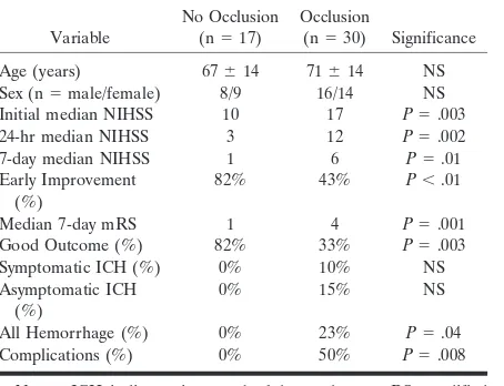
Overall, the location of occlusion had a significant effect on the NIHSS scores at onset, 24 hours and 7 days (P ⬍ .02,P ⫽ .01, P ⫽ .05, respectively) (see Table 1). The stroke subtype based on TOAST crite-ria was associated with median NIHSS scores at onset (cardioembolic 18 versus large artery atherosclerosis 12 versus lacunar 6,P⫽ .008) but not 7-day NIHSS (P ⫽ .1). The presence or absence of major arterial occlusion had a significant relationship to many of the outcome variables (see Table 2). Despite the large range of overlap in initial NIHSS scores between patients with occlusion (NIHSS range 8 –27) and without visualized occlusion (NIHSS range 5–27), there was a statistically significant difference in the median initial NIHSS (17 versus 10, respectively,P⫽ .003). Infarction territories in those patients without visualized occlusion on CTA were distributed as la-cunar (n⫽3), ACA (n⫽1), M1 (n⫽1), M2 (n⫽5) and M3/M4 (n⫽7).
[image:3.585.57.531.71.123.2]In a univariate analysis of predictors of early im-provement, only the presence of occlusion (OR 6.1, 95% confidence interval 1.4 to 25.8) and time to treatment (OR 0.98, CI 0.96 – 0.99, P ⫽ .04) were significant (see Fig 1). Increasing time to treatment was correlated with a worse 24-hour NIHSS (rho⫽ 0.375,P⫽.01). However, after adjustment for initial NIHSS, age, and time to treatment, only the absence
TABLE 2: Impact of the presence or absence of visualized arterial occlusion on CTA
Variable
No Occlusion
(n⫽17)
Occlusion
(n⫽30) Significance
Age (years) 67⫾14 71⫾14 NS
Sex (n⫽male/female) 8/9 16/14 NS
Initial median NIHSS 10 17 P⫽.003
24-hr median NIHSS 3 12 P⫽.002
7-day median NIHSS 1 6 P⫽.01
Early Improvement (%)
82% 43% P⬍.01
Median 7-day mRS 1 4 P⫽.001
Good Outcome (%) 82% 33% P⫽.003
Symptomatic ICH (%) 0% 10% NS
Asymptomatic ICH (%)
0% 15% NS
All Hemorrhage (%) 0% 23% P⫽.04
Complications (%) 0% 50% P⫽.008
Note.—ICH indicates intracerebral hemorrhage; mRS, modified Rankin Scale; NIHSS, National Institutes of Health Stroke Scale. TABLE 1: Location of arterial occlusion and median NIHSS scores
Absent n⫽17 M2 n⫽19 M1 n⫽7 Multiple n⫽4 Significance
Initial NIHSS 10 (5–27) 15 (8–27) 18 (2–27) 18 (14–22) P⬍.02
24-hr NIHSS 3 (0–16) 12 (1–27) 12 (4–32) 8 (1–17) P⫽.01
7-day NIHSS 1 (0–10) 7 (1–27) 8 (0–22) NM P⫽.05
[image:3.585.307.534.174.348.2]of occlusion remained associated with early improve-ment (OR 5.0, CI 1.1 –23.3, P⫽.04).
In a univariate analysis of predictors of a seven-day good outcome (mRS ⱕ 2), both initial NIHSS (OR 1.14, 95% CI 1.0 –1.3,P⫽.006) and age (OR 1.06, CI 1.01 –1.12, P⫽.03) were associated with a small but significantly worse seven-day outcome. In this small cohort, we found no effect of time to treatment or stroke subtypes on the seven-day good outcome. Whereas, absence of occlusion was associated with a significantly better seven-day outcome (OR 9.3, 95% CI 2.2 to 40.1,P⫽.003) (see Fig 2). After adjustment for initial NIHSS, age, and time to treatment, only the absence of occlusion remained associated with a good seven-day outcome (OR 6.8, CI 1.3 –34.6, P⫽.02).
Complications, which included all intracranial hemorrhages, systemic hemorrhages or death, were not statistically associated with age, time to treat-ment, initial NIHSS score, site of occlusion or stroke subtypes. Only the presence of occlusion was signifi-cantly associated with complications (see Table 2). Symptomatic intracranial hemorrhage occurred in 6.4% of all cases. However, none of the above
vari-ables reached statistical significance in association with symptomatic hemorrhages. Patients without vi-sualized occlusion had a significantly lower preva-lence of combined asymptomatic and symptomatic intracranial hemorrhages (P ⬍.04). No patient with symptomatic or asymptomatic hemorrhage had a good seven-day outcome (P⫽.003). Unlike the pres-ence of occlusion, initial NIHSS score was not a statistically significant risk factor for combined intra-cranial hemorrhage.
Discussion
Because of the risk of systemic and intracerebral hemorrhage (1– 6) and the narrow therapeutic win-dow (1), the widespread use of IV tPA as a treatment for acute stroke ischemic stroke remains challenging. We believe CT angiography identifies a subgroup of patients without occlusion of the large proximal ar-teries of the circle of Willis who have a significantly better outcome and less hemorrhagic risk after treat-ment with IV tPA than those with a demonstrated arterial occlusion proximal to the M3 branches. The clinical suspicion of ischemia as the cause of the neurologic deficits was very high and all patients had evidence of infarct on subsequent MR imaging or examination. Therefore CTA was not used in our practice as a means to separate those who would or would not be treated with IV tPA. Instead it was initially implemented to identify patients who might benefit from subsequent intraarterial lysis. CTA is immediately performed following the emergency un-enhanced CT scan. The time to acquire CTA images is approximately five minutes and has not dramati-cally affected our time to treatment, which is lower than other published reports (16, 17).
Our outcomes data are in agreement with Verro et al (18) who reported that occlusion on CTA por-tended a worse NIHSS at discharge in acute stroke patients who did not receive thrombolytic therapy. Unlike the study by Verro et al, all of our patients were treated with IV rt-PA within the three hours from stroke onset and had imaging preceding throm-bolysis. It is important to note that our data do not offer information on the efficacy of IV tPA in patients without visualized major arterial occlusion because there is no nontreated comparison group. Efficacy studies would require randomization of patients with and without visualized occlusion to a treatment arm and an observation arm. In fact one might argue that tPA is incidental to recovery in those patients with patent vasculature as evidenced by Toni et al (19), who demonstrated in nonthrombolysis patients that patent vasculature on conventional angiography was associated with improved outcome. The difficulty with interpreting the data from Toni et al is that their angiographic imaging was delayed up to 48 hours thereby precluding the potential application for acute treatment algorithms and the good outcome may have only identified those with spontaneous recana-lization, lacunes or stroke mimics.
In retrospect, it may appear obvious that patients
FIG 1. Site of occlusion predicts early improvement at 24
hours. Those patients without CTA-visualized occlusion proxi-mal to the M3 division have significantly better early improve-ment (P⬍.02).
without visualized occlusion will have better out-comes than those with occlusion, however it is not possible to reliably determine this by clinical exami-nation and a standard CT scan alone. As illustrated, two patients (one with occlusion vs. one without) in our cohort were indistinguishable by NIHSS scores (both scores of 27), unenhanced CT or clinical exam-ination. However the patient without occlusion had early improvement and a 7-day NIHSS of four com-pared with the other patient with occlusion who had no change and a 7-day NIHSS of 27. Although there are many reasons for a patient without visualized occlusion to have a large stroke score, the problem remains that the clinical examination and standard CT do not yield adequate information for appropriate understanding of the complex temporal events that occur during ischemic insult. A patient who has patent extracranial and large intracranial arteries and a persistent deficit may fall into five pathophysiolog-ical states: 1) large arterial recanalization before CTA with persistent penumbral tissue that will return to normal function; 2) large arterial recanalization be-fore CTA with completed infarct; 3) small vessel lacunar infarct (not visible on CTA); 4) multiple dis-tal embolic occlusion beyond the reliable resolution of CTA; 5) stroke mimics such as migraine and sei-zures. In examples one, two and five IV tPA is un-likely to be helpful and could potentially be harmful. Whereas, cases three and four are possibly most re-sponsive to IV tPA.
Pilot data from the IV tPA study group (8) dem-onstrated that IV tPA was more effective in achieving vascular recanalization in stroke due to small distal MCA occlusions then in MCA stem, ICA or basilar occlusions. These results were further validated by Wolpert et al (7) who demonstrated an 8% recanali-zation rate for extracranial artery occlusion compared with 41% recanalization in intracranial arteries. The effect of thrombus location on recanalization is also supported by Linfante et al (20) who demonstrated that MCA occlusions were more likely to recanalize compared with ICA occlusions. It is possible that some patients with higher NIHSS without major prox-imal arterial occlusion had experienced spontaneous recanalization before the CTA despite all imaging done within 3 hours of onset. At least six patients without occlusion proximal to M3 had infarction in the M1 and M2 territories despite no evidence of clot in these territories supporting the concept of hyper-acute recanalization. Since all patients in this study had evidence of infarct on follow-up, none of these patients represented stroke mimics or complete early reperfusion TIAs and only three patients without CTA occlusion had lacunar stroke subtypes. The small sample size is a limitation of the present retro-spective analysis. However, we believe this cohort is representative of larger studies given the similar me-dian initial NIHSS score and prevalence of symptom-atic hemorrhage compared with the NINDS study cohort (1). In the absence of visualized occlusion proximal to the M3, none of the patients developed intracerebral hemorrhage. The lack of hemorrhagic
complications may be due to smaller areas of isch-emic tissue damage, better collateral flow in patients with distal branch occlusion, more rapid thrombolysis or avoidance of delayed reperfusion injury (9, 10). The presence of occlusion itself may be another risk factor for intracranial hemorrhage (2, 21). In the NINDS study (1) lower hemorrhage rates were seen in patients with better NIHSS. Although the patients in our cohort with absence of visualized occlusion had lower initial NIHSS scores, we did not demonstrate an effect of initial NIHSS scores on hemorrhage. In fact, presence or absence of visualized occlusion was the only variable that was statistically significant in our small study. If this finding is not due to chance then it suggests that it may play a larger role or select for a more homogenous pathophysiology than previ-ously reported risk factors such as age, sex, hyperten-sion or NIHSS score (2, 21). Of course other possi-bilities include inadequate power to detect an effect or that there may have been other factors which contributed to outcome and hemorrhage which we did not evaluate.
Conclusion
We believe that CTA can cheaply, safely and rap-idly support prospective stratification of stroke patients based on location, degree and presence of vascular occlusion. For patients receiving IV throm-bolysis, CTA with patent vasculature or occult arterial occlusion identifies a subgroup that is likely to have good clinical outcomes with low risk of hemorrhagic complications. In addition, CTA may be important in creating more homogeneous patient cohorts for clin-ical trials who might expect a differential response to treatment based on site of occlusion. Coupled with CT perfusion imaging based on bolus tracking meth-ods, contrast CT study in acute stroke patients can also provide insight into the temporal window of ischemic injury (22–24). The demonstrated efficacy of intraarterial thrombolysis for patients with M1-M2 occlusion (25) and the increasing body of literature suggesting that the level of arterial occlusion corre-lates with tPA efficacy and outcomes supports the concept of treatment stratification based on the site of vascular occlusion. Further studies are needed to validate our findings and to confirm the utility of CTA as a means of patient selection for acute intervention.
References
1. The National Institute of Neurological Disorders and Stroke rt-PA
stroke study group.Tissue plasminogen activator for acute
isch-emic stroke.N Engl J Med1995;333:1581–1587
2. The NINDS t-PA stroke study group.Intracerebral hemorrhage
after intravenous t-PA therapy for ischemic stroke. Stroke.
1997;28:2109 –2118
3. Hacke W, Kaste M, Fieschi C, et al.Intravenous thrombolysis with
recombinant tissue plasminogen activator for acute hemispheric stroke. The European cooperative acute stroke study (ECASS).
JAMA.1995;274:1017–1025
4. Hacke W, Kaste M, Fieschi C, et al.Randomised double-blind
alteplase in acute ischaemic stroke (ECASS II). Second European-Australasian acute stroke study investigators. Lancet.1998;352: 1245–1251
5. Clark WM, Wissman S, Albers GW, Jhamandas JH, Madden KP,
Hamilton S.Recombinant tissue-type plasminogen activator
(alte-plase) for ischemic stroke 3 to 5 hours after symptom onset. The ATLANTIS study: A randomized controlled trial.JAMA.1999;282: 2019 –2026
6. Katzan IL, Furlan AJ, Lloyd LE, et al.Use of tissue-type
plasmin-ogen activator for acute ischemic stroke: The Cleveland area expe-rience.JAMA.2000;283:1151–1158
7. Wolpert SM, Bruckmann H, Greenlee R, Wechsler L, Pessin MS,
del Zoppo GJ.Neuroradiologic evaluation of patients with acute
stroke treated with recombinant tissue plasminogen activator. The rt-PA acute stroke study group. AJNR Am J Neuroradiol.
1993;14:3–13
8. del Zoppo GJ, Poeck K, Pessin MS, et al. Recombinant tissue
plasminogen activator in acute thrombotic and embolic stroke.Ann Neurol.1992;32:78 – 86
9. Heo JH, Kim SH, Lee KY, Kim EH, Chu CK, Nam JM.Increase
in plasma matrix metalloproteinase-9 in acute stroke patients with thrombolysis failure.Stroke.2003;34:48 –50
10. Montaner J, Molina CA, Monasterio J, et al.Matrix
metallopro-teinase-9 pretreatment level predicts intracranial hemorrhagic complications after thrombolysis in human stroke. Circulation.
2003;107:598 – 603
11. Knauth M, von Kummer R, Jansen O, Hahnel S, Dorfler A, Sartor K.Potential of CT angiography in acute ischemic stroke. AJNR Am J Neuroradiol.1997;18:1001–1010
12. Lev MH, Farkas J, Rodriguez VR, et al.CT angiography in the
rapid triage of patients with hyperacute stroke to intraarterial thrombolysis: Accuracy in the detection of large vessel thrombus.
J Comput Assist Tomogr.2001;25:520 –528
13. Shrier DA, Tanaka H, Numaguchi Y, Konno S, Patel U, Shibata D. CT angiography in the evaluation of acute stroke.AJNR Am J Neuroradiol.1997;18:1011–1020
14. Haley EC, Jr., Lewandowski C, Tilley BC.Myths regarding the
NINDS rt-PA stroke trial: Setting the record straight.Ann Emerg
Med.1997;30:676 – 682
15. Adams HP, Jr., Bendixen BH, Kappelle LJ, et al.Classification of
subtype of acute ischemic stroke. Definitions for use in a multi-center clinical trial. TOAST. Trial of org 10172 in acute stroke treatment.Stroke.1993;24:35– 41
16. Koennecke HC, Nohr R, Leistner S, Marx P.Intravenous tPA for
ischemic stroke team performance over time, safety, and efficacy in a single-center, 2-year experience.Stroke.2001;32:1074 –1078 17. Albers GW, Bates VE, Clark WM, Bell R, Verro P, Hamilton SA.
Intravenous tissue-type plasminogen activator for treatment of acute stroke: The standard treatment with alteplase to reverse stroke (STARS) study.JAMA.2000;283:1145–1150
18. Verro P, Tanenbaum LN, Borden NM, Sen S, Eshkar N. CT
angiography in acute ischemic stroke: Preliminary results.Stroke.
2002;33:276 –278
19. Toni D, Fiorelli M, Bastianello S, et al.Acute ischemic strokes
improving during the first 48 hours of onset: Predictability, out-come, and possible mechanisms. A comparison with early deterio-rating strokes.Stroke.1997;28:10 –14
20. Linfante I, Llinas RH, Selim M, et al.Clinical and vascular
out-come in internal carotid artery versus middle cerebral artery oc-clusions after intravenous tissue plasminogen activator. Stroke.
2002;33:2066 –2071
21. Kent DM, Ruthazer R, Selker HP.Are some patients likely to
benefit from recombinant tissue-type plasminogen activator for acute ischemic stroke even beyond 3 hours from symptom onset?
Stroke.2003;34:464 – 467
22. Cullen SP, Symons SP, Hunter G, et al.Dynamic
contrast-en-hanced computed tomography of acute ischemic stroke: CTA and CTP.Semin Roentgenol.2002;37:192–205
23. Lev MH, Segal AZ, Farkas J, et al.Utility of perfusion-weighted CT
imaging in acute middle cerebral artery stroke treated with intra-arterial thrombolysis: Prediction of final infarct volume and clin-ical outcome.Stroke.2001;32:2021–2028
24. Wintermark M, Reichhart M, Thiran JP, et al.Prognostic accuracy
of cerebral blood flow measurement by perfusion computed tomog-raphy, at the time of emergency room admission, in acute stroke patients.Ann Neurol.2002;51:417– 432
25. Furlan A, Higashida R, Wechsler L, et al.Intra-arterial
prouroki-nase for acute ischemic stroke. The PROACT II study: A random-ized controlled trial. Prolyse in acute cerebral thromboembolism.