Magnetic Resonance Imaging of the Cavernous Sinus
Full text
Figure
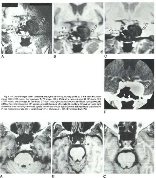
![Fig. 1.-Pituitary section. (Reprinted from [7].) gland and anterior part of cavernous sinus](https://thumb-us.123doks.com/thumbv2/123dok_us/1179479.640636/2.615.57.563.85.504/fig-pituitary-section-reprinted-gland-anterior-cavernous-sinus.webp)

Related documents
Thus, in this study, we compared the difference of clinic-pathologic characteristics, especially tumor and pathology molecular markers, and 5-year overall survival rate between MGC
Moreover, patients with ductal carcinoma but not those with lobular carcinoma had a decreased anti-ABCC3 IgG level compared with control subjects (adjusted P=0.0006); the levels
The two synthetic compounds 15b and 15h is tested for their ability to prevent cervical cancer using HeLa cell line.. The study shows
Figure 1-2: Sperm dynamics in cauda epididymis after endosulfan treatment in rats. Sperm motility is affected by altered enzymatic activities of
21 formalin-fixed and paraffin-embedded (FFPE) tissue specimens of heart valves from 18 BCNE patients were subjected to hematoxylin-eosin (HE) staining and Periodic acid-Schiff
We calculated the biologically effective dose (BED) of each radiation regimen to compare the effects of the treatments. This observation may account for the poor survival
reuteri tested for antibacterial activity the unconcentrated filtrate showed no effect on bacterial isolates the same results obtained with one-fold concentrated
Trope nominalism, of the sparse, a posteriori type, combined with a dependence- based bundle theory of more substantial particulars, a process ontology, a non- maximalist
![Bis[tris(pyrazol 1 yl)methane κ2N,N′]platinum(II) bis(hexafluorophosphate) nitromethane solvate](data:image/gif;base64,R0lGODlhAQABAIAAAP///wAAACH5BAEAAAAALAAAAAABAAEAAAICRAEAOw==)