Robert M. Quencer1 Berta M. Montalvo 1
This article appears in the September/Octo-ber 1984 issue of AJNR and the December 1984 issue of AJR.
Received November 2, 1983; accepted January 4,1984.
, Department of Radiology R-130, University of Miami School of Medicine, University of Miami/ Jackson Memorial Medical Center, P.O. Box 016960, Miami, FL 331 01. Address reprint requests to R. M. Quencer.
AJNR 5:501-505, September/October 1984 0195-6108/84/0505-0501
© American Roentgen Ray Society
501
Normal Intraoperative Spinal
Sonography
The normal intraoperative sonographic features of the spinal canal, spinal cord, conus medullaris, and cauda equina are described and illustrated. Important observations concerning the normal spinal cord include its highly reflective dorsal and ventral surfaces, its uniform hypoechogenicity, and the presence of a central echo. Other easily identified structures within the spinal canal include the dura-arachnoid layer, subarach-noid space, denticulate ligament, dorsal arachsubarach-noid septations, and the roots of the cauda equina. In addition the sonographic appearance of commonly encountered iatro-genically introduced material including Gelfoam, Pantopaque, cottonoid pledgets, suture material, Harrington rods, and freeze-dried dura is also demonstrated. These normal images can serve as a baseline for the interpretation of various pathologic conditions of the spinal canal and its contents as seen with intraoperative spinal sonography.
With the widespread use of computed tomography and water-soluble myelo-graphic contrast agents, the ability to more precisely assess the spine and its contents has improved significantly over the past few years. Magnetic resonance imaging of the spine offers an even more promising method of spine evaluation without subjecting the patient to the risk and discomfort of myelography. Sonog-raphy has also been reported as a pre- and postoperative method of imaging the spine in postlaminectomy patients [1] and in screening infants with spinal dysraph-ism [2-5].
Despite these advances in preoperative diagnoses, until recently [6, 7] there has not been a satisfactory method of imaging the spine and its contents during surgery. Such a method would enable structures beyond the immediate operative field to be visualized, and abnormalities within the dura or spinal cord could be identified and localized before being subjected to operative intervention. The recent development of a high-resolution, portable, real-time sonographic scanner that can be used in the operating room allows not only the visualization of intraspinal abnormalities but permits the surgeon and radiologist to assess the progress and final result of surgery before the operation is completed. We have termed this procedure intraoperative spinal sonography (lOSS).
A basic understanding of the normal sonographic anatomy of the spine is essential before full advantage of this technique can be taken. It is our object to describe and illustrate the normal intraoperative sonographic anatomy of the spinal canal, spinal cord, and cauda equina, so that this information can serve as a foundation for analyzing various pathologic states of the spinal canal and its contents. The sonographic characteristics of a number of iatrogenically introduced materials will also be described and shown.
Materials and Methods
502
QUENCER AND MONTALVO
AJNR:5, Sept/Oct 1984A
8
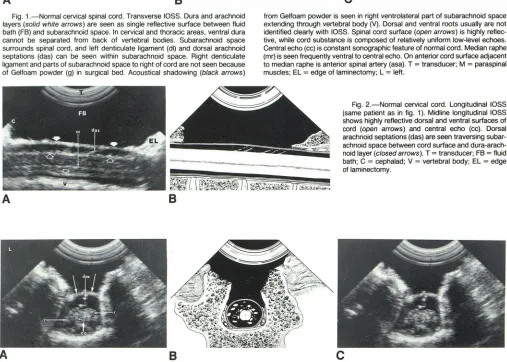
Fig. 1. -Normal cervical spinal cord. Transverse lOSS. Dura and arachnoid
layers (solid white arrows) are seen as single reflective surface between fluid
bath (FB) and subarachnoid space. In cervical and thoracic areas, ventral dura
cannot be separated from back of vertebral bodies. Subarachnoid space surrounds spinal cord, and left denticulate ligament (dl) and dorsal arachnoid
sept at ions (das) can be seen within subarachnoid space. Right denticulate ligament and parts of subarachnoid space to right of cord are not seen because
of Gelfoam powder (g) in surgical bed. Acoustical shadowing (black arrows)
A
8
from Gelfoam powder is seen in right ventrolateral part of subarachnoid space extending through vertebral body (V). Dorsal and ventral roots usually are not
identified clearly with lOSS. Spinal cord surface (open arrows) is highly reflec-tive, while cord substance is composed of relatively uniform low-level echoes. Central echo (cc) is constant sonographic feature of normal cord. Median raphe
(mr) is seen frequently ventral to central echo. On anterior cord surface adjacent
to median raphe is anterior spinal artery (asa). T = transducer; M = paraspinal muscles; EL = edge of laminectomy; L = left.
Fig. 2.-Normal cervical cord. Longitudinal lOSS
(same patient as in fig. 1). Midline longitudinal lOSS
shows highly reflective dorsal and ventral surfaces of cord (open arrows) and central echo (ee). Dorsal arachnoid septations (das) are seen traversing subar-achnoid space between cord surface and dura-arach-noid layer (closed arrows). T = transducer; FB = fluid bath; C = cephalad; V = vertebral body; EL = edge
of laminectomy.
Fig. 3.-Normal conus medullaris. Transverse lOSS. Dura-arachnoid layer (long arrows), dorsal arachnoid septations (das), multiple roots (r) surrounding conus
medullaris, highly reflective surfaces of normal conus (short arrows), and central echo (ee) are well seen. L = left. with resultant bone, disk and/or bullet fragments within the canal,
posttraumatic intramedullary or subarachnoid cysts, spondylosis, and herniated disks, had been analyzed sonographically. Our ability to distinguish normal from abnormal findings in the canal and spinal cord on lOSS comes from this clinical experience and the correlation we have made with the findings during surgery. Normal lOSS images are shown in figures 1-7, and accompanying drawings highlight the important features. The sonographic images are appropriately la-beled, but the matched drawings are self-explanatory and are free of any overlying labels. Also, in figures 1 and 3, where many labels were used, a duplicate sonographic image is shown for clarity.
The images shown here were obtained with the ATL NeuroSectOR portable real-time unit (Advanced Technology Laboratories, Bellevue,
[image:2.612.61.558.82.223.2] [image:2.612.53.560.218.580.2]AJNR:5, Sept/Oct 1984 NORMAL INTRAOPERATIVE SPINAL SONOGRAPHY
503
Fig. 4.-Normal conus medullaris. Longitudinal lOSS (same patient as in fig. 3). Midline longitudinal sonogram shows typical tapering of conus medullaris and highly reflective nature of cord surface (open arrows). Central echo (cc) is seen extending to tip of conus. Dorsal arachnoid septations (das) extend from surface of conus to dura-arachnoid layer (short solid arrows). Roots distal to tip of conus are poorly seen because they are at periphery of sound beam. Echo-genic band (long straight arrows) perpendicular to vertebral body edge is intervertebral disk material. Multiple scattered air bubbles (curved arrows) can be seen within fluid bath. C = cephalad.
Fig. 5.-Normal proximal cauda equina. Trans-verse lOSS. Individual roots (r) of proximal cauda equina can be seen forming nearly an x-shaped pat-tern, a configuration noted often by us on transverse lOSS in this area. Single midline structure may rep-resent filum terminale (ft). Slight separation can be seen between ventral dura-arachnoid layers (arrow) and back of vertebral body (v). Probably this is lumbar epidural space, which may become even more prom-inent in distal lumbar canal. L = left.
Fig. 6.-Normal proximal cauda equina. Longitu-dinal lOSS (same patient as in fig. 5). Along central acoustical axis, higher level echoes are seen when compared with more peripheral parts of sonogram. Roots of cauda equina are identified clearly. C = cephalad.
A
A
A
after a corpectomy (i.e., partial or total removal of a vertebral body). The transducer is placed in a sterile latex sheath and is coupled to it
via a sterile gel. Any air bubbles trapped within the latex sheath are
smoothed out, and rubber bands are placed over the latex sheath to
secure it firmly to the transducer.
lOSS is performed in both longitudinal and transverse planes. Multiple images are obtained in each plane by gradually moving the transducer across the operative field. The entire procedure is
re-corded on videotape, and in addition filmed static images are obtained
throughout the study. On the longitudinal sonograms our convention is to have the most cephalad area to the viewer's left. The transverse
sonograms are labeled just as the surgeon sees the operative field,
namely, after a laminectomy, the patient's left will be to the viewer's
B
B
left. Conversely, following a corpectomy the patient's left will be to
the viewer's right.
Observations
504
QUENCER AND MONTALVO AJNR:5, Sept/Oct 19847
8
9
10
11
12
sound beam, and as a result the reflectivity pattern in the
center of the image is the strongest (e.g., fig. 6).
Conse-quently, structures of the spinal canal are seen most clearly
when they are in line with the central acoustical axis.
There-fore, before an area of the spinal cord is called abnormal, the
examiner must be assured that the area under question is
centered to the transducer. Bleeding can be observed on
real-time sonography as finely echogenic uniform material of
relatively high intensity that swirls and mixes with the fluid
bath. This fresh blood usually then layers along the dorsal
surface of the dura and forms a fluid layer (fig. 8), but
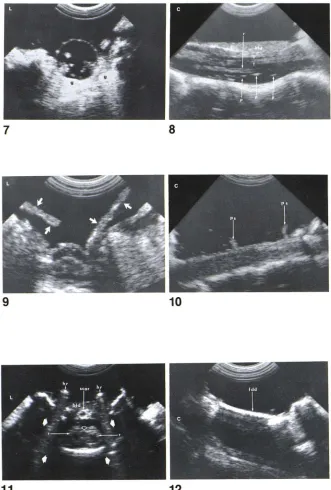
Fig. 7.-Normal distal cauda equina. Transverse lOSS. Distal roots of cauda equina are no longer seen to be arranged in x-shaped pattern as was seen in proximal cauda equina (see fig. 5) but instead are more widely spread throughout subarachnoid space. Thick, highly echogenic areas surrounding thecal sac ventrally and laterally represent Gelfoam (g). Gelfoam
blends with and acoustically shadows ventral epidural space and adjacent vertebral body. L
=
left.Fig. 8.- Pantopaque in subarachnoid space. Lon
-gitudinal lOSS. Roots (r) of cauda equina are well seen within subarachnoid space. When perpendicular
to sound beam, each root is seen as parallel echo-genic lines (arrows) with central hypoechogenic
re-gion. In addition isolated, highly reflective echogenic structures (p) represent Pantopaque droplets within cerebrospinal fluid. Blood (bid) is seen layering along
dorsal dural surface. C = cephalad.
Fig. 9.-Cottonoid pled gets in laminectomy de -fect. Transverse lOSS. After posterior decompression for cervical stenosis and cervical spondylosis, c
otto-noid pled gets (arrows) were placed in surgical site. These pledglets are less echogenic than Gelfoam
powder and are not associated with significant acoustical shadowing (cf. figs. 1 and 7). As in fig. 1,
dura-arachnoid layer, ventral and dorsal subarachnoid space, dorsal arachnoid septations, and highly reflec -tive surface of cervical cord are seen well. L = left.
Fig. 10.-Suture material used to sew dura to
-gether. Longitudinal lOSS. After opening of dura, needle biopsy of thoracic intramedullary mass was
performed. Subarachnoid space has collapsed after dura and arachnoid were entered. Prolene sutures
(ps), used to sew dura back together, are seen along
dorsal dural surface projecting into fluid bath. Sutures
are creating no shadowing artifacts. Central echo is
not visualized, which we attribute to infiltrating spinal
cord glioma. C = cephalad.
Fig. 11.-Harrington rods. Transverse lOSS. Pa
s-sage of sound waves through Harrington rods (hr) results in typical sonographic reverberations (closed
arrows) that obscure all anatomic details in its path.
Layering of blood (bid) and densely echogenic scar
(scar) are seen along dorsal dural surface. Echogenic
surface of conus (open arrow) and roots of cauda
equina surrounding conus (r). L
=
left.Fig. 12.-Dural graft. Longitudinal lOSS. After
posterior C l-C2 decompression and excision of dura
for treatment of Chiari I malformation, freeze-dried
dura (fdd) was used as dural graft. This echogenic
material transmits sound waves poorly, and as a
result no significant sonographic information behind this material can be obtained. Note the lack of visual -ization of spinal cord. C = cephalad.
occasionally it can appear as clumps of finely echogenic
material dorsal to the dura. Constant features outside of the
spinal canal seen with lOSS at all levels examined are labeled
only in figures 1 and 2; these include the transducer, the fluid
bath in which the transducer sits, dorsal paraspinal muscles,
edge of the laminectomy, and the vertebral body. Normal
lOSS of the spinal cord is shown and described later (figs,
1-7). Iatrogenically introduced substances commonly seen with
lOSS include Gelfoam, Pantopaque, cottonoid pledgets, su
-tures, Harrington rods, and freeze-dried dura. These are
[image:4.612.53.384.87.577.2] [image:4.612.56.386.90.213.2]AJNR:5, Sept/Oct 1984 NORMAL INTRAOPERATIVe SPINAL SONOGRAPHY 505
Spinal Cord
In figures 1 and 2 intraopoerative sonography of a normal cervical cord after a midcervical laminectomy is shown in the transverse (fig. 1) and longitudinal (fig. 2) planes. The only sonographic difference we have noticed between the cervical and thoracic levels is that the thoracic cord has a rounder
shape and the anterior spinal artery is less clearly defined in the thoracic area. The important structures are clearly labeled
and illustrated; however, a few features deserve special
com-ment. The dorsal· and ventral surfaces of the spinal cord are seen as highly reflective structures because the change in physical density from cerebrospinal fluid to spinal cord tissue creates an acoustical interface. The spinal cord itself is com
-posed of relatively uniform, low-level echoes, and this hypo-echogenicity is best appreciated when the spinal cord is compared with the echogenic properties of the surrounding paraspinal muscles. The linear highly reflective structure in the mid to ventral part of the spinal cord is the central echo.
These three features-the highly reflective cord surfaces, the uniform hypoechogenicity of the cord, and the central echo-should be looked for carefully, because in our experience [6] their absence indicates the presence of a pathologic process affecting the spinal cord. The dorsal and ventral rootlets surrounding the cervical and thoracic. cord are usually not
clearly delineated; however, the dorsal arachnoid septations and the denticulate ligaments are commonly seen within the
subarachnoid space. It is important not to mistake these
normal structures for either aberrantly coursing nerve roots
or fibrotic bands or scars. Constant pulsations of the cord that reflect the normal heartbeat can be observed with real-time lOSS. Occasionally, pulsations of the anterior spinal artery itself can be seen.
Conus Medullaris
The characteristic tapering of the conus medullaris is well seen on longitudinal lOSS (fig. 4), and on midline sections the central echo can be followed to its tip. On transverse lOSS (fig. 3) the highly reflective surface of the conus is more difficult to identify than is the cord surface in the thoracic or cervical area because the conus is surrounded by nerve roots
that tend to blend in with the surface of the conus. However, careful observation will show the individual roots to be sepa-rable from the conus itself.
Cauda Equina
Individual roots of the cauda equina area seen both in the upper (figs. 5 and 6) and lower (fig. 7) lumbar canal. Fewer roots are seen in the distal sac since the upper lumbar roots have already exited. We have noted that in the upper cauda equina the roots assume nearly an X pattern when viewed transversely (fig. 5). In the lumbar sac there is often a
widening of the ventral epidural space because of the
pres-ence of fat and connective tissue. This may be seen so no-graphically as a space between the dura-arachnoid layer and
the edge of the adjacent vertebral body (fig. 5). This space is
not seen as well in the cervical and thoracic spine since the ventral epidural space is normally very small in those areas.
Iatrogenically Introduced Substances
During an operative procedure, materials may be used that may cause a confusing sonographic image and/or may o
b-scure many of the anatomic details within the spinal canal. A
number of these substances are shown in figs. 7-12. Gelfoam
powder used in the surgical wound to diminish bleeding is highly echogenic (fig. 7) and will obscure many of the details within the spinal canal. Pantopaque droplets (fig. 8) within the
subarachoid space are seen as highly reflective structures, without shadowing, not in continuity with the surrounding nerve roots. Cottonoid pled gets (fig. 9) have a typical
·squared-off" appearance with echogenic properties quite
different from those of Gelfoam powder. The pledgets are less echogenic and do not have the acoustical shadowing we have noted with Gelfoam powder. Suture material commonly
used to sew the dura (prolene sutures in fig. 10) causes no shadowing artifacts in the sonographic field. Harrington rods (fig. 11) are clearly identified with lOSS and, like other metallic
foreign bodies, give off a characteristic reflective shadow in response to the effect of the sound waves striking the metallic
rods. Freeze-dried dura (fig. 12) acts as an effective acoustical
barrier, and as a result no useful sonographic information is obtained distal to this material after it has been sewed in place.
ACKNOWLEDGMENTS
We thank Barth A. Green and Frank J. Eismont for providing the patients whose sonograms appear in this article.
REFERENCES
1. Braun IF, Ragahavendra BN, Kricheff II. Spinal cord imaging
using real-time high-resolution ultrasound. Radiology 1983;
147:459-465
2. James HE, Scheible W, Kerber C, Hilton SVW. Comparison of high-resolution real-time ultrasonography and high-resolution
computed tomography in an infant with spinal dysraphism.
Neu-rosurgery 1983;13:301-305
3. Scheible W, James HE, leopold GR, Hilton SVW. Occult spinal dysraphism in infants: screening with high-resolution reaJ..tme ultrasound. Radiology 1983;146:743-746
4. Ragahavendra BN, Epstein FJ, Pinto RS, Subramanyam BR,
Greenberg J, Mitnick JS. The tethered spinal cord: diagnosis by high-resolution real-time ultrasound. Radiology 1983;149:1 23-128
5. Naidich TP, Mclone DG, Shkolnik A, Fembach SK. Sonographic evaluation of caudal spine anomalies in children. A1
1983;4:661-664
6. Quencer RM, Morse BMM, Green BA, Eismant FJ, Brost P.
Intraoperative spinal sonography: adjunct to metrizBmide CT in the assessment and surgical decompression of posttraumatic
spinal cord cysts. AJNR 1984;5: 71-79. AIR 1984;1 :593-601 7. Rubin JM, Dohrmann GJ. Work in progress: intraoperatMt""