David M. Yousem, Laurie A. Loevner, Jennifer D. Tobey, Rena J. Geckle, Warren B. Bilker, and Ara A. Chalian
PURPOSE: To determine the factors that correspond to adenoidal hypertrophy, often prominent in human immunodeficiency virus (HIV)-positive patients. METHODS: The sagittal T1-weighted MR images of 21 HIV-positive patients (age range, 25 to 50 years; mean, 37 years) and 21 healthy control subjects (age range, 24 to 55 years; mean, 35 years) were reviewed blindly and indepen-dently by two radiologists who measured the maximal dimension of the nasopharyngeal lymphoid tissue. Twenty-six additional HIV-positive patients were combined with the original 21 HIV-positive patients, and the hematologic studies of these 47 patients were compared with the adenoidal measurements to assess whether a relationship existed between nasopharyngeal prominence and hematocrit, white blood cell count, and CD4 count. RESULTS: Mean adenoidal width was 6.76 mm (SD, 5.82) in the HIV-positive population, but was only 3.36 mm (SD, 2.48) in the age-matched control group. Age and HIV status correlated with nasopharyngeal width measurements. No relationship between adenoidal width and hematocrit, CD4 count, or white blood cell count was evident. CONCLUSION: After correcting for age, we found that adenoidal lymphoid tissue is more abundant in HIV-positive persons than in control subjects. The hematologic ramifications of this finding remain uncertain.
Index terms: Acquired immunodeficiency syndrome (AIDS); Neck, inflammation
AJNR Am J Neuroradiol18:1721–1725, October 1997
Head and neck lesions in patients with ac-quired immunodeficiency syndrome (AIDS), AIDS-related complex, and human immunode-ficiency virus (HIV) positivity are common, with a reported prevalence in the range of 40% to 70% (1– 4). On sagittal magnetic resonance (MR) images of the head and on the scout views of computed tomographic (CT) scans, promi-nence of the nasopharyngeal adenoidal tissue is present in 35% of patients who are HIV positive (1). In fact, the constellation of nasopharyngeal adenoidal tissue prominence, abnormal bone
marrow signal (5), posterior triangle lymphad-enopathy (6), and intraparotid cysts and or nodes (6, 7) strongly suggests HIV positivity. All of these findings may be seen on sagittal scout views of MR images of the brain; the axial MR or CT studies may not necessarily include the pa-rotid glands or posterior triangle lymph node regions. The purpose of this investigation was to examine the relationship between nasopharyn-geal adenoidal width seen on MR images and hematologic factors associated with HIV infec-tion.
Materials and Methods
Twenty-one HIV-positive patients and 21 HIV-negative persons had MR imaging between July 1995 and July 1996. (The HIV testing was performed as part of an enroll-ment procedure for a different project involving MR spec-troscopy.) The HIV-negative subjects, who formed the control group, had no risk factors for AIDS. The ages of the HIV-positive patients ranged from 25 to 50 years, with a mean age of 37 years; the age range of the healthy volun-teers was from 24 to 53 years, with a mean age of 35 years. Standard deviations were 6.6 years for the HIV-positive patients and 8 years for the HIV-negative subjects.
Received January 30, 1997; accepted after revision April 28. Dr Geckle was supported by research grant 5 PO1 DC 00161–15 from the National Institute on Deafness and Other Communication Disorders, National Institutes of Health.
From the Departments of Radiology (D.M.Y., L.A.L., R.J.G.), Otorhino-laryngology: Head and Neck Surgery (D.M.Y., A.A.C.), and Clinical Epide-miology and Biostatistics (W.B.B.), University of Pennsylvania Medical Center, and the Department of Radiology, Thomas Jefferson University Hospital (J.D.T.), Philadelphia, Pa.
Address reprint requests to David M. Yousem, MD, Department of Radiology, University of Pennsylvania Medical Center, 3400 Spruce St, Philadelphia, PA 19104.
AJNR 18:1721–1725, Oct 1997 0195-6108/97/1809 –1721
©American Society of Neuroradiology
The sagittal T1-weighted (500 – 600/11–17/1 [repeti-tion time/echo time/excita[repeti-tions], 256 3 192 matrix, 24-cm field of view) MR images of the 42 subjects were reviewed blindly and independently by two neuroradiolo-gists. The readers measured the maximal dimension of the nasopharyngeal lymphoid tissue at a plane roughly per-pendicular to the skull base using the scales provided with the images (Fig 1). Measurements were rounded to the nearest millimeter. The mean value of the two neuroradi-ologists’ measurements was correlated with patients’ age and HIV status. MR images from an additional group of HIV-positive patients (n526) obtained between February 1991 and April 1992 were evaluated in an identical man-ner and were combined with the first set to assess the relationship between nasopharyngeal width and CD4 count, hematocrit, and white blood cell count at the time the MR examination was performed. Fifty-six MR studies were performed in the 47 HIV-positive persons (21 from the 1995–96 group and the 26 additional patients). A hematologic study including either a hematocrit (n556), white blood cell count (n5 52), or CD4 count (n5 21) was performed within 3 weeks before or after the MR examination in every patient.
The relationships between age, nasopharyngeal width, and HIV status were evaluated initially with Spearman’s rank correlation coefficients and with an exact stratified Wilcoxon-Mann-Whitney test (stratified for age) to assess for a difference in nasopharyngeal width in HIV-positive and HIV-negative persons, adjusted for age. The values of the hematologic studies (hematocrit, white blood cell count, and CD4 count) were compared with mean naso-pharyngeal adenoidal width using a Spearman’s correla-tion coefficient and partial Spearman’s correlacorrela-tion coeffi-cients corrected for age.
The nasopharyngeal width measurements between the two observers were equal in 34 of the 77 readings and were within 1 mm of each other in 33 cases. The maximum discrepancy between readings was 4 mm (n 5 1). An intraclass correlation coefficient, calculated to assess in-terobserver agreement between values, was .984 (95% CI, .974, .993) indicating excellent agreement.
Results
The mean adenoidal width was 6.76 mm (SD, 5.82 mm) in the 21 HIV-positive patients. The values ranged from 1.5 mm to 20 mm, with a median value of 5.0 mm. For the control sub-jects, the mean nasopharyngeal adenoidal width was 3.36 mm (SD, 2.48 mm). The mini-mum value was 1 mm and the maximini-mum value was 10 mm (median, 2.5 mm). Only three of the healthy volunteers had nasopharyngeal ad-enoidal widths greater than 4.5 mm (Fig 2). Using Spearman’s rank correlation coefficient, we found an inverse relationship between pa-tients’ age and nasopharyngeal adenoidal tissue width in the 21 HIV-positive patients (P5.045), but not in the 21 control subjects (P 5 .18). After adjustment for age, using an exact strati-fied Wilcoxon-Mann-Whitney test stratistrati-fied for age, we found the HIV-positive subjects had sig-nificantly wider nasopharyngeal tissue (P 5
.01) than did the HIV-negative subjects.
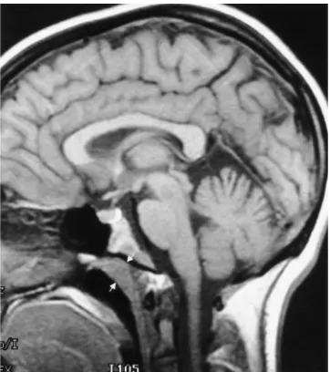
Spearman’s rank correlation coefficient showed no significant relationship between ad-Fig 1. Sagittal T1-weighted MR image shows the technique
[image:2.587.79.260.83.287.2]used to measure maximum adenoidal width perpendicular to the skull base (betweenarrows). In some cases the measurement was anteroposterior in direction, depending on the configuration of the adenoidal tissue.
[image:2.587.305.548.84.277.2]enoidal tissue width and CD4 count (P 5 .80, n521 samples), hematocrit (P 5.63, n5 56 samples), or white blood cell count (P 5 .58, n552 samples) in the 47 HIV-positive patients. We then performed a partial Spearman’s corre-lation analysis adjusting for the age of the HIV-positive patients. This analysis showed no rela-tionship between hematocrit, white blood cell count, and CD4 count and nasopharyngeal width (all P values..60).
No patient had nasopharyngeal lymphoma at the time of the examination or at follow-up by clinical report.
Discussion
That the nasopharyngeal adenoidal tissue may become hypertrophied in patients who are HIV positive is well established (1, 8). France et al (8) found adenoidal hypertrophy in 33 (60%) of 55 HIV positive patients in their series. In the vast majority of cases, the enlargement is at-tributable to reactive lymphoid hyperplasia, al-though histopathologic proof of this finding has been limited to just a few studies (1, 9 –12). Among HIV-infected persons, lymphoid hyper-plasia may occur in this location even in those who have had an adenoidectomy (13).
Histopathologically, one finds follicular hy-perplasia with large and irregular germinal cen-ters (9, 14, 15) and thinning of the mantle zones (11). Particles of the HIV virus, HIV antigens, or markers for its RNA can be detected within these germinal centers or on the mucosal sur-face of the adenoids (14, 16, 17). One study found granulomas although no microorganisms were seen in the specimens (15). Nasopharyn-geal adenoidal hypertrophy is commonly asso-ciated with hyperplastic lymph nodes that also have follicular hyperplasia within them (11, 12, 14). Fox et al (17) postulated that the lymphoid tissue in the nodes and adenoids may serve as a filter for the virus and associated immune com-plexes. However, the retained immune complex antigens on the dendritic surface of the follicular lymphoid cells may pass onto any cell that traverses the germinal center, thereby causing greater dissemination of the HIV virus (17, 18). Thus, the lymphoid tissue serves as a reservoir of HIV RNA, and the continued stimulus of the virus may induce hyperplasia.
Although lymphoid hyperplasia is the most common cause of adenoidal enlargement in this patient population, numerous reports of
un-usual pathogens that may produce similar find-ings have been published. Organisms such as
Rhinosporidium seeberi (1, 19), Microsporida
(20, 21) organisms, Staphylococcus aureus
(22), Bordetella pertussis (23), and Pneumo-cystis carinii (13) have been cultured from na-sopharyngeal aspirates in HIV-infected patients. Additionally, one must be concerned about the possibility of lymphoma (1, 10), Kaposi sar-coma (1), minor salivary gland neoplasms, or squamous cell carcinoma (1) when one identi-fies an abnormality in this region. Symptoms of nasal stuffiness, ear congestion, nasal bleeding, and hearing loss (with or without associated cervical or parotid lymphadenopathy) (9, 15, 24) may be present with any of the entities listed above. Only when there is asymmetry of the lymphoid tissue, deep invasion, obliteration of the parapharyngeal and/or retropharyngeal fat, or obliteration of the distinction between the tensor and levator veli palatini muscles should one suspect a more aggressive process. One report of malignant transformation of nasopha-ryngeal lymphoid hyperplasia into a diffuse large-cell immunoblastic lymphoma has been published, documented with serial radiographs and biopsy findings (10).
difference between the CD4 count of the patients with normal adenoidal depth and that of the group with moderate hypotrophy, the patients with less adenoidal tissue (intense hypotrophy) had a sig-nificantly lower CD4 count than did those with normal adenoidal tissue or moderate hypotrophy. The difference in the two studies may be due to the different methods used to measure the ade-noidal tissue, to the use of clustered groups in Zagdanski’s study, to the use of controlled data for age in our study, or to other mitigating factors that may affect the immunologic status of an HIV-positive person. In fact, our HIV-HIV-positive patient with the lowest CD4 count (CD4 count53) had an adenoidal width (2 mm) similar to that of our patient with the highest CD4 count (adenoidal width53 mm, CD4 count5655).
The relationship between advancing age and declining nasopharyngeal width (P5.049) was not unexpected, since regression of this tissue may occur into the fourth and fifth decades. The relationship between HIV status and adenoidal measures was independent of the age factor (P 5.0046).
A number of factors that could conceivably affect adenoidal enlargement were not exam-ined in this study. Some of the medications commonly taken by HIV-positive patients to bolster their immune system presumably might affect the lymphoid proliferation. We did not solicit histories of adenoidectomy, active or past oral candidiasis infection, or presence of ongoing odontogenic infections in our subjects, and, since this was a retrospective study, most of the patients have died in the interval between 1991 and the completion of the study. Our in-stitution has only recently been quantifying viral load in HIV-positive patients; this is a potential source of fruitful evaluation if the adenoids are truly a reservoir for HIV antigens.
In summary, HIV-positive patients have ade-noidal enlargement when compared with age-and sex-matched control subjects, although the range of adenoidal hypertrophy is wide. Factors other than hematocrit, total white blood cell count, and CD4 count should be explored to ascertain what, if any, relationship there is be-tween adenoidal width and stage of disease.
Acknowledgment
We acknowledge the immense help of Zoraida Morris of the Laboratory Information Systems, Department of
Pa-thology and Laboratory Medicine, in obtaining hemato-logic results in the study patients.
References
1. Olsen WL, Jeffrey RB Jr, Sooy CD, Lynch MA, Dillon WP. Lesions of the head and neck in patients with AIDS: CT and MR findings.
AJR Am J Roentgenol1988;151:785–790
2. Rosenberg RA, Schneider KL, Cohen NL. Head and neck presen-tations of acquired immunodeficiency syndrome. Otolaryngol Head Neck Surg1985;93:700 –705
3. Rosenberg RA, Schneider KL, Cohen NL. Head and neck presen-tations of acquired immunodeficiency syndrome.Laryngoscope
1984;94:642– 646
4. Marcusen DC, Sooy CD. Otolaryngologic and head and neck manifestations of acquired immunodeficiency syndrome (AIDS).
Laryngoscope1985;95:401– 405
5. Eustace S, McGrath D, Albrecht M, Fogt F, Buff B, Longmaid HE. Clival marrow changes in AIDS: findings at MR imaging. Radiol-ogy1994;193:623– 627
6. Shugar JM, Som PM, Jacobson AL, Ryan JR, Bernard PJ, Dick-man SH. Multicentric parotid cysts and cervical adenopathy in AIDS patients: a newly recognized entity. CT and MR manifesta-tions.Laryngoscope1988;98:772–775
7. Holliday RA, Cohen WA, Schinella RA, et al. Benign lymphoepi-thelial parotid cysts and hyperplastic cervical adenopathy in AIDS-risk patients: a new CT appearance.Radiology1988;168: 439 – 441
8. France AJ, Kean DM, Douglas RH, et al. Adenoidal hypertrophy in HIV-infected patients (letter).Lancet1988;2:1076
9. Shahab I, Osborne BM, Butler JJ. Nasopharyngeal lymphoid tis-sue masses in patients with human immunodeficiency virus-1: histologic findings and clinical correlation.Cancer1994;74:3083– 3088
10. Kieserman SP, Stern J. Malignant transformation of nasopharyn-geal lymphoid hyperplasia.Otolaryngol Head Neck Surg1995; 113:474 – 476
11. Barzan L, Carbone A, Tirelli U, et al. Nasopharyngeal lymphatic tissue in patients infected with human immunodeficiency virus: a prospective clinicopathologic study.Arch Otolaryngol Head Neck Surg1990;116:928 –931
12. Barzan L, Carbone A, Saracchini S, Vaccher G, Tirelli U, Comor-etto R. Nasopharyngeal lymphatic tissue hypertrophy in HIV-in-fected patients (letter).Lancet1989;1:42– 43
13. Biavati MJ, Khan A, Kessler C. DisseminatedPneumocystis cari-nii infection involving the neck and nasopharynx. Otolaryngol Head Neck Surg1993;109:773–776
14. Carbone A, Gloghini A, Vaccher E, Barzan L, Tirelli U. Nasopha-ryngeal lymphoid tissue masses in patients with human immuno-deficiency virus-1 (letter).Cancer1995;76:527–528
15. Stern JC, Lin PT, Lucente FE. Benign nasopharyngeal masses and human immunodeficiency virus infection.Arch Otolaryngol Head Neck Surg1990;116:206 –208
16. Pantaleo G, Graziosi C, Demarest JF, et al. HIV infection is active and progressive in lymphoid tissue during the clinically latent stage of disease [see comments].Nature1993;362:355–358 17. Fox CH, Tenner-Racz K, Racz P, Firpo A, Pizzo PA, Fauci AS.
Lymphoid germinal centers are reservoirs of human immunode-ficiency virus type 1 RNA [published erratum appears inJ Infect Dis1992;165:1161].J Infect Dis1991;164:1051–1057
19. Gori S, Scasso A. Cytologic and differential diagnosis of rhino-sporidiosis.Acta Cytol1994;38:361–366
20. Doultree JC, Maerz AL, Ryan NJ, et al. In vitro growth of the microsporidian Septata intestinalis from an AIDS patient with disseminated illness.J Clin Microbiol1995;33:463– 470 21. Ryan NJ, Sutherland G, Coughlan K, et al. A new
trichrome-blue stain for detection of microsporidial species in urine, stool, and nasopharyngeal specimens. J Clin Microbiol
1993;31:3264 –3269
22. Amir M, Paul J, Batchelor B, et al. Nasopharyngeal carriage of Staphylococcus aureus and carriage of tetracycline-resistant strains associated with HIV-seropositivity.Eur J Clin Microbiol Infect Dis1995;14:34 – 40
23. Cohn SE, Knorr KL, Gilligan PH, Smiley ML, Weber DJ. Pertussis is rare in human immunodeficiency virus disease.Am Rev Respir Dis1993;147:411– 413
24. Chow JH, Stern JC, Kaul A, Pincus RL, Gromisch DS. Head and neck manifestations of the acquired immunodeficiency syndrome in children.Ear Nose Throat J1990;69:416 – 419, 422– 413 25. Davis BR, Zauli G. Effect of human immunodeficiency virus
in-fection on haematopoiesis.Baillieres Clin Haematol1995;8:113– 130