FORUM
Editor's note: From time to time a particularly provoc-ative Letter-to-the-Editor is received. Rather than include such letters as part of the "Letters to the Editor" feature, occasionally we will elect to have a "Neuroradiology Forum," in which the original comment, responses, and additional remarks solicited by the Editor are published. The following forum was stimulated by a comment con-tributed by Dr Howard Yonas et al.
Internal Carotid Balloon Test Occlusion Does
Require Quantitative CBF
We would like to respond to the three articles in the Nov /Dec issue of AJNR ( 1-3) concerning the use of cere-bral blood flow (CBF) studies with Tc-99m HMPAO SPECT as an aid for reducing the associated risk of stroke following acute internal carotid artery (ICA) occlusion. Risk assess-ment for acute ICA occlusion has been an area of great interest at the University of Pittsburgh. Over 400 balloon test occlusions (BTO) have been performed in conjunction with xenon-enhanced tomographic (Xe/CT) CBF measure-ment as an aid to guide decision making in patients who may require carotid occlusion. Although not consistently stated in all previous articles, our criterion for identification
Group I
no baseline asymmetry, BTO asymmetry CBF
a)
c)
baseline
b)
• 30ml/100g/mln
occluded side nonoccluded side
Fig. 1. Schematic illustration of CBF response in 42 patients, who presented with symmetric baseline CBF values, but devel-oped hemispheric asymmetries due to BTO. Bilateral (a) increase, (b) decrease, and (c) increase on nonoccluded side with simulta-neous decrease on occluded side have been observed.
of patients with high risk for developing cerebral infarction metry (Fig .. 1): 1) In 5.1% (eight/156) of patients, asym-has been asymmetrically reduced blood flow on the side of metry developed due to an asymmetrical
increase in
CBF occlusion with the development of CBF values below 30 on both sides during BTO. Considering that this group mL/1 00 g/min (4). We believe that using only the devel- presented with average CBF values of 64 (45-119) mL/ opment of hemispheric asymmetry due to BTO is too 100 g/min and 76 (52-137) mL/1 00 g/min on the occluded sensitive and lacks adequate specificity for distinguishing and the nonoccluded side, respectively, these patients a group of patients at elevated risk of hemodynamic cannot be considered to be at increased risk of developinginfarction. ischemia. 2) In 10.9% (17 /156) of patients, asymmetry
Based on the quantitative analysis of CBF response to developed during BTO by decreasing CBF more on the BTO in a randomly selected group of patients (n = 156), . occluded and the nonoccluded side. Only 11.8% (two/17) we would like to support, but also advise caution, regarding of this group dropped CBF values below 30 mL/100 g/ some of the conclusions of the AJNR articles. Our concerns min due to BTO. 3) In 10.9% (17 /156) of patients, CBF are based on our observation, which is similar to the results dropped on the occluded side and increased on the non-of Monsein et al (3), that, immediately following BTO, CBF occluded side, resulting in asymmetry. Only 17.6% (three/ may rise differentially either on the side of occlusion or on 17) of patients in this group exhibited flow values below the contralateral side. Without absolute flow values, asym- 30 mL/1 00 g/min. Thus, while asymmetry alone would metries at normal or even elevated CBF levels may be identify 26.9% (42/156) of patients to be at risk, our criteria given unwarranted significance. (4) would identify only 3.2% (five/156).
We agree with Peterman et al (1) and Moody et al (2) Like Moody et al (3) we are concerned about the group that, regardless of the technique utilized, having an ICA- (9.6% of our series, 15/156) who presented with baseline occluded CBF study increases the sensitivity for detecting (preocclusion) asymmetry and had an increased asymme-patients at higher risk for hemodynamically induced stroke try during BTO (Fig. 2): 1) In 7.1% (11/156) of patients, after permanent internal carotid artery occlusion, compared CBF fell 19% on the occluded side, but CBF fell below 30 with exclusive reliance on neurologic examinations during mL/1 00 g/min only in two of these patients. The average BTO. Because qualitative SPECT studies are dependent on asymmetry of these 11 patients increased from 17% to the development of asymmetry, it is important to appre- 32%. 2) In two of these 15 patients, CBF increased bilat-ciate that 26.9% (42/156) of our patients who revealed no erally, resulting in increased asymmetry (12%-21 %). This asymmetry prior to BTO developed asymmetry (contralat- group of patients were not likely to have an increased eral-ipsilateral/average >10%) during BTO. We identified hemodynamic risk from carotid occlusion because they three different types of CBF responses leading to asym- exhibited CBF values between 53.4 and 77.0 mL/1 00 g/
1148
Group II
baseline asymmetry, rised BTO asymmetry
CBF
c 30ml/100g/mln
8
occluded side
b)
c)
baseline a)
nonoccluded side
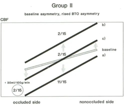
Fig. 2. Schematic illustration of CBF response in 15 patients, who showed asymmetry prior to BTO and increased asymmetry during BTO. Bilateral (a) decrease, (b) increase, and (c) increase on nonoccluded side with simultaneous decrease on occluded side have been observed.
min during BTO. 3) The remaining two from the group of 15 patients demonstrated decreased CBF on the occluded side and increased CBF on the nonoccluded side. Asym-metry in this group increased from 19% to 31% during BTO and CBF values ranged between 35.8 and 63.7 mL/ 100 g/min during BTO.
Between September 1982 and June 1991, 34 patients underwent acute therapeutic ICA occlusion guided by our Xe/CT CBF criteria. Fifteen (44%) of these patients would not have met the criteria of symmetry (contralateral-ipsi-lateral/average< 10%) considered to be the limit of normal by Risberg et al (5).
While quantitative Tc-99m HMPAO SPECT imaging in combination with clinical BTO may be sensitive enough to identify most patients at risk for hemodynamic stroke after ICA sacrifice, it lacks the specificity available from quanti-tative CBF studies. Excluding patients from therapy based on qualitative Tc-99m HMPAO SPECT will reduce one's stroke rate. Unfortunately a significant number of patients who could safely tolerate ICA sacrifice, and in whom ICA sacrifice might be the best therapeutic choice, will also be excluded.
Although Xe/CT is a demanding technology, it has been proven to be a safe and reliable clinical study. The tech-nique is totally noninvasive and because studies which require 6 minutes of data acquisition can be repeated within 20 minutes, baseline and BTO studies can be both com-pleted within an hour. Even though we and others have been concerned that xenon inhalation can alter CBF, recent studies have shown that this has no significant effect on the Xe/CT CBF-derived flow calculation (6, 7). In the near future, the availability of Xe/CT systems that are cost effective and independent of CT manufacturers should overcome earlier problems of limited distribution.
References
AJNR: 13, July/ August 1992
Howard Yonas, MD*·t
Mark Linskey, MD* David W. Johnson, MDt Joseph A. Horton, MDt lvo P. Janecka, MD~
J.-Peter Witt* Charles Jungreis, MDt William L. Hirsch, MDt Laligam N. Sekhar, MD*
* Department
of Neurological Surgery t Department of Radiology and ~ Department of Otolaryngology University of Pittsburgh School of Medicine Pittsburgh, PA1. Peterman SB, Taylor A, Hoffman JC. Improved detection of cerebral hypoperfusion with internal carotid balloon test occlusion and 99
m Tc-HMPAO cerebral perfusion SPECT imaging. AJNR 1991; 12:
1035-1041
2. Moody EB, Dawson RC, Sandler MP. 99
mTc-HMPAO SPECT imaging in interventional neuroradiology: validation of balloon test occlusion. AJNR 1991;12:1043-1044
3. Monsein LH, Jeffrey PJ, van Heerden BB, et al. Assessing adequacy of collateral circulation during balloon test occlusion of the internal carotid artery with 99
mTc-HMPAO SPECT. AJNR 1991;12: 1045-1051
4. Linskey ME, Sekhar LN, Horton JA, Hirsch WL, Yonas H. Aneurysms of the intracavernous carotid artery: a multidisciplinary approach to treatment. J Neurosurg 1991; 75:525-534
5. Risberg J, Halsey JH, Wills EL, Wilson EM. Hemispheric specialization in normal man studied by bilateral measurement of the regional cerebral blood flow. Brain 1975;98:511-524
6. Good W, Gur D. Xenon enhanced CT of the brain: effect of flow activation on derived cerebral blood flow measurement. AJNR 1991; 12:83-85
7. Witt JP, Holl K, Heissler HE, Dietz H. Stable xenon CT CBF: effects of blood flow alterations on CBF calculations during inhalation of 33% stable xenon. AJNR 1991; 12:973-975
Editor's note: This comment by Yonas et al was sent to the authors of the three articles cited within it. Their responses follow.
Reply
Assessment of Collateral Cerebral Circulation
during Test Occlusion of the Carotid Artery
A number of direct and indirect techniques are available that allow assessment of cerebral perfusion. In the context of cerebral test occlusion studies, a methodology that has a high enough sensitivity to exclude those that will have inadequate collateral circulation, and thus be spared the risk of hemiplegia, is necessary. Stable xenon/CT, Xe133 external probe, Xe133 SPECT, H2
0
15
[image:2.614.55.302.78.288.2]AJNR: 13, July/ August 1992
who might have benefited from carotid occlusion might be unnecessarily rejected and required to face the hazards of
their original disease. As long as the specificity of the test is also high, however, a high sensitivity can be tolerated.
Tc-99m HMPAO SPECT test occlusion studies as
de-scribed by Peterman et al (1 ), Moody et al (2), and Eckard
and Purdy (3) have a high sensitivity but suffer from a low
specificity because they are nonquantitative. This becomes
a dangerous situation because the sensitivity of such tests can be easily altered by changing the acquisition, display,
and interpretation of the information gathered. The impor-tance of this principle is demonstrated by our own data.
We have noted subtle changes with Tc-99m HMPAO SPECT and TCD (unpublished data) between baseline and test occlusion studies on almost every test occlusion study we have done. We could, therefore, potentially exclude all
patients from having permanent occlusion of the carotid artery.
On the other hand, when tolerance of an occlusion test of the carotid artery is based upon accurate cerebral blood flow (CBF) data, the specificity of the methodology is increased, resulting in fewer people being inappropriately
prevented from having permanent occlusion. For this rea-son, we enthusiastically agree with Yonas et al (see letter) that, at this time, decisions about the adequacy of cerebral test occlusion during occlusion tests are optimally based upon quantitative measurements of CBF.
Although Dr Yonas's group has demonstrated the safety and utility of the stable xenon/CT balloon test occlusion methodology, we have some concerns. Although Dr Yonas claims, "The technique is totally non invasive . . . " his group (4) has reported an incidence of 3.7% complications
of which the predominance was dissections of the carotid artery. We have never experienced this complication and believe that transporting a patient to a CT scanner with a catheter in the carotid artery and inflating and deflating the balloon in a blind fashion can only increase the potential of these untoward events. Furthermore, compared to SPECT, the equipment continues to be expensive and less available, the spatial resolution is relatively poor, and the patient receives a higher radiation dose.
For these reasons, we believe that a methodology that provides high-resolution quantitative CBF information and
is either portable or allows transport of the patient to the
instrument after the balloon has been removed will be the most useful in the long run. At the present time, further improvements in attenuation and scatter correction, sen-sitivity and spatial resolution, tracers, and blood sampling or input function acquisition are required before SPECT will fulfill this need.
Before any particular level of CBF is universally accepted
as critical for maintenance of normal cerebral function in
humans, further validation is required. Furthermore, several
other issues need to be addressed. For how long should
the test be performed and how many CBF measurements should be obtained? Should patient activity, blood pressure, or other physiological indicators be considered during or after the test occlusion? Should the auto regulatory
capac-1149
ity of the cerebral circulation be assessed in conjunction
with test occlusion of the carotid?
References
Lee Monsein, MD Johns Hopkins Hospital Baltimore, MD
1. Peterman SB, Taylor A, Hoffman JC. Improved detection of cerebral hypoperfusion with internal carotid balloon test occlusion and 99mTc
-HMPAO cerebral perfusion SPECT imaging. AJNR 1991;12:
1035-1041
2. Moody EB, Dawson RC, Sandier MP. 99mTc-HMPAO SPECT imaging
in interventional neuroradiology: validation of balloon test occlusion. AJNR 1991; 12:1043-1044
3. Eckard DA, Purdy PD. Temporary balloon occlusion of the carotid artery combined with brain blood flow imaging as a test to predict tolerance prior to permanent carotid sacifice. Proceedings of the 29th Annual Meeting of the American Society of Neuroradiology. 1991:
160
4. Tarr RW et al. Complications of preoperative balloon test occlusion of the internal carotid arteries: experience in 100 cases. Proceedings
of the 29th Annual Meeting of the American Society of Neuroradiol-ogy. 1991:138
Reply
We reviewed with interest the comments by Yonas et
al, stressing the need for true quantitative information when assessing asymmetries of cerebral perfusion that develop during balloon test occlusion of an internal carotid artery.
Their data suggest that the evaluation of SPECT images using semi-quantitative methods may have a much higher
false positive rate than those seen in truly quantitative
methods such as Xe-CT. We acknowledge that quantitative
methods are preferable, but do not agree that a false positive asymmetry discovered on SPECT imaging neces -sarily results in withholding of treatment. In many cases,
patients developing perfusion asymmetries during balloon test occlusion are suitable candidates for vascular graft to
augment blood flow to the ipsilateral hemisphere. The
action taken following a false positive study is more likely to be a modified surgical approach rather than withholding
of treatment. Given the high stroke morbidity associated
with permanent internal carotid artery sacrifice, the per-formance of occasional "unnecessary" grafting procedures
may result in lower overall surgical morbidity.
Although all of their data is not available for review, the 3.2% of patients identified to be "at risk" due to perfusion
asymmetry is well below the reported incidence of stroke
associated with permanent carotid sacrifice. We would be interested to hear their explanation for this. A flow
thresh-old that is too low will increase the number of false negative
studies and increase the number of postoperative strokes.
The authors have considerable experience (over 400
cases) with balloon test occlusion. At many institutions,
test occlusion is an infrequently performed procedure and
would not warrant the purchase of Xe-CT equipment.
SPECT imaging offers the advantage of requiring no equip
1150
departments, being less labor intensive, and having no need for a second balloon inflation during imaging. The cost effectiveness and availability of SPECT imaging make it an attractive alternative for institutions where balloon test occlusion is an infrequently performed procedure. The accompanying relatively high false positive rate should reduce the incidence of stroke associated with the proce-dure. The cost for this is that it may increase the number of vascular graft procedures performed, and, in rare cases, result in withholding of treatment. However, we think that semi-quantiative analysis of SPECT images provides a significant improvement over reliance on neurologic testing alone.
With the proliferation of PET imaging centers, the future of quantitative cerebral blood flow imaging during balloon test occlusion probably rests in this modality. The radio-pharmaceuticals 13-NH3 and 11-C-Nicotine have suitable half lives for the identification of flow abnormalities during test occlusion and we suspect that in the future PET imaging will play an important role in the assessment of cerebral hemodynamics during interventional procedures.
Reply
Edward B. Moody, MD Robert C. Dawson, MD*
Martin P. Sandler, MD Department of Radiology and Radiological Sciences Vanderbilt University Medical Center Nashville, Tennessee
*
Currently with the Department of RadiologyEmory University Hospital
Atlanta, GA
We agree with Yonas et al that quantification of regional cerebral blood flow (rCBF) probably would increase the specificity of Tc-99m HMPAO cerebral perfusion SPECT and internal carotid balloon test occlusion (BTO) in detect-ing clinically silent cerebral hypoperfusion, identifying pa-tients at higher risk for infarction post-permanent occlu-sion. Like xenon-enhanced tomographic (Xe/CT) cerebral imaging with BTO, Tc-99m HMPAO with BTO is evolving from rCBF pattern identification to rCBF quantification (1-4). At present, there is no generally accepted technique for
quantitating rCBF in mL/100 g/min using Tc-99m HMPAO
SPECT imaging, although Monsein et al have reported
early work in this area (5). Even without quantification, our experience does not suggest that a significant decrease in specificity occurs. We believe that Tc-99m HMPAO SPECT scanning is more suitable than Xe/CT in evaluating rCBF during BTO. However, no study to date has reported enough patients to show statistical evidence that any type of cerebral blood flow imaging with BTO is beneficial.
We would expect the rate of clinically silent hypoper-fusion during BTO to match the 5%-20% reported rate of infarctions post-permanent occlusion in patients with clin -ically negative BTO (2, 3, 6). In our report, two of 17 patients ( 11%) met our criteria of clinically silent, reversible, asymmetric hypoperfusion (7). We have now studied a
AJNR: 13, July/ August 1992
total of 32 patients, five of whom met our criteria (16%). These rates are well within the expected 5%-20% rate.
Thus, our method of Tc-99m HMPAO rCBF pattern iden-tification does not seem to be overly sensitive.
Monsein et al had a higher rate of asymmetric Tc-99m HMPAO scans with BTO than would be expected, perhaps related to the increased sensitivity of their four-headed SPECT camera (5). They began to quantify rCBF using a ratio of brain SPECT scan to peripheral arterial blood counts. With further development of this technique, rCBF may be routinely quantified with Tc-99m HMPAO SPECT scans.
Yonas et al argue in favor of Xe/CT with BTO, citing their unpublished data of 156 patients. If the rCBF dropped below 30 mL/100 g/min during BTO, their patients were categorized as moderate risk for infarction post-permanent occlusion; the rCBF pattern did not matter. New hemi-spheric asymmetry developed in 42/156 patients; pre- and post-BTO asymmetry was present in 15/156 patients. Five of the 156 patients (3.2%) met their criteria for moderate risk for infarction post-permanent occlusion. Thirty-four patients, 15 of which had asymmetry, underwent perma-nent carotid occlusion.
These data raise several questions. The 3.2% moderate risk rate is lower than the expected 5%-20% rate; however the 5%-20% rate, based on small studies, has a large variance (2, 3, 6). It is unclear how many patients with asymmetry and rCBF below 30 mL/100 g/min underwent permanent carotid occlusion. Patient outcome post-per-manent occlusion is not mentioned; we do not know if moderate risk patients were indeed at risk for infarction
post-permanent occlusion.
Yonas et al describe Xe/CT as noninvasive and safe. Both Xe/CT and Tc-99m HMPAO SPECT scans are non-invasive; BTO, which is performed in both, is an invasive procedure. Xe/CT with BTO has a reported complication rate of 3.7%, most of which are arterial dissections (8). With an uninflated balloon catheter in the internal carotid artery, the patient is transferred to the CT department. The balloon is then inflated without fluoroscopy. We have had no complications other than a groin hematoma. The Tc-99m HMPAO is injected intravenously while the balloon catheter is inflated under fluoroscopy in the angiography suite. The pharmacokinetics then allow the balloon catheter to be deflated and removed before the patient is transported to the nuclear medicine department for the SPECT scan.
We would agree with Yonas et al that Xe/CT is a demanding technology that is still not widely available. Most nuclear medicine departments with a SPECT camera
can perform a Tc-99m HMPAO SPECT scan. Thus, BTO
with Tc-99m HMPAO is available in most large institutions with active neurosurgery, otolaryngology, neuroradiology,
and nuclear medicine departments.
AJNR: 13, July/August 1992
more widely available technology. Tc-99m HMPAO rCBF
quantification, currently under development, probably
would increase BTO and Tc-99m HMPAO specificity. Since the risk of infarction following permanent occlusion in patients who have clinically passed BTO is relatively low (5%-20%), a large study under controlled circumstances is required to evaluate the predictive value of a positive test. It is unlikely that a single center will be able to acquire enough patients with permanent carotid occlusion, so we are pursuing a multicenter study. Finally, moderate risk patients need not be excluded from permanent carotid
occlusion; arterial bypass may be performed in addition to permanent occlusion to prevent infarction postocclusion.
References
Susan Brothers Peterman, MD Andrew Taylor, Jr., MD James
C.
Hoffman, Jr., MDDepartment of Radiology Emory University School of Medicine Atlanta, GA 30322
1. Erba SM, Horton JA, Latchaw RE, et al. Balloon test occlusion of the internal carotid artery with stable xenon/CT cerebral blood flow imaging. AJNR: Am J Neuroradio/1988;9:533-538
2. de Vries EJ, Sekhar LN, Horton JA, et al. A new method to predict safe resection of the internal carotid artery. Laryngoscope 1990; 100:85-88
3. Steed DL, Webster MW, de Vries EJ, et al. Clinical observations on the effect of carotid artery occlusion on cerebral blood flow mapped by xenon computed tomography and its correlation with carotid artery back pressure. J Vase Surg 1990; 11 :38-44
4. Linskey ME, Sekhar LN, Horton JA, Hirsch WL, Yonas H. Aneurysms
of the intracavernous carotid artery: a multidisciplinary approach to
treatment. J Neurosurg 1991;75:525-534
5. Monsein LH, Jeffery PJ, van Heerden BB, et al. Assessing adequacy
of collateral circulation during balloon test occlusion of the internal carotid artery with 99
mTc-HMPAO SPECT. AJNR: Am J Neuroradio/
1991 ;12:1045-1051
6. Gonzalez CF, Moret J. Balloon occlusion of the carotid artery prior to surgery for neck tumors. AJNR: Am J Neuroradiol 1990; 11: 649-652
7. Peterman SB, Taylor A, Hoffman JC. Improved detection of cerebral hypoperfusion with internal carotid balloon test occlusion and 99
mT
c-HMPAO cerebral perfusion SPECT imaging. AJNR: Am J Neuroradiol
1991 ;12:1035-1041
8. Tarr RW et al. Paper presented at the annual meeting of the American Society of Neuroradiology, Washington, DC, June 1991
Editor's note: Dr Yonas' comment was also sent to Richard
Frackowiak, MA, MD, FRCP, Professor of Clinical Neurol-ogy and Assistant Director of the MRC Cyclotron Unit, Hammersmith Hospital, London. Dr Frackowiak is a rec-ognized authority on cerebral blood flow and agreed to respond although he admits to little experience in either
Tc-99m HMPAO SPECT scanning or xenon CT scanning.
His commentary follows.
Reply
Since I have neither major experience in Tc-99m HMPAO SPECT scanning nor in xenon CT scanning, I can
1151
only look at this issue from the perspective of a practicing
vascular neurologist and someone with more than a passing
interest in cerebrovascular hemodynamics.
The first issue is that clinical practice, before high technology investigations became available, clearly
dem-onstrated that stroke could occur following occlusion of
the internal carotid artery for therapeutic purposes, not
only on the operating table, but also in a delayed fashion,
up to a number of days later. The reason for this was not
entirely clear at the time although various proposals were
raised, including 1) hemodynamic compromise of a critical
nature that required some further insult to tip the brain into ischemia and 2) the possibility that clots might form
proximally or distally to the occlusion and act as the site
of origin of investigations of cerebral hemodynamics have only limited value. Having said that, embolization into a
region with impaired hemodynamics should, on a priori grounds, be more dangerous than that into an area where
perfusion and vascular reserve mechanisms are normal. The three recent papers are of great interest (1, 2, 3). I think it is clear that asymmetry of perfusion may occur for a number of reasons before, during, and after balloon occlusion. Before occlusion, such asymmetry may repre-sent areas of previous ischemia and infarction that may also have been clinically silent. During occlusion, they may
indicate areas of hemodynamic compromise, and following
ischemia, they may represent differential reactive
hyper-perfusion, or ischemic damage caused by the balloon
occlusion, which may itself have remained clinically silent.
I think that the point, made forcefully in the commentary
from Pittsburgh, that absolute levels of blood flow would be helpful to make these distinctions, is well taken. It should, however, be remembered that hypoperfusion in response to vascular occlusion is a comparatively late phenomenon suggesting critical hemodynamics. The initial response to decreased perfusion pressure is a reactive
vasodilation that maintains perfusion. Indeed, it is the " dis-obliteration" of such occlusion with reperfusion into a
dilated vasculature that may be responsible for some of
the postocclusive hyperperfusion. It is not until this
vasa-dilating reserve is exhausted that perfusion begins to fall in response to further falls in perfusion pressure and, at this stage, function and metabolism are maintained by an
increasing fractional extraction of energy substrates.
This brings me back to the issue of absolute levels of
blood flow. Clearly, one must perform an estimation prior to balloon occlusion given that the test itself might co
n-ceivably result in some permanent neuronal loss. Secondly,
because the circle of Willis distributes the perfusion
pres-sure from each carotid artery to both sides of the brain,
occlusion on one side may have effects on both.
Asym-metries may therefore be small and this suggests that
absolute falls in blood flow are as important to detect as
any asymmetry. This point is again made by the group
from Pittsburgh. They appear to have the largest experi
-ence and certainly their results would seem to confirm
conclusions that could be drawn from theory. This phe
1152
by Moody et al (2) is unable to help us empirically in this
respect as only six cases are described. The most powerful
argument produced for Tc-99m SPECT method is that it
is easy to perform and gives a snapshot measurement of
cerebral blood flow which can be obtained by making the
tracer injection at the time of occlusion itself. The article
by Peterman and her colleagues (1) refers to 17 patients,
which is also rather a small number. The results of this
article are marred by the fact that the control scan was
performed after the balloon inclusion. It is also unfortunate
that one of 15 with symmetric perfusion subsequently
developed a stroke. The fact that this stroke occurred
during intraoperative hypotension and was ipsilateral to the occluded carotid strongly suggests a hemodynamic mech-anism and, therefore, impaired hemodynamics on that side
compared to the contralateral side. It seems that, in this
case, the symmetrical SPECT scan failed to reveal a
differ-ential and compromised hemodynamic environment.
The Pittsburgh group's experience is certainly much
larger than anyone elses. However, there are considerable
reservations regarding the xenon CT method, largely
be-cause the sensitivity to change in xenon concentration in
the brain is really rather poor using Hounsefield units. The suggestion that a CBF of 30 ml/100 g/min is a significant
threshold also seems rather arbitrary. One fact missing
from this commentary is the outcome of ICA occlusion in
the 34 patients who underwent this treatment between
September 1982 and June 1991. This would be particularly
interesting in the 44% of patients who would not have met
the criterion of symmetry considered to be the limit by
Risberg et al (4).
AJNR: 13, July/ August 1992
In summary, it is difficult not to be left with the impres-sion that the clinical research articles concerning this rare problem, which is usually found in the context of an
aneurysm of the internal carotid that is inoperable, are
determined largely by loyalty to a technique than any other
considerations. This does seem to be an area where pooling
of rare material and careful thought regarding the
under-lying pathophysiology before appropriate measurements are made would lead to a much more satisfactory answer to the question than the reporting of small groups of patients and the championing of locally available tech-niques.
References
Richard Frackowiak, MA, MD, FRCP Professor of Clinical Neurology
MRC Cyclotron Unit Hammersmith Hospital London, England
1. Peterman SB, Taylor A, Hoffman JC. Improved detection of cerebral
hypoperfusion with internal carotid balloon test occlusion and 99
m Tc-HMPAO cerebral perfusion SPECT imaging. AJNR 1991;12:
1035-1041
2. Moody EB, Dawson RC, Sandler MP. 99
mTc-HMPAO SPECT imaging
in interventional neuroradiology: validation of balloon test occlusion.
AJNR 1991;12:1043-1044
3. Monsein LH, Jeffrey PJ, van Heerden BB, et al. Assessing adequacy of collateral circulation during balloon test occlusion of the internal carotid artery with 99
mTc-HMPAO SPECT. AJNR 1991; 12: 1045-1051
4. Risberg J, Halsey JH, Wills EL, Wilson EM. Hemispheric specialization
in normal man studied by bilateral measurement of the regional