Shusuke Sone
1Tokuro Higashihara
1Shizuo Morimoto
1Junpei Ikezoe
1M itsuto Nakatsukasa
1Jun Arisawa
1Hiromitsu Tamaki
2Received June 9.1981; accepted after revision October 14. 1981 .
Presented at the XVth International Congress of Radiology. Brussels. Belgium. June 1981.
, Department of Radiology. Osaka University Medical School. Fukushima-ku. Osaka. 553. Ja-pan. Address reprint requests to S. Sone.
2 Department of Otorhinolaryngology. Osaka
University Medical School. Osaka. Japan. AJNR 3:143-147, March/April 1982 0195-6108/82/0302-0143 $00.00 © American Roentgen Ray Society
143
CT of Parotid Tumors
Computed tomographic (CT) appearances of 18 surgically proven parotid tumors were studied. Digital radiography (GE, ScoutView) was performed to select the desired gantry angulations and then plain CT and CT combined with sialography were per-formed. Plain CT clearly showed parotid tumors in nine of the patients. CT combined with sialography was more helpful than plain CT for delineating the tumors in the other patients who had either tumors in high-density parotid glands or tumors of low density. CT allows more accurate evaluation of tumor location and extent in the parotid gland than any other method and helps distinguish malignant from benign lesions. The normal CT appearances of the parotid region are necessary for precise interpretation of CT images in this region.
In evaluating tumors of the parotid region, it is important to know whether they are intrinsic or extrinsic to the parotid gland and to establish the extent of the tumor in relation to the adjacent structures, especially the facial nerve. We
recently performed digital radiography (GE, ScoutView) followed by plain
com-puted tomography (CT) and CT combined with sialography for clarification of these diagnostic problems. We briefly describe our technique, discuss the normal CT appearances of the parotid region, especially the appearances of the su r-rounding muscles, and report a series of 18 patients with parotid tumors on
whom CT combined with sialography was performed.
Materials and Methods
Since March 1979, we have examined tumors of the parotid gland using the GE CT /T 8800 scanner. We investigated 18 surgically proven cases, 01 which 10 were pleomorphic adenomas, five malignant lesions (a case each of mucoepidermoid tumor, adenocarcinoma, undifferentiated carcinoma, acinic cell tumor, and malignant oncocytoma), and three were benign lesions (a case each of Warth in tumor, tubular adenoma, and parotid cyst).
With our technique, a lateral ScoutView image of the skull is obtained initially with two metal markers taped onto the skin; a straight marker is placed along a presumed course of the parotid duct from the tragus to the middle of the upper lip and a second marker outlines the contour of the palpable mass. A selection of CT scan locations is made on this
ScoutView on the video display using a movable cursor. The preferable gantry angulation to obtain the full length image of the parotid duct on one slice, and to make additional scans at 1.0 or 1.5 cm intervals parallel to the duct. is determined on the ScoutView by lining up the cursor on the straight marker. The superior and inlerior slice locations can be determined by the marker that outlines the mass. When metal dentures interfere with the cursor, another gantry angulation may be used so as to avoid artifacts but to cover the
remaining part of the pathologic area (fig. 1). Scans are obtained with a slice thickness 01
5 or 10 mm.
144 SONE ET AL. AJNR:3, Marchi April 1982
Fig. 1 .- Lateral digital film (GE, ScoutView) of skull with two metal markers taped onto skin: one delineates tumor, the other parotid ducL A movable cursor is lined up with straight marker on video display to select desired gantry angulation in order to make contiguous CT images paralleling parotid ducL
2
3
Fig. 2.- Musculatures deep to parotid bed. lateral view. M = masseter, SM = sternomastoid, DG = digastric, 1 = stylohyoid, 2 = styloglossus, 3
= stylopharyngeus. Medial and lateral pterygoids running from pterygoid
plate to ramus of mandible are omitted.
Fig. 3.- Normal CT of parotid region. PG = parotid gland, M = masseter,
SM = sternomastoid, DG = dipastric, RMV = retromandibular vein, EC =
65% Angiografin) was injected in order to obtain an adequate parenchymal filling with least discomfort to the patient. However, it should be noted that some of the injected contrast medium occa -sionally leaked back around the tip of the cannula during injection and also during the subsequent scans. The contrast medium was tolerated without substantial discomfort, but the injection was te r-minated when the patient felt pain. When Ethiodol from previous conventional sialography, performed either days or weeks before, remained in the parotid tissues and was seen on the CT images, no additional sialography using Angiografin was carried out. In pe r-forming conventional sialography before CT scanning, we now avoid the use of Ethiodol when a mass lesion is suspected.
In our series, 14 patients had both plain CT and parotid CT sialography using Angiografin. The other four patients had previous Ethiodol injected and had no further CT sialography using Angio -grafin. Of 18 patients, CT scans were also obtained during the intravenous infusion of contrast medium in 12.
Anatomy
The parotid gland is seen as a lower density area than the surrounding muscles and a denser area than the fatty tissue on the CT images. The main part of the parotid gland lies immediately below and in front of the external ear posterior to the angle of the jaw. It is partly separated by the facial nerve into superficial and deep parts, which are connected by an isthmus. The superficial part rests anteriorly on the posterior third of the masseter muscle. The posterior part, behind the mandible, extends medially to form the deep retromandibular part, which lies posterior to the pterygoid muscles. The posterior surface of the parotid gland reaches the external acoustic meatus and the mastoid process, inferiorly ex-tends over the sternomastoid muscle and medially over the posterior belly of the digastric muscle and the transverse process of the atlas. The posteromedial aspect of the parotid gland rests on the styloid process and the three muscles (the stylohyoid, the styloglossus,
I J I C EC
4
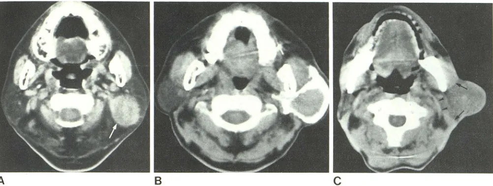
[image:2.612.52.554.462.680.2]AJNR:3, March! April 1982 CT OF PAROTID TUMORS 145
A
B
Fig. 5.- CT appearances of parotid tumors. A, Pleomorphic adenoma with slightly irregular margin in posteroinferior part of left parotid gland. On plain CT. mass (arrow) in low density parotid tissue is clearly shown. B, Warthin tumor in superficial part of left parotid gland. Slightly lobulated margin of tumor on CT sialogram. Mass displaces masseter muscle forward and ster
-and the stylopharyngeus muscles) that are attached to it, and, more anteriorly, the gland extends toward the parapharyngeal space
(figs. 2 and 3) [1-3].
The internal jugular vein, internal carotid artery, and the last four
cranial nerves pass deeply to the digastric muscle and the styloid
process. The external carotid artery is separated from the internal
carotid artery by the styloid process in the upper part of the neck
(fig. 4) [1]
The parotid duct takes a course between the tragus and the middle part of the upper lip. At the anterior border of the masseter
muscle, it turns medially and opens on the mucosal surface of the
cheek [2].
The facial nerve descends from the stylomastoid foramen, runs
in front of the digastric muscle and lateral to the styloid process, and enters the parotid gland. Within the gland, it takes a forward course and becomes more superficial. It then divides into peripheral branches and lies superficial to the retromandibular vein [1 , 2]. The
origin of the posterior belly of the digastric muscle lies posterior to the facial nerve. The more distal part of the posterior part of the digastric muscle is medial to the nerve. The retromandibular vein is
usually located medial to and is crossed by the facial nerve. Th
ere-fore, the locations of the posterior belly of the digastric muscle and
the retromandibular vein provide landmarks for the estimation of the course of the nerve.
Results
The density of the parotid gland on the CT images varies
considerably from patient to patient. It ranges between a
slightly higher density than the fatty tissue to a slightly lower density than Hle muscle. The parotid gland of the uninvolved
side showed a low density (slightly denser than the fatty
tissue) in nine of the 18 patients in this series, intermediate density in three patients, and a relatively high density (slightly less dense than the muscule) in six patients. No
definite relation was observed between the density of the
parotid gland and the patient's advancing age. The factors
c
nomastoid muscle backward. Normal filling of deep part of parotid gland. C, Adenocarcinoma of left parotid. Mass with indistinct margins displaces
mas-seter muscle forward and sternomastoid muscle backward (arrows). Digastric muscle also slightly displaced medially (arrowheads).
associated with the variable density of the gland need to be studied further.
Parotid tumors usually showed a slightly higher attenua -tion value than the gland. They may easily be seen when the density of the surrounding normal gland tissue is low,
but not when the density is high. In six of the nine patients with low density parotid glands, the parotid tumors were
clearly shown. In the other three patients (two with pl eo-morphic adenomas and one with a tubular adenoma), the tumors were faintly shown due to the low density of the tumor itself. In two of the three patients with intermediate density parotid glands, the tumors were easily recognized,
and, in the other patient with a parotid cyst, the margin of the mass could not be well delineated on the plain CT images, probably due to the low CT density of the mass (23 Hounsfield units). The cyst became clearly outlined on the
intravenous postenhancement CT scan because of the
en-hancement of the surrounding normal parotid gland. In five of the six patients with relatively high density parotid glands, the mass could not be identified in the enlarged glands. In the other patient with an oxyphilic adenoma, identification of the mass was not difficult because the mass was huge
with an irregular margin and a density higher than the parotid gland and approximately equal to the muscle tissue. There were 10 patients with pleomorphic adenomas. Of
these, nine patients had plain CT scanning and one had
only CT after sialography. One of the nine adenomas dem-onstrated a smooth and sharply marginated mass. Five showed a slightly irregular outline (fig. 5A). The margins of the adenomas in the other three patients, who had relatively dense parotid glands, were rather indistinct. The Warthin tumor showed a moderately lobulated margin (fig. 58). In all five patients with malignant lesions, irregular or poorly
de-fined margins of the tumors were shown (fig. 5C).
[image:3.612.55.556.80.269.2]146 SONE ET AL. AJNR:3, Marchi April 1982
A
B
t rnomastoid muscle posteriorly and the posterior belly of th digastric posteromedially. Anterior lesions displaced the post rior part of the masseter muscle anteriorly. The tumors with r tropharyngeal extension passed through the stylo -m ndibul r tunnel and occupied the space between the
m
di I pterygoid muscle and the stylomandibular ligament, th styloid process, and the three muscles attached to the proc s, The low density fatty area surrounding these mus-cl nd th major vessels IIlat lie medial to the styloid
proc b came narrowed or indistinct. Comparing these tructur s with thos on the opposite side helped to detect
minor bnormality.
Involv ment of the facial nerve by a parotid tumor could b pr dicted with a high degree of confidence when the ccupi d an upper part of the gland, intimately as
-d with the tip of the mastoid and the stylomastoid f r m n. Wh n the tumor was located in a lower part of the
I n, th r tromandibular vein was used to evaluate the r I tion of the tumor to the facial nerves.
CT combin with ialography showed the course of the t n n duct and its major branches, and the parenchymal
flilin of th parotid gl nd atter a sufficient amount of con -tr t m dium h d been injected. This method was useful in
ho~ ing the size and e tent of the parotid tumor not clearly id ntifi d on plain CT because the tumor was located in a p rotid gl nd of a r I ti ely high density. The site of origin f th tumor 'I ithin the parotid gland could be determined po ition of the filling defects in the parotid paren -(fig. 58) and/or by the di placement of the main
I ter II or
m
diall (fig. 6). The tumors arising from up dici I lob di pi ced the main duct medially and/h 'I d norm I p r nch m I filling deep to the mass. The tumor
in
th r p rt di plac d the duct laterally and/r th I filling up rfici I to the rna s as ob
-Th
tum
n
ion did not ftect the main ducts.
Fig. 5.- Displacement of parotid duct on pa -rotid CT sialography. A, (Same case as in fig. 5C.) Medially displaced main duct (arrow) suggests tumor arises from superficial lobe. Poor parenchy
-mal filling because of painful injection. B, Pleo -morphic adenoma. Mass with slightly irregular
margin. Lateral displacement of parotid duct (
ar-row) suggests tumor arises from deep lobe. Poor parenchymal filling because of leakage of contrast into oral cavity.
used to obtain more precise mapping than plain CT can
provide imaging of the gland's parenchyma and of tumor filling defects [4-8].
In nine patients in our series, the parotid tumors were clearly seen on plain CT images and almost all of them had
relatively high density contrasting the low density parotid
gland. In the other nine patients, the tumor could not be clearly identified on the plain CT images because of the high density of the surrounding parotid glands or due to the low density of the tumor itself. For these patients, CT combined with sialography was valuable to delineate filling defects within the glands.
Our technique using a ScoutView localization radiograph permits easy selection of CT scan locations to obtain the full length image of the parotid duct on one slice and with the other slices effectively covering the tumor. The image of the parotid duct thus obtained provides additional informa
-tion concerning the tumor location. CT combined with sial -ography can determine, by lateral or medial displacement of the main ducts, the site of origin of any tumor located near the junction of the superficial and deep lobes. When a tumor was clearly shown on CT images, CT combined with sialography was not required, especially for posteriorly lo
-cated tumors because these tumors usually do not affect the main ducts.
CT was helpful in distinguishing malignant from benign lesions. Irregular or poorly defined margins suggested ma -lignancy and smooth or slightly irregular margins benignity.
In estimating extension of the tumor. displacement or de -formity of the muscles such as the masseter the sterno -mastoid, and the posterior belly of the digastric was one of the most important findings. Facial nerve involvement could be predk;ted using the mastoid tip and styloid process for superior tumors and the retromandibular vein for inferior tumors.
CT during intravenous infusion of contrast medium was useful only in the case of the parotid cyst, which was clearly delineated as a lower density area within the contrast-en
-hanced normal gland tissue. In other patients with parotid
tumor , however, little additional important information was
AJNR:3, Marchi April 1982 CT OF PAROTID TUMORS 147
ACKNOWLEDGMENT
We thank Satoko Matsuura for assistance in manuscript prepa-ration.
REFERENCES
1. Boileau Grant JC. Grant's atlas of anatomy, 5th ed. Baltimore: Williams & Wilkins, 1966:540-552,572-574
2. Rankow RM, Polayes 1M. Surgical treatment of salivary gland tumors. In: Diseases of the salivary glands. Philadelphia:
Saun-deffi, 1976:156-161, 239-250
3. Fluur E. Parapharyngeal tumors. Arch Otolaryngol 1964;
80:557-565
4. Carter BL, Karmody CS. Computed tomography of the face and neck. Semin RoentgenoI1978;13:257-266
5. Mancuso AA, Rice 0, Hanafee W. Computed tomography of
the parotid gland during contrast sialography. Radiology
1979;132:211-213
6. Som PM, Biller HF. The combined CT-sialogram. Radiology
1980; 135: 387 -390
7. Mancuso AA, Bohman L, Hanafee W, Maxwell O. Computed
tomography of the nasopharynx: normal and variants of normal.
Radiology 1980; 137: 113-1 21
8. Stone ON, Mancuso AA, Rice 0, Hanafee WN. Parotid CT