With 10 figures Printed in Great Britain
FEED-BACK MODULATION OF CONE SYNAPSES BY
L-HORIZONTAL CELLS OF TURTLE RETINA
H. M. GERSCHENFELD,* M. PICCOLINO.f AND J. NEYTON*
• Laboratoire de Neurobiologie, Ecole Normale Superieure, 46, rue d'Ulm, 75230 Paris Cedex 05, France, and -\Istituto di Neurofisiologia del C.N.R., Via San Zeno 51,
56100 Pisa, Italy
SUMMARY
Light stimulation of the periphery of the receptive field of turtle cones can evoke both transient and sustained increases of the cone Ca24" conductance, which may become regenerative. Such increase in the cone Ca2+ conductance evoked by peripheral illumination results from the activation of a poly-synaptic pathway involving a feed-back connexion from the L-horizontal cells (L-HC) to the cones. Thus the hyperpolarization of a L-HC by inward current injection can evoke a Ca2* conductance increase in neigh-bouring cones. The cone Ca2* channels thus activated are likely located at its synaptic endings and probably intervene in the cone transmitter release. Therefore the feed-back connexion between L-HC and cones by modifying the Ca2* conductance of cones could actually modulate the transmitter release from cone synapses. Such feed-back modulation of cone synapses plays a role in the organization of the colour-coded responses of the chroma-ticity type-horizontal cells and probably of other second order neurones, post-synaptic to the cones. The mechanisms operating the feed-back connexion from L-HC to cones are discussed.
Introductory remarks
Cones are the main class of photoreceptors in the retina of the red-eared turtle (Pseudemys scripta elegans). According to their spectral properties and morphology they are classified as either red, green, blue and double cones (Baylor & Hodgkin, 1973; Richter & Simon, 1974). Green cones are much more sensitive to green lights than to deep red lights, while red cones are moderately more sensitive to deep red lights than to green ones. When turtle cones are impaled by a microelectrode they show, in the dark, membrane potentials of 25-35 m^ (dark potential). Photoisomerization of their pigment by light impinging on them induces a graded hyperpolarization which may reach amplitudes up to 15-25 mV according to the light intensity (Fig. 1 a).
178 H. M. GERSCHENFELD AND OTHERS
(Werblin & Dowling, 1969; for the turtle retina see Schwartz, 1974; Richter & Simonl
1973)-Horizontal cells play an interneuronal role in the outer plexiform layer of turtle retina (see below). They respond to light stimuli with graded potentials and are characterized by the great extension of their receptive field. According to their spectral properties two main classes of horizontal cells have been recognized (Svaetichin & MacNichol, 1958): the 'luminosity' horizontal cells (L-HC), maximally sensitive to red lights, which respond by graded hyperpolarizing responses to light stimuli of any wavelength, and the ' chromaticity' horizontal cells, which respond to light by either a depolarization or a hyperpolarization according to the wavelength of the stimulus.
Synoptic transmission from cones to second order cells
In contrast with the majority of synapses known in the central and peripheral nervous system of vertebrates and invertebrates, the synapses connecting cones to second-order cells are of the tonic type. In the dark they release transmitter con-tinuously, whilst light (the physiological stimulus) hyperpolarizes the cone, thus de-pressing or supde-pressing the transmitter release (Trifonov, 1968). The sustained trans-mitter release in the dark is Ca2+-dependent: when the ionic content of the extra-cellular medium is altered by removing Ca2"1" or adding Co2"*" or other divalent cations which block Ca5^ conductance, the transmitter release from the cone synapses is blocked. Therefore Ca24" channel-blocking agents mimic the effects of light stimula-tion on the second-order neurones. This has been thoroughly demonstrated in turtle retina (Cervetto & Piccolino, 1974) as well as in other vertebrate retinas for both cones and rod synapses (Dowling & Ripps, 1973; Kaneko & Shimazaki, 1975; Dacheux & Miller, 1976).
When transmitter release from cone synapses is suppressed by light hyperpolariza-tion different effects are observed in the second order neurones: the L-horizontal cells are hyperpolarized as a result of a decrease in their membrane conductance (Trifonov, Byzov & Chailahian, 1974) probably to Na+ ions (Waloga & Pak, 1978), the hyperpolarizing bipolar cells are also hyperpolarized and their membrane con-ductance i9 also decreased, the depolarizing bipolar cells are depolarized and their membrane conductance increased (Nelson, 1973; Toyoda, 1973; Werblin, 1977; Saito, Hondo & Toyoda, 1979). The effect of the cone transmitter on the C-horizontal cells will be discussed in the last section of this paper. The synaptic transmitter (or transmitters?) of the cones has (have ?) not yet been identified. However, the cone transmitter(s) should be expected to promote effects opposite to those observed when the transmitter release is suppressed. Thus, the synaptic transmitter(s) released from the cones could be expected to (a) depolarize the horizontal cells by increasing their membrane conductance, (b) depolarize the hyperpolarizing bipolar cells by increasing their membrane conductance and (c) hyperpolarize the depolarizing bipolar cells by decreasing their membrane conductance.
Antagonistic organization of the turtle cone receptive field
tthe cone response to a centred spot of a given intensity gradually increases in ampli-tude as the diameter of the spot increases up to 120 fim (Baylor, Fuortes & O'Bryan, 1971). This signifies that a rather large population of neighbouring cones contributes to the response to each single cone through electrotonic coupling (Baylor et al. 1971) across gap-junctions (Raviola & Gilula, 1975; see Raviola, 1976). This electrical coupling is very specific, the red cones are exclusively coupled to other red cones, the green cones to other green ones, etc. (Baylor & Hodgkin, 1973; Detwiler & Hodgkin,
IO
-79)-Spot Annulus -79)-Spot + annulus
(a) (b) (c)
10mV|
[image:3.451.56.409.172.292.2]2s
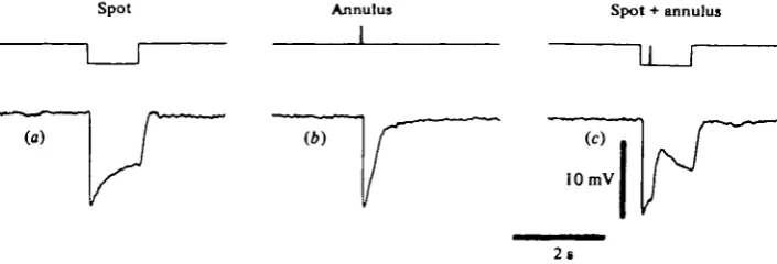
Fig. 1. Depolarizing response of a turtle red cone to peripheral stimulus, (a) Hyperpolarizing response of the cone to a centred spot of white light (250 fim diameter, attenuation —2-3 log units). (A) Fast hyperpolarization evoked by a white light annulus flash (inner diameter 430 fim, attenuation —0-3 log units), due to scattering of light from the annulus to the centre of the receptive field, (c) Feed-back depolarizing potential evoked by the annulus flash when properly combined with the spot stimulation. In these experiments, as well in those shown in the other figures, the external diameter of the light annuli used was 3600 fim. In all the experiments the flux density of the white unattenuated light on the retina, measured inside the limits of the visual range, was about 2'5~5 x 1 o~* /
Light stimuli covering areas of diameters larger than 120 /im can exert an antagon-istic effect on the cone response, which may result in both a reduction of the amplitude and a change in the time course of their response or in the appearance of a de-polarizing potential. An example of this antagonistic effect is shown in the cone of Fig. 1, which responded by a classical hyperpolarizing response to a centred spot of light (Fig. 1 a). The flashing of a large annulus of intense white light, concentric to the spot, also evoked a fast hyperpolarization due to a direct response of the cone to light scattered from the annulus to the centre of its receptive field (Fig. 1 b). When the same annulus was flashed during the stimulation of the cone by a centred spot (Fig. 1 c) it evoked a depolarizing potential.
Such depolarizing responses to large light stimuli were discovered by Baylof et al: (1971) who showed that they were due to the activation of a polysynaptic circuit: a large population of peripheral cones feed their signals to L-horizontal cells which are electronically connected between them (Kaneko, 1971; see Raviola, 1976) and which feed-back their signals to the central cones, depolarizing them. This circuit, therefore, involves a feed-back connexion between the L-HC processes and the cones, opposite in direction to the synapse between the cones and the second-order cells.
i 8 o H. M. GERSCHENFELD AND OTHERS
the cone. These authors interpreted this depolarization as a synaptic potential resulting from the activation of the feed-back connexion between L-HC and cones.™ O'Bryan (1973) found that following the depolarizing potential evoked in cones by peripheral stimuli the membrane resistance was decreased. These observations further supported the view that depolarizing potentials could be classical synaptic potentials involving an increase in membrane conductance.
Spot 155 340 960 1600
IT
T
T
T
10 mV
1 s
¥
T
T
T
Ann 960 430 280
Fig. 2. Stimulation of the periphery of the receptive field evokes spike responses in a red turtle cone of an untreated retina. The figures above the upper records correspond to the diameters of the unattenuated light spot stimuli whereas the figures below the lower records are the inner diameters of the unattenuated light annuli (in /im).
Spikes evoked by peripheral illumination
Some useful indications for the understanding of the nature of the peripherally evoked depolarizing potentials in cones resulted from the study of particular responses to peripheral illumination that showed the characteristics of action potentials. Fuortes, Schwartz & Simon (1973) first observed that stimulation of large retinal areas with red light evoked in green cones a spike-like response which these authors attributed to the activation of the feed-back connexion from L-HC to green cones. Such spike responses were also observed by O'Bryan (1973) when stimulating the periphery of the receptive field of a red cone with annuli of bright white light. We have recently analysed the properties, ionic basis and the possible synaptic mechanisms involved in the generation of these spikes which were observed in a 20% of the cones studied in our experiments (Piccolino & Gerschenfeld, 1978, 1980; Gerschenfeld & Piccolino,
/unction. But as with other exaggerated manifestations of activity evoked in various cells by stimuli at the limits of the physiological range, their study gave new informa-tion on certain synaptic phenomena taking place at the cone endings. Thus, these spikes were shown to result from a regenerative increase of cone membrane conduct-ance to Ca^ ions. We will summarize in the next sections the experimental evidence supporting this view and will discuss the functional implications of our findings.
Ann.
n
(a)
r
2s
ib)
Spot *• A n n .
1 1
h
10 mV
Ann. 0
Ann.
n
\(c)
1
1
[image:5.451.40.412.184.328.2]id)
f
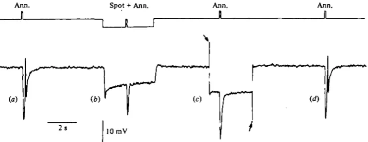
Fig. 3. Hyperpolarization block of the spike evoked by peripheral illumination of a turtle cone, (a) Stimulation with a light annulus (430 m inner diameter, unattenuated) evokes a spike. (6) Background illumination with a centred spot (250 fim diameter, attenuation —22 log units) evokes a hyperpolarization which blocks the spike, (c) The spike is also blocked by hyperpolarizing the cone by injecting inward current (between the arrows, io~lc A), through the recording microelectrode. (rf) Control recording showing the persistence of the spike.
Properties of cone spikes evoked by peripheral stimulation
As already mentioned, Fig. 2 shows that stimulation with bright light of the periphery of the receptive field of cones evokes spike responses. In Fig. z{c, b) the stimulation with centred spots of 340 and 960 jim already elicits, following the direct hyperpolarizing response, depolarizing potentials that increased in amplitude with the increase in the spot diameter. Spots of diameters beyond 1 mm evoked spike-like responses (Fig. zd). When annuli of bright light were used (in all cases their external diameter was 3-6 mm), they also evoked a direct hyperpolarizing response due to the light scattered to the centre of the receptive field, which in all cases was followed by a spike response (Fig. ze, h).
182 H. M. GERSCHENFELD AND OTHERS
Ionic mechanism of the spikes evoked in cones by peripheral light stimulation
In the majority of excitable cells two main ion species have been shown to carry the currents responsible for the action potentials: Na+ and Ca2+. It was therefore natural to investigate if one of these cations was involved in the generation of the cone
Spot Ann. Spot Spot + Ann
1 n r
Control (a) (b) (c)
if) is)
10 mV
ih)
2 s
+ nicotine ' " (m)
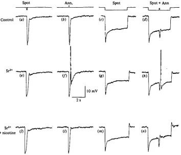
Fig. 4. (a-d) Responses of a cone bathed in normal saline to different light stimuli as indicated in the upper traces, (e-h) responses of the same cone to the same sequence of stimuli recorded during bath application of iomM-SrCl|. Note the discharge of spikes following the annuli stimulation, (t-n) The spikes evoked by peripheral stimulation are blocked by nicotine (5 mtu) added to the Sr*+-containing medium. Stimulation parameters: (a, e, i) centred spot, 250 fan diameter, attenuation —0-5 log units; (6,/,/) concentric annulus, 430/*m inner diameter, unattenuated; (c,g, m) centred spot, 2 5 0 / m diameter, attenuation —1-5 log units; (d, h, n) combination of a spot such as in (c) with an annulus like in (b). (Piccolino & Gerschenfeld, 1978.)
spikes responses to peripheral illumination. Tetrodotoxin (TTX), a poison known to block Na+ channels involved in spike generation, did not affect cone spikes even after prolonged extracellular applications at concentrations up to io/iM.
[image:6.451.35.390.141.443.2]in the responses to light (Bertrand, Fuortes & Pochobradsky, 1974). Increasing extracellular Ca2"*" concentration was however shown to facilitate the spike evoked by peripheral illumination, before the hyperpolarization blocked them. More interest-ing, in the presence of a high Ca2+ environment (15-20 mitf), cones which did not previously respond with either a spike or a depolarizing potential to peripheral stimuli responded by an action potential to light annuli until the hyperpolarizing effect of high Ca2* media blocked them.
- 0 5 - 1 - 5 -2-5 -3-5
[image:7.451.57.403.171.333.2]10 mV
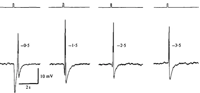
Fig. 5. Spike responses to peripheral stimulation in a Sr^-treated cone stimulated with annuli (inner diameter 430 /im) of decreasing light intensity. The figures besides the responses indicate the light attenuation in log units.
Fortunately, Sr2"1", a divalent cation known to permeate Ca2"1" channels and to replace Ca2"1" in synaptic transmission (Fatt & Ginsborg, 1958; Miledi, 1966; Katz & Miledi, 1969 a; Dodge, Miledi & Rahamimoff, 1969; Meiri & Rahamimoff, 1971) affects neither the cone membrane potential nor the synaptic transmission between cones and second order cells (Piccolino, 1976). In the presence of 4-10 mM-Sr2"1" in the extra-cellular environment, spikes evoked in the cones by peripheral illumination became increased in amplitude. Moreover, every cone in the turtle retina gave a spike response to an annulus flash in the presence of Sr2"1" ions in the extracellular medium. Fig. 4 shows an example of a cone which did not show any depolarizing response in a normal medium, when flashing an annulus either alone (Fig. 46) or during a period of central illumination (Fig. \d). After bathing the retina in 10 mM-Sr2"1" for some minutes, the annulus stimulation by itself (Fig. 4/) or in combination with a centred spot (Fig. 4A) evoked a spike. Stimulation with a small centred spot (100-300/im) was not able to evoke a spike in Sr^-treated retinas (Fig. \e, g). In Sr2+-containing media, a dim light annulus could produce a spike without any preceding hyper-polarization, so that the spike arose at the dark potential (Fig. $d). In such cases the latency of the spike was found to be ca. 100 ms.
184 H. M. GERSCHENFELD AND OTHERS
or in the presence of Sr2+ or Baa+ were all blocked by agents known to block Ca2+ channels such as Co2*, Mg2"1" and D-600.
These results show that light stimulation of the periphery of the receptive field evoked in turtle cones spike responses due to a regenerative increase in Ca2+ con-ductance. Moreover, the experiments with Ba2"1" and Sr24" show that every cone of the turtle retina is able to respond to peripheral illumination with a spike response.
Control
Ba2+ 3 mM
10 mV Cone
[image:8.451.90.376.154.408.2]L-HC—1
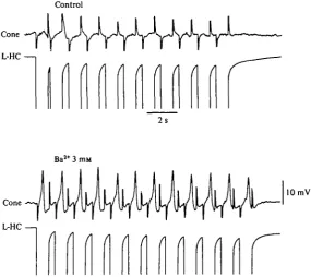
Fig. 6. Simultaneous recoding with two independent single micropipettes from both a cone and a neighbouring L-horizontal cell. T h e two upper records were obtained when the retina preparation was superfused with normal saline. Using a bridge circuit, pulses of inward current (20 nA) were injected through the recording micropipette impaling the L - H C . N o effect is observed in the cone, except for the capacitative artifacts. T h e two lower traces are recordings from the same two cells obtained during the superfusion of the preparation with a saline containing 3 mM-BaCl,. At this time, each inward current pulse injected in the L - H C evokes a spike in the cone.
Synaptic mechanisms involved in the generation of the spikes evoked by peripheral stimulation
The experiments of Fuortes et al. (1973) and of O'Bryan (1973) already mentioned, strongly suggested that the spikes evoked in cones by peripheral stimulation involved the activation of the same feed-back connexion between L-HC and cones described by Baylor et al. (1971).
logical agents on the cone membrane itself. A new series of experiments have brought more direct evidence on the participation of the feed-back connexion of L-HC to cones in the generation of the cone spikes. In these experiments, as in Fig. 6, we followed a protocol similar to that of Baylor et al. (1971) commented on above. We impaled both a cone and a neighbouring L-HC with independent microelectrodes and injected inward current pulses to directly hyperpolarize the L-HC.
Cone
10 mV
r
[image:9.451.90.350.168.433.2]200 ms
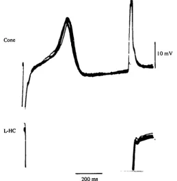
Fig. 7. Superimposed recordings of the responses of the L-HC and the cone as in Fig. 6. Each pulse of inward current injected in the L-HC evoked a constant latency spike in the cone.
In four experiments in which no apparent effects of the L-HC hyperpolarization were observed in the cones when bathed in a normal medium (Fig. 6, top), after addition of either 4 mM-Sr2"1" or 3 mM-Ba2"1" the inward current-induced hyper-polarization of the L-HC evoked in the cone the discharge of a spike (Fig. 6, bottom). The superimposed recordings from both cells in Fig. 7 show that all the cone spikes obtained in these conditions showed a constant latency of less of 100 ms and un-doubtedly resulted from the L-HC hyperpolarization, since removing the pipette from the L-HC and passing the same current through it had no effect on the cone membrane potential.
186 H. M. GERSCHENFELD AND OTHERS
Prolonged effects of peripheral illumination on the cone membrane conductance
From experiments as in Figs. 6 and 7 it follows that under appropriate ionic condi-tions there is a direct relacondi-tionship between the L-HC hyperpolarization and the cone Ca2"1" spike. What is the important signal across the feed-back connexion? Is it the variation of the L-HC potential? Or is it the attainment of a certain level of hyper-polarization of the L-HC? In other words, are the effects of L-HC hyperhyper-polarization on the cone membrane conductance transient or sustained ?
Spot-on -i
Ann. Spot-off
Ann.
J
(W
*<* I*
Spot
(c)
r
10 mV 2s
Fig. 8. Sustained effects of peripheral stimulation on the membrane conductance of a turtle cone, (a) Prolonged stimulation with a central light spot (250 /tm diameter, attenuation — 1 log unit) is combined with peripheral illumination by a concentric annulus (430 /tm inner dia-meter, unattenuated light). A sustained depolarization is observed. Square inward current pulses (io~10 A) were passed through the cone membrane to measure the membrane resistance, which becomes markedly decreased during the depolarization.
(b) Recordings from another cone bathed in 10 miu-SrCl.; prolonged stimulation with a light
annulus (150 fun inner diameter, attenuation — 1 log units) evokes a repetitive discharge of spikes, whereas in (c) the stimulation with a centred spot (250/tm, attenuation —1.3 log units), which evokes a peak hyperpolarization similar to the one elicited by the annulus, does not induce any spike.
t
\ is evident that the depolarization evoked by the annulus stimulation was associated ith a sustained increase in membrane conductance. In other cases (see Gerschenfeld & Piccolino, 1980) different patterns of responses to sustained activation of the feed-back connexion were observed. In all these cases, as well as in cones that did not show manifest effects of peripheral stimulation, addition of Sr2"1" ions to the extracellular medium induced the appearance of a repetitive discharge of spikes. An example of such an experiment is shown in Fig. 8(6) for a cone which did not show a feed-back response, in a normal medium. The discharge of spikes could be observed as long as the peripheral stimulus was applied. Such repetitive spike discharge never appeared in response to prolonged stimulation with a centred spot of light.These and other effects of prolonged peripheral stimulation of the cones (see Gerschenfeld & Piccolino, 1980) demonstrate that stimuli able to keep the L-HC hyperpolarized for prolonged periods of time induce sustained increases in the cone membrane conductance to Ca2+ ions.
Mechanism of the feed-back connexion between L-horizontal cells and cones
From the evidence presented above it appears that a feed-back connexion between L-HC and cones, opposite in direction to that from cones to L-HC, is able to modify in a sustained way the Ca2+ conductance of the cone membrane. Where are located the Ca2+ channels affected by this connexion? How does the connexion operate?
The contacts between L-HC processes and cones take place at the synaptic regions of the cone pedicle. In invertebrate photoreceptors (see Ross & Stuart, 1978) it has been shown that Ca2"*" channels located in the synaptic areas can generate action po-tentials. It is not therefore too far-fetched to assume that the Ca2+ channels involved in the cone depolarizations evoked through the feed-back connexion of the L-HC are situated in the synaptic endings of the cone and possibly are the same Ca2+ channels intervening in the transmitter release from the cone synapse. If so, the feed-back connexion from the L-CH would actually modulate the transmitter release from cone endings.
Two main hypotheses have been considered for the mechanism by which the feed-back connexion operates. The first proposes an electrical mechanism (Byzov & Golubtzov, 1977; Byzov, 1979). According to such model the current generated at the membranes of the hyperpolarized L-HC processes would partly flow across the mem-brane of the cone synaptic ending and generate a potential drop across, depolarizing it. No particular membrane junctions would be necessary for such interaction to take place, the particular geometry of the L-HC processes inside the invaginated cone synapses being highly favourable to such electric field interactions.
The second model postulates the existence of a chemical feed-back synapse between L-HC processes and cones (Gerschenfeld & Piccolino, 1980). If such would be the case it is likely that the L-HC, as the cones, releases its transmitter continuously in the dark. Light stimuli evoking an hyperpolarization of the L-HC would decrease or suppress this transmitter release. In such case it can be postulated that the effects of the chemical transmitter released from the L-HC would be opposite from the de-scribed feed-back effects, (i.e. it would directly or indirectly turn-off Ca2+ channels in the cone endings),
188 H. M. GERSCHENFELD AND OTHERS
channels. Thus it has been found that invertebrate heart acetylcholine and muscarinjj agonists cause a decrease in the duration of the action potential. In the frog atriar muscle fibres it has been found that such effect is associated with a decrease in Ca2+ conductance (Giles & Noble, 1976; Garnier et al. 1978). Moreover, in dorsal root ganglion cells in culture, Dunap & Fishbach (1979) have also recently observed that GABA, serotonin decreased the Ca^ component of the action potential.
However, also other possible chemical synaptic mechanisms could result in a de-crease of cone Ca2"1" conductance. For instance, the L-HC transmitter when released in the dark could induce an increase in a K+ conductance (as inhibitory transmitters do in the CNS) or a dependent K-conductance, thus keeping the voltage-dependent Ca conductance of the cone ending under control, thus impeding re-generative increase. The decrease in the L-HC transmitter release evoked by peripheral light stimulation could turn-off these K+ channels, the Ca2* conductance thus increasing and becoming regenerative. In relation with such an idea it may be remembered that Katz & Miledi (1969) observed Ca2* regenerative responses in the TTX-treated squid giant synapses endings after blocking K+ conductance.
It is difficult to conclude, at present, which mechanism operate the feed-back con-nexion between L-HC and cones. Electron-microscope studies have not been very helpful in this direction. Neither in the turtle retina (Lasansky, 1971; Schaeffer & Raviola, 1975) nor in those of other lower vertebrates (see for example Dowling & Werblin, 1969; Stell, 1976) synaptic specialization or synaptic vesicles have been observed in relation with the membrane of the L-HC processes facing the cone endings. Moreover, in contrast with classical synapses of the 'phasic* type in which, either manipulation of the extracellular Ca2+ content or Ca2* blocking agents can help to clarify the chemical nature of a connexion, these procedures cannot be used in the study of the feed-back connexion because more than one step involving Ca** ions intervene in the feed-back circuit.
L-HC have been recently shown to be able to take up GABA through a high affinity mechanism (Marc ct al. 1978) and experiments by Lam, Lasater & Naka (1978) suggest that bicuculine, a GABA antagonist, blocks some effects of the L-HC feed-back on the cones. Picrotoxin, another GABA antagonist, has been shown to block some of the possible repercusions of the L-HC feed-back mechanism on second order neurones (Djamgoz & Ruddock, 1979).
Further pharmacological experiments on the depolarizations evoked on the cones by the feed-back connexion from the L-HC may help to give support to the chemical hypothesis.
Physiological consequences of the feed-back modulation of the cone transmitter release
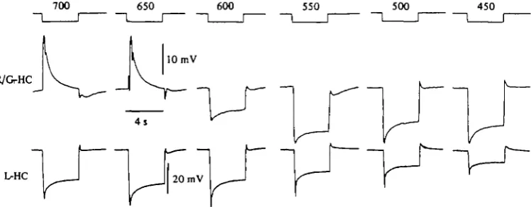
If through its feed-back connexion with the cone the L-HC modulates the trans-mitter release from the cone, and, if such modulation operates in all the cones, it can be expected that the responses of all the second-order neurones in the retina should be affected by this feed-back mechanism. We have recently explored such hypothesis on one of the second-order neurones - the red/green horizontal cell (R/G-HC), a chromaticity type horizontal cell (see above) that shows colour-coded responses.
«
pend on the wavelength of the stimulus. These cells are hyperpolarized by red muli (Fig. 9, top records, 700 and 650 nm) and hyperpolarized by stimuli of shorter wavelength.Fuortes & Simon (1974) postulated that the different polarity of the responses of the R/G-HC could be accounted for by the following mechanism: (a) R/G-HC receive a direct input from green cones but not from red cones (this has been confirmed by
700 650 600 550 500 450
R/GrHC
10 mV
[image:13.451.38.413.148.294.2]4 s
Fig. 9. Spectral sensitivity of horizontal cells of turtle retina. Top recordings, responses of a red/green chromaticity type horizontal cell (R/G-HC) to large spots (3600 /im diameter) of light of different wavelength. The wavelengths are indicated over the timing traces in nm. Bottom recordings, responses of a L-HC to same series of monochromatic stimuli.
morphological studies, see Leeper, 1978); (b) thence, green light which hyperpolar-izes green cones also evokes the hyperpolarization of the R/G-HC, (c) green cones are unaffected by red light impinging on them, (d) red illumination of a large area of the retina hyperpolarizes the red cones that feed their signal on the L-HC, which, in turn, feed-back their signal on the green cones depolarizing them, (e) as a con-sequence of the feed-back depolarization of green cones the R/G-HC are depolarized in response to red light.
However, this hypothesis does not appear too convincing in its original formulation since no correlation is generally observed between the amplitude of the depolarizing responses to red light in the R/G-HC (up to 25-30 mV) and the amplitude of the responses of the green cones to such stimuli (which are usually of small amplitude and, in some cones-, even undetectable). Moreover, in most green cones, prolonged stimulation with red lights induces only a depolarizing transient at the onset of the stimulus while the R/G-HC responses to the same stimulus generally show a sustained component.
However, this apparent lack of correlation between the effects of red light on green cones and R/G-HC can be discarded when the results of our own experiments are taken into account. Thus we can assume: (1) that red light stimuli evoke an increase of transmitter release from the green cone due to an increase of Ca2"1" conductance in their endings due to the activation of the feed-back connexion from L-HC on green cones; (2) this transmitter release would evoke a depolarization in the R/G-HC even ^fcen only small changes in the green cones potential would be detected, and (3) when
190 H. M. GERSCHENFELD AND OTHERS
using prolonged red light stimulation, the increase in Ca2+ conductance and consequent increase in transmitter release from green cones would be sustained.
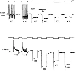
To test such assumptions, experiments were performed in retinas bathed in either Sr2"1"- or Ba^-containing media. We knew that if in such conditions a prolonged red light stimulation depolarized the green cones through the feed-back connexion from L-HC, we should observe a repetitive discharge of spikes in green cones. That was in
Green
cone i,
700
10 mV
10s
650 600
r
J~ur
550 500 450
R/G-HC
700 650
600
(
' 5 5 0
500
[image:14.451.59.366.149.435.2]450
Fig. 10. Recordings from both a green cone and a R / G - H C obtained from different retinas bathed in a medium containing 6 mM-SrCl). T h e wavelength of the light stimuli in nm are indicated below each response. T h e quantum flux for all the light stimuli was 1 7 X 10* photons /tm-'.s"1.
fact what we observed as illustrated in Fig. 10, top records, where red light stimuli (700 and 650 nm) actually evoked a repetitive discharge of spikes in a green cone of a Sr^-treated retina. What was the repercussion of such phasic increases of conduct-ance across Ca^ channels in the green cones on the R/G-HC of the same retina? It could be expected that such repetitive Ca2"1" spikes would evoke repeated phasic releases of transmitter from the green cone. Indeed, when a R/G-HC was recorded from the same retina (Fig. 10, bottom) red light stimuli that evoked Ca2"1" spikes in the green cones also evoked depolarizing potentials in the R/G-HC. The depolarizing po-tentials in the R/G-HC were found to show the properties of depolarizing synaptic potentials (Piccolino et al. 1980): they increase in amplitude when the R/G-HC is hyperpolarized, they become blocked when the spikes in the green cones are blocked by hyperpolarization evoked by intense green light stimulation.
«
|74) hypothesis in our modified formulation. They clearly show that stimuli which polarize the green cones through the feed-back connexion from L-HC to those cones, actually modulate the Ca2"*" conductance, evoking an additional release of trans-mitter which depolarizes the R/G-HC. These results, therefore, support the idea that the feed-back modulation of the cone Ca24" conductance by the feed-back connexion from the L-HC does actually play a physiological role in the generation of responses of the second-order neurones, postsynaptic to the turtle cones.The work of the authors was supported by grants of the Centre National de la Recherche Scientifique (CNRS), France, Delegation a la Recherche Scientifique et Technique, France, and Institut National de la Sante1 et de la Recherche Me'dicale, France. M. Piccolino was supported by the Consiglio Nazionale delle Ricerche, Italy, and the CNRS.
REFERENCES
BAYLOR, D. A., FUORTES, M. G. F. & O'BRYAN, P. M. (1973). Receptive fields of cones in the retina of the turtle. J. Physiol., Land. 214, 265—294.
BAYLOR, D. A. & HODCKIN, A. L. (1973). Detection and resolution of visual stimuli by turtle photo-receptors. J. Phytiol., Land. 234, 163-198.
BERTRAND, D., FUORTES, M. G. F. & POCHOBRADSKY, J. (1978). Actions of EGTA and high calcium in the cones in the turtle retina. J. Physiol., Lond. 275, 419—437.
BYZOV, A. L. (1979). Origin of non-linearity of voltage-current relationships of turtle cones. Vision Res.
19,
469-477-Bizov, A. L., GOLUBTZOV, A. V. & TRIFONOV, J. (1977). The model of feed-back between horizontal cells and photoreceptors in vertebrate retina. In Vertebrate Photoreception (ed. H. B. Barlow and P. Fatt), pp. 265-274. London, New York, San Francisco: Academic Press.
CERVETTO, L. & PICCOLINO, M. (1974). Synaptic transmission between photoreceptors and horizontal cells in the turtle retina. Science, N.Y. 183, 417-419.
DACHEUX, R. F. & MILLER, R. F. (1976). Photoreceptor-bipolar transmission in the perfused retina-eyecup of the mudpuppy. Science, N.Y. 191, 963—964.
DBTWILER, P. B. & HODCKIN, A. L. (1979). Electrical coupling between cones in turtle retina. J. Physiol.,
Lond. 291, 75-100.
DJAMCOZ, M. B. A. & RUDDOCK, K. H. (1979). Effects of picrotoxin and strychnine on the fish S-potentials: evidence for inhibitory control of depolarizing responses. Neurosci. Lett, ia, 329-334. DODGE, F. A., MII.EDI, R. & RAHAMINOFF (1969). Strontium and quanta! release of transmitter at the
neuromuscular junction. J. Physiol., Lond. 200, 267—283.
DOWLING, J. E. & RIPPS, J. H. (1973). Effect of magnesium on horizontal cell activity in the skate retina. Nature, Lond. 242, 101-103.
DOWLINC, J. E. & WERBLIN, F. S. (1969). Organization of the retina of the mudpuppy, Necturus
maculostis. II. Synaptic structure. J. Neitrophysiol. 32, 315—338.
DUNLAP, K. & FISCHBACH, G. D. (1978). Neurotransmitter decrease the calcium component of sensory neurone action potentials. Nature, Lond. 276, 837-839.
FATT, P. & GINSBORC, B. L. (1958). The ionic requirements for the production of action potentials in crustacean muscle fibers. J. Physiol. Lond. 142, 516—543.
FUORTES, M. G. F., SCHWARTZ, E. A. & SIMON, E. J. (1973). Colour dependence of cone responses in turtle retinas. J. Physiol., Lond. 34, 199—216.
FUORTES, M. G. F. & SIMON, E. J. (1974). Interactions leading to horizontal cell responses in the turtle. J. Physiol. Lond. 240, 177-198.
GARNIER, D., NARGEOT, J., OJEDA, C. & ROUCIER, O. (1978). The action of acetylcholine on background conductance in frog atrial trabeculae. J. Physiol., Lond. 274, 381-396.
GERSCHENFELD, H. M. & PICCOLINO, M. (1979). Sustained feed-back effects of L-horizontal cells on turtle cones. Proc. R. Soc. Lond. B 206, 465-480.
GILES, W. & NOBLE, S. J. (1976). Changes in membrane currents in bullfrog atrium produced by acetyl-choline. J. Physiol., Lond. 261, 113—123.
KANEKO, A. (1971). Electrical connexions between horizontal cells in the dogfish retina. J. Physiol.,
d. 213, 95-105.
O, A. & SHIMAZAKI, H. (1975). Effects of external ions on the synaptic transmission from photo-to horizontal cells in carp retina. J. Physiol., Lond. 252, 509-522.
192 H. M. GERSCHENFELD AND OTHERS
KATZ, B. &MILEDI, R. (1969a). The effect of divalent cations on transmission in the squid giantsynaj^B
Publ. Staz. zool. Napoliyj, 303-310. ^ B
KATZ, B. & MILEDI, R. (10696). Tetrodotoxin resistent electrical activity in presynaptic terminals.
J. Physiol., Land. 303, 459-487.
LAM, D. M. K., LASATER, E. & NAKA, K. I. (1978). y-aminobutyric acid: a neurotransmitter for cone horizontal cells of the catfish retina. Pioc. natn. Acad. Sci. U.S.A. 75, 6310-6313.
LASANSKY, A. (1971). Synaptic organization of cone cells in the turtle retina. Pltil. Trans. R. Soc. Lond. B
262, 365-381.
LEEPER, H. F. (1978). Horizontal cells of the turtle retina. II. Analysis of interconnections between photoreceptor cells and horizontal cells by light microscopy. J. Comp. Neurol. 182, 795—810. MARC, R. E., STELL, W. K., BOK, D. & LAM, D. M. K. (1978). GABA-ergic pathways in the goldfish
retina. J. comp. Neirrol. 183, 231-246.
MEIRI, U. & RAHAMINOFF, R. (1971). Activation of transmitter release by strontium and calcium ions at the neuromuscular junction. J. Physiol., Lond. 315, 709-726.
MILEDI, R. (1966). Strontium as a substitute for calcium in the process of transmitter release. Nature
Lond. 212, 1233-1234.
NELSON, R. (1973). A comparison of electrical properties of neurons in Necturus retina. J. Neuropkysiol. 36,
5I9-535-O'BRYAN, P. M. (1973). Properties of the depolarizing synaptic potential evoked by peripheral illumi-nation on cones of the turtle retina. J. Physiol., Lond. 235, 207-223.
PICCOLINO, M. (1976). Strontium-calcium substitution in synaptic transmission in turtle retina. Nature,
Lond. z6i,
554-555-PICCOLINO, M. & GERSCHENFELD, H. M. (1978). Activation of a regenerative calcium conductance in turtle cones by peripheral stimulation. Proc. R. Soc. Lond. B 301, 300-315.
PICCOLINO, M. & GERSCHENFELD, H. M. (1979). Characteristics and ionic processes involved in feed-back spikes in turtle cones. Proc. R. Soc. Lond. B 306, 439-463.
PICCOLINO, M., NEYTON, J. & GERSCHENFELD, H. M. (1980). Synaptic mechanisms involved in the responses of the chromaticity horizontal cells of turtle retina. Nature, Lond. 284, 58-60.
RAVIOLA, E. (1976). Intercellular junctions in the outer plexiform layer of the retina. Invest. Ophthalmol. 11, 881-894.
RAVIOLA, E. & GILULA, N. B. (1975). Intramembrane organization of specialized contacts in the outer plexiform layer of the retina. A freeze-fracture study. J. Cell Biol. 65, 190-222.
REUTER, H. (1973). Divalent cations as charge carriers in excitable membranes. Progr. Biophyt. Mol.
Biol. 36, 1-43.
RICHTER, A. & SIMON, E. J. (1974). Electrical responses of double cones in the turtle retina. .7. Physiol.,
Lond. 242, 673-683.
RICHTER, A. & SIMON, E. J. (1975). Properties of centre hyperpolarizing red-sensitive bipolar cells in the turtle retina. Journal of Pkytiol. Lond., 248, 317-334.
Ross, W. N. & STUART, A. E. (1978). Voltage sensitive calcium channels in the presynaptic terminals of a decrementally conducting photoreceptor. J. Physiol., Lond. 274, 173—191.
SAITO, T., HONDO, H. & TOYODA, T. (1979). Ionic mechanisms of two types of on-center bipolar cells in the carp retina. I. The responses to central illumination. J. gen. Physiol. 73, 73—90.
SCHAEFFER, S. F. & RAVIOLA, E. (1975). Ultrastructural analysis of junctional changes in the synaptic endings of turtle cone cells. Cold Spring Harb. Symp. quant. Biol. 40, 521-528.
SCHAEFFER, S. F. & RAVIOLA, E. (1978). Membrane recycling in the cone cell endings of the turtle retina. J. Cell Biol. 79, 802-825.
SCHWARTZ, E. A. (1974). Responses of bipolar cells in the retina of the turtle. J. Physiol., Lond. 236, 211-224.
STELL, W. (1976). Functional polarization of horizontal cell dendrites in goldfish retina. Invest.
Ophthal-mol. 15, 895-908.
SVAETICHIN, G. & MACNICHOL, E. F. (1958). Retinal mechanisms for chromatic and achromatic vision.
Ann. N.Y. Acad. Sci. 74, 385-404.
TOYODA, J. (1973). Membrane resistance changes underlying the bipolar cell response in the carp retina.
Vision Res. 313, 283-294.
TRIFONOV, Y. U. (1968). Study of synaptic transmission between photoreceptors and horizontal cells by means of electrical stimulation of the retina. (In Russian.) Biofizika 13, 809-817.
TRIFONOV, Y. A., BYZOV, A. L. & CHAILAHIAN, L. (1974). Electrical properties of subsynaptic and non-synaptic membranes of horizontal cells in fish retina. Vision Res. 14, 220-241.
WALOGA, G. & PAK, W. L. (1978). Ionic mechanism for the generation of horizontal cell potentials in isolated axolotl retina. J. gen. Physiol. 71, 69—92.
WERBLIN, F. S. (1977). Synaptic interactions mediating bipolar response in the retina of the tiger salamander. In Vertebrate Photoreception (ed. H. B. Barlow and P. Fatt).
WERBLIN, F. S. & DOWLING, J. E. (1969). Organization of the retina of the mudpuppy,