J. exp. Bhl. 106, 143-161 (1983) 1 4 3 ed in Great Britain © The Company of Bwlogists Limited 1983
CP-STIMULATED ADENOSINE TRIPHOSPHATASE:
EXISTENCE, LOCATION AND FUNCTION
BY GEORGE A. GERENCSER AND SOON-HO LEE
Department of Physiology, College of Medicine, University of Florida, Gainesville, Florida 32610 U.SA. and Laboratory of Pharmacology,
National Institute of Environmental Health Sciences, Cornelius V. Whitney Laboratory, St. Augustine, Florida 32084 U.SA.
SUMMARY
The three universally accepted mechanisms of chloride transport across plasma membranes are: (i) sodium-coupled symport; (ii) anion-coupled antiport; and (iii) coupling to primary ion transport through electrical and/or chemical mechanisms. No direct evidence has been provided for primary chloride transport despite numerous reports of cellular, anion-stimulated ATPases and of chloride transport that cannot be accounted for by the three well-accepted chloride transport processes. Anion-stimulated ATPases are of mitochondrial origin and are a ubiquitous property of prac-tically all animal cells. It also appears that there are other subcellular sites of anion-stimulated ATPase activity, especially the plasma membranes. Recent studies have provided indirect evidence (through parallel studies on the same tissue of anion-stimulated ATPase activity and chloride fluxes) which suggests a possible involvement of ATPase in net movement of chloride up its electrochemical gradient across plasma membranes. Further studies are required to substantiate a direct transport function to Cl~-stimulated ATPases located in the plasma membrane.
INTRODUCTION
The electrical activity of isolated epithelia has been a source of intense interest and much physiological study since the early reports of DuBois-Reymond (1848) and Galeotti (1904). However, it was not until the brilliant and innovative studies of Ussing and his collaborators (Ussing & Zerahn, 1951) on frog skin in the mid 1950's and, later, those of Leaf (1965) and his co-workers on toad urinary bladder that the nature of the bioelectric potential across isolated epithelia was defined. The defined interrelationship between bioelectric potential and active Na+ transport ushered in the modern era of the study of ion transport by epithelia. Skou (1965) defined in molecular terms the nature of Na+ transport with his ingenious work on the (Na+ + K+)-stimulated ATPase enzyme. For years thereafter active Na+ transport across epithelia was intensively studied, with Cl~ assuming a secondary role of passive counter-ion. However, recently there has been an explosive interest in transepithelial Cl~ movement, primarily because Cl~ has been found to be moved actively in a wide range of species (Frizzell, Field & Schultz, 1979; Gerencser, 1983a).
144 G. A. GERENCSER AND S.-H. LEE
In the last several years three general mechanisms of transepithelial Cl ^ ^ have been reasonably well established. The first of these is a strictly passive means of Cl~ transport coupled electrically and/or chemically to primary active Na+ transport, and is exemplified by isolated frog skin (Ussing, 1960) and toad urinary bladder (Leaf, 1965). The second well accepted Cl~ transport process is active by nature and is thought to be effected through an electrically neutral Na+-coupled carrier mechanism which drives Cl~ uphill into epithelial cells via the inward flow of Na+ down a favourable electrochemical potential gradient. This NaCl co-transport process is located within the mucosal membrane if Cl~ is actively absorbed by the epithelium or is located within the basolateral membrane if Cl~ is actively secreted. Extrusion of Na+ from the cell, and therefore maintenance of the favourable Na+ electrochemical potential gradient, occurs by the ouabain-sensitive (Na+ + K+)-stimulated ATPase (i.e., primary active Na+ transport) located within the basolateral membrane. Epithelia which exemplify NaCl co-transport absorption include prawn intestine (Ahearn, Maginnis, Song & Tornquist, 1977), flounder intestine (Field et al. 1978; Duffy, Thompson, Frizzell & Schultz, 1979), sculpin intestine (House & Green, 1965), marine eel intestine (Skadhauge, 1974), flounder urinary bladder (Renfro, 1977), trout urinary bladder (Lahlou & Fossat, 1971), Necturus gallbladder (Graf & Giebisch, 1979; Ericson & Spring, 1982), Necturus proximal tubule (Spring & Kimura, 1978), bullfrog small intestine (Quay & Armstrong, 1969; Armstrong, Suh & Gerencser, 1972), frog skin (Watlington, Jessee & Baldwin, 1977; Nagel, Garcia-Diaz & Armstrong, 1981), bovine rumen (Chien & Stevens, 1972), rat colon (Binder & Rawlins, 1973), rabbit gallbladder (Frizzell, Dugas & Schultz, 1975), rabbit ileum (Nellans, Frizzell & Schultz, 1973) and human intestine (Turnberg, Bieberdorf, Morawaki & Fordtran, 1970). Epithelia in which Na+-coupled Cl~ secretion has been demonstrated include killifish operculum (Degnan, Karnaky & Zadunaisky, 1977), pinfish gills (Farmer & Evans, 1981), shark rectal gland (Silva et al. 1977), frog stomach (Sachs, Spenney & Lewin, 1978), frog cornea (Candia, 1972; Zadunaisky, 1972), rabbit ileum (Nellens et al. 1974) and dog trachea (Al-Bazzaz & Al-Awqati, 1979). In these systems Na+ is thought to be actively recycled at the basolateral membrane by the Na+ pump, while Cl~ moves energetically downhill from cytosol to the mucosa via a cAMP-enhanced Cl~ conductance (Klyce & Wong, 1977). The third widely accepted epithelial Cl~ transport process involves C1~/HCO3~ counter-transport or exchange and is found, for example, in anal papillae of mosquito larvae (Stobbart, 1967), fish gills (Maetz & Garcia-Romeu, 1964; DeRenzis & Maetz, 1973; Kerstetter & Kirshner, 1972), frog skin (Watlington et al. 1977), urodele intestine (Gunter-Smith & White, 1979), turtle bladder (Leslie, Schwartz &Steinmetz, 1973), rat intestine (Hubel, 1968), rabbit colon (Frizzell, Koch & Schultz, 1976) and human small intestine (Turnberget al. 1970). The energy source for this process is unknown, but it has been suggested that uphill Cl~ transport is energized by a favourable downhill electrochemical potential gradient for HCCb" (Frizzell et al. 1979).
Cl 'stimulated ATPase 145
^ have provided evidence for an electrogenic Cl~ accumulative mechanism located in the mucosal membrane of locust rectal epithelium. This mechanism is activated and stimulated directly by K+ and is also independent of Na+ and HC0"3~. Gerencser (19836) has presented results that are consistent with an active Cl~ ex-trusion process which exists in the basolateral membrane of Aplysia intestinal epithelia. This mechanism is electrogenic and is independent of Na+. It is also, most probably, independent of HCCb" from a counter-transport perspective.
Even though Frizzell et al. (1979) and Schultz (1979) have stated that there is no compelling evidence for primary active Cl~ transport in animal epithelia, the possibil-ity so exists because of transport data as exemplified, in part, by Amphiuma intestinal epithelia (White, 1980), locust rectal epithelia (Hanrahan & Phillips, 1983), Aplysia gut epithelia (Gerencser, 19836), bullfrog intestinal epithelia (Armstrong et al.
1972), and, also, because of the numerous ever-increasing reports of anion-stimulated ATPase sensitive to Cl~ in epithelial systems known to transport Cl~ (DePont & Bonting, 1981; Gerencser, 1983a). Therefore the present review of Cl~-stimulated ATPases will focus on the existence of the enzyme, its location within the micro-architecture of epithelial cells, and its possible role in Cl~ transport, if any. Indeed, the speculation by Frizzell et al. (1979) and Schultz (1979) that anion-stimulated ATPases are not involved in animal epithelial Cl~ transport may have been too presumptuous and premature considering the recent ground-swell of possible evidence to the contrary.
EXISTENCE AND PROPERTIES
Since the time Durbin & Kasbekar (1965) demonstrated anion-stimulated ATPase activity in a microsomal fraction of frog gastric mucosa there has been little question as to the existence of the enzyme. The distribution of anion-stimulated ATPase activity seems to be as wide throughout the animal kingdom as the number of different animals studied (DePont & Bonting, 1981; Schuurmans Stekhoven & Bonting, 1981). It also appears that most extra-mitochondrial, anion-stimulated ATPase activity resides in asymmetrical (epithelial) cell systems; (DePont & Bonting, 1981); therefore conferring a directionality or vectorial component to the function of the enzyme, which possibly, provides a clue as to its role in cellular homeostasis.
Anion-stimulated ATPase activity has been demonstrated in both microsomal and mitochondrial fractions of many tissues (Table 1) in which HCO"3~, Cl~ or H+ transport occur, suggesting a transport function for this enzyme. DeRenzis & Bornan-cin (1977) demonstrated the existence of a Cl~/HCO"3~-stimulated ATPase in gold-fish gill epithelia. It was not until this observation that HCO3~-stimulated ATPase activity was linked with possible primary active Cl~ transport, because Cl~ stimula-tion of this enzyme had not been previously demonstrated.
146
G. A. GERENCSER AND S.-H. LEETable 1. Some vertebrate and invertebrate tissues in which anion-stimulatedATPdse
activity has been demonstrated
TISSUE Brain Gastric mucosa Gill Intestine Midgut Rectum Kidney Liver Pancreas Pancreatic islets Placenta Salivary gland Seminiferous tubules Uterus SPECIES Rat Dog Frog Lizard Nectunu Rabbit Rat Blue Crab Eel Fiddler Crab Trout Eel Rat Sea hare Moth Desert locust Larval dragonfly Dog Frog Mouse Rabbit Rat Rat Cat Dog Rat Rat Human Dog Rabbit Rat Rat Rat REFERENCE
Kimelberg & Bourke (1973) Sachs et al. (1972) Durbin&Kasbekar(196S) DePont, Hansen & Bonting (1972) Wiebelhaus et al. (1971)
Van Amelsvoort et al. (1977a, b)
Soumarmon, Lewin, Cheret & Bonfils (1974) Lee (1982)
Solomon, Silva, Bend & Epstein (1975) DePew&Towle(1979)
Bornancin, DeRenzis & Naon (1980) Morisawa&Utida(1976)
Humphreys & Chou (1979) Gerencser & Lee (1983)
Turbeck, Nedergaard & Kruse (1968) Herrerae/a/. (1978)
Gassner & Komnick (1982) Iyengar, Mailman & Sachs (1982) Gassner & Komnick (1982) Gassner & Komnick (1982) Liang & Sacktor (1976) Kinne-Saffran & Kinne (1974) Izutsu & Siegel (197S) Simon & Thomas (1972) Simon, Kinne & Sachs (19726) Van Amelsvoort, Jansen, DePont &
Bonting (1978a)
Sener, Valverde & Malaisse (1979) Boyd & Chipperfield (1980) Izutsu & Siegel (1972) Simon, Kinne & Knauf (1972a) Wais&Knauf (1975)
Setchell, Smith & Munn (1972) Iritani& Wells (1976)
sulphate and sulphite (Blum et al. 1971; Turbeck, Nedergaard & Kruse, 1968; Simon, Kinne & Knauf, 1972a; Simon, Kinne & Sachs, 1972; Wais & Knauf, 1975). There are considerable differences in effectiveness of the various anions in different tissues (Van Amelsvoort, DePont & Bonting, 1977a). As an extreme example, glucaronate stimulates ATPase activity in lizard gastric mucosa (DePont, Hansen & Bonting, 1972) while it inhibits, presumably, the same enzyme in frog gastric mucosa (Kasbekar, Durbin & Lindley, 1965). As emphasized by Schuurmans Stekhoven & Bonting (1981) this species and tissue variability may very well be caused by affinity differences of the various anions for the enzyme.
Cl '-stimulated ATPase 147
ratio ranging from 0-5 to 2-0 (Simon & Thomas, 1972; Tanisawa & rte, 1971; Van Amelsvoort et al. 1977a). GTP and ITP are less preferred sub-strates than ATP for the anion-stimulated ATPase, whereas UTP and CTP are slightly hydrolysed or not hydrolysed at all by the enzyme (Blum et al. 1971; Simon & Thomas, 1972).
The divalent cation Mg2"1" is absolutely required for anion-stimulated ATPase activity, but inhibits at high concentrations (Kasbekar et al. 1965), as is also the case for the cation-stimulated enzymes: (Na++ K+)-ATPase and (Ca2+ + Mg2 *)-ATPase. Mn2+ can substitute for Mg2"1" in the gastric mucosal enzyme (Sachs, Mitch & Hirschowitz, 1965) but does so to a lesser extent in the pancreatic enzyme (Simon & Thomas, 1972). Generally Na+ or K+ have little or no effect on the activity (Kasbekar et al. 1965; Simon & Thomas, 1972) but K+ was shown to have a stimulat-ory effect on the enzyme in rat salivary glands (Wais & Knauf, 1975). The NH4"1" ion appears to inhibit anion-stimulated ATPase activity (Sachs et al. 1965).
LOCATION
The most controversial issue regarding Cl~-stimulated ATPase activity is its site or anatomical localization within the microarchitecture of cells. It appears that Cl~-stimulated ATPase activity resides in both mitochondrial and microsomal fractions (DePont & Bonting, 1981) of cell homogenates. However, DePont & Bonting (1981) and Schuurmans Stekhoven & Bonting (1981) have categorically stated that microsomal or plasma membrane localization of this enzyme is entirely due to mitochondrial contamination. Hence the dispute. If Cl~-stimulated ATPase activity is exclusively of mitochondrial origin it is extremely difficult to conceive how it could drive net Cl~ movement across plasma membranes. Therefore the Cl~-stimulated ATPase should play no direct role in transcellular Cl~ transport, but could function, in some capacity, in intracellular Cl~ transport. On the other hand, if the Cl~-stimulated ATPase is located in the plasma membrane then primary Cl~ transport by this enzyme would be analogous to the (Na+ + K+)-stimulated ATPase which mediates net transport of Na+ and K+ across plasma membranes (Skou, 1965).
148 G. A. GERENCSER AND S.-H. L E E
activity is part of the (Ca2+ + Mg2"1")-stimulated ATPase of the red cell m e m b r ^ p rather than representing a separate, functional anion-stimulated ATPase (Van Amels^ voort, Van Hoof, DePont & Bonting, 19786). Au (1979) confirmed these findings by showing that calmodulin stimulated, in parallel, both red cell (Ca2+ + Mg2+)-ATPase and anion-stimulated ATPase activity and that the activities of both enzymes were depressed by an inhibitory protein present in pig red cells. These results suggest that the observed anion-stimulated ATPase activity in red cells is either part of another enzyme system that also requires divalent cations or that this enzyme is structurally and functionally different from those CP-stimulated ATPases described previously (DePont & Bonting, 1981).
Another perplexing example of Cl~-stimulated ATPase activity possibly co-existing with another enzyme system is that demonstrated in rat enterocyte plasma membranes (Humphreys & Chou, 1979) and the plasma membranes from human placental epithelial cells (Boyd & Chipperfield, 1980). The CP-stimulated ATPase activity in brush border membranes of these two tissues was relatively low compared to the enzyme activity in other tissues (DePont & Bonting, 1981). Specifically, the Cl~-stimulated ATPase in intestinal brush-border membranes could be inhibited by L-phenylalanine and L-cysteine (specific inhibitors of alkaline phosphatase), which suggested that this anion-stimulated enzymatic activity and alkaline phosphatase activity originate from a single, functional enzyme (Humphreys & Chou, 1979). Supporting this hypothesis was the additional observation that the CP-stimulated ATPase showed a pH optimum of 8-5, commensurate with pH optima of alkaline phosphatase preparations (Humphreys & Chou, 1979), and widely different from the pH optima of 7-5—7-6 of Cl~-stimulated ATPase activity found in other tissues (Schuurmans Stekhoven & Bonting, 1981). Similar results and a like conclusion were reached for the Cl~-stimulated ATPase found in human placental brush border mem-branes (Boyd & Chipperfield, 1981). It appears, most likely, that the Cl~-stimulated ATPase activity from these two brush border membrane preparations is a coexistent property of the alkaline phosphatase enzyme system.
Without question, the primary location of anion (specifically Cl~) stimulated ATPase activity within animal cells appears to be in the mitochondria. Even though Grisolia & Mendelson (1974) presented evidence that anion-stimulated ATPase activ-ity is located within the outer membrane of mitochondria, the major portion is prob-ably located within the inner membrane and is possibly identical to the ATPase involved in oxidative phosphorylation (Racker, 1962; Lambeth & Hardy, 1971). Obviously, the key question that remains is: what is the origin of the Cl~-stimulated ATPase activity of non-mitochondrial organelles? Is it as Van Amelsvoort et al. (1977a) have so strongly stated that all non-mitochondrial organelles which exhibit Cl~-stimulated ATPase activity have been contaminated with the mitochondrial-based enzyme or is there a true, separate Cl~-stimulated ATPase that is localized within the cellular plasma membranes and, therefore, can possibly act as the prime effector of net Cl~ movement between the intracellular and extracellular milieu?
Cr-stimulated ATPase 149
^Hm-stimulated ATPase localization found in other studies (Simon et al. \97Za,b; m i s & Knauf, 1975; Kerstetter & Kirschner, 1974) on the basis that the results from these studies were possibly artefactual due to improper homogenization and density gradient centrifugation techniques. They stated that excessive or 'drastic' homogenization may inactivate the mitochondrial anion-stimulated ATPase by release of the endogenous mitochondrial inhibitory protein (Chan & Barbour, 1976); therefore this effect would amplify, in a relative sense, mitochondrial contamination observed in non-mitochondrial organelles. However, they did not comment why the mitochondrial inhibitory protein would not also inactivate the mitochondrial contaminant, anion-stimulated ATPase found in non-mitochondrial organelles. Sur-prisingly, in the same study, Van Amelsvoortef al. (1977a) observed low cytochrome oxidase activity in presumed mitochondrial-rich fractions of rabbit kidney and stated that cytochrome oxidase was either specifically inactivated, or that loss of the mitochondrial inhibitory protein led to an exaggerated anion-stimulated ATPase activity in these fractions. They did not present data nor did they speculate on how these mechanisms were actuated in the light of the apparent contradiction based on the argument that they put forth for 'drastic' homogenization effects. They also stated that 'drastic' homogenization techniques may yield extremely small submitochondrial particles, which may not reach their equilibrium position in normal empirically deter-mined times of density gradient centrifugation, which could also account for erroneous plasma membrane localization of anion-stimulated ATPase activity.
Several other reports have supported the contention that Cl~ (anion)-stimulated ATPase activity resides exclusively in the mitochondria (Ho & Chan, 1981; Grisolia &Mendelson, 1974; Van Amelsvoort ef al. 19776; Kimelberg&Bourke, 1973; Izutsu & Siegel, 1972, 1975; Iritani & Wells, 1976). This is an absolute possibility in tissues whose sole function is utilizing the anion-stimulated ATPase in the production of energy for cellular maintenance. However, through adaptational demands, other specialized groups of cells (tissues) may possibly need the Cl~-stimulated ATPase for other cellular functions such as transducing metabolic energy into net osmotic (Cl~) movement between the intracellular and extracellular milieu in order to maintain cellular homeostasis. This supposition necessitates the plasma membrane localization of the Cl~ transport process. As suggested earlier (vide supra) there are numerous examples of those tissues that transport Cl~ whose processes of transfer have been modelled mechanistically, but thermodynamically have not been rigorously defined nor tested. Invoking a cellular active Cl~ transport mechanism on energetic grounds justifies the search for such a process in the one cellular organelle that regulates the transfer of material and information (Cl~) between the external world and intracellular contents, the plasma membrane.
150 G. A. GERENCSER AND S.-H. LEE
luminal membrane of rat proximal tubule epithelial cells contains a ^ ^ stimulated ATPase. Similar conclusions were reached by Liang & Sacktor (1976) for brush-border membrane preparations from rabbit kidney cortex.
However, because of valid, stringent, criticism of these experiments by Van Amels-voort et al. (1977a), who championed the contention that mitochondrial contamina-tion of brush border anion-stimulated ATPase could not be ruled out, Kinne-Saffran & Kinne (1979) re-investigated the problem of a non-mitochondrial anion-stimulated ATPase in rat kidney. This investigation proved to be the hallmark study in defining the plasma membrane existence of non-mitochondrial anion-stimulated ATPase activity.
Kinne-Saffran & Kinne (1979) proceeded to isolate simultaneously under identical conditions both a plasma membrane fraction, rich in brush border membranes, and a mitochondrial fraction. This was done to avoid different types of chemical and/or physical perturbations of the enzyme activities in the two fractions. Since it was observed that both mitochondrial and brush border membrane fractions contained a Mgz+-ATPase which could be stimulated by both Cl~ and HCO3~, the critical ques-tion of whether the ATPase activity observed in the brush border membrane fracques-tion could be accounted for by part of the total ATPase activity in the mitochondria was answered by the following results: (i) the specific activity of the brush border mem-brane (Mg2* + anion) -ATPase was two-to-three times that of the mitochondrial enzyme, and (ii) there was a direct relationship between the enrichment of the ATPase and the reduction of succinic dehydrogenase in the membrane fraction. This disparity became greater when the mitochondrial inhibitory protein (Chan & Bar-bour, 1976) was added to each fraction and, it was strikingly clear that only the mitochondrial enzyme was inhibited, whereas no effect was observed on the brush border membrane enzyme. Calculations by Kinne-Saffran & Kinne (1979) revealed that 2 % mitochondrial contaminant anion-stimulated ATPase contributed to the total anion-stimulated ATPase activity observed in the brush border membrane frac-tion.
L-p-bromotetramisole, an inhibitor of alkaline phosphatase activity, was added to both brush border membrane and mitochondrial fractions (Kinne-Saffran & Kinne, 1979). All alkaline phosphatase activity in the brush border fraction was inhibited; however, virtually no effect was observed on the anion-stimulated ATPase activities of either the plasma membrane or mitochondrial fractions. This observation made it quite unlikely that the measured activity of anion-stimulated ATPase in the brush border was due to a hydrolytic action of alkaline phosphatase on ATP.
Cl 'stimulated ATPase
151Table 2. The effect of carboxy-atractyloside on the Mg+-ATPase and the (Mgi+ + HC03~) -ATPase activity in the mitochondrial fraction and in the brush border
mem-brane fraction
Mg2+
MITOCHONDRIAL FRACTION
Control
50 fta carboxy-atractyloside
100 ftu carboxy-atractyloside
15-910-8 7-610-5 7-610-5 Added ions % of control 100 47-8 47-8
BRUSH BORDER MEMBRANE FRACTION
Control
50 fM carboxy-atractyloside
IOO^M carboxy-atractyloside 57-311-9 57-112-0 68-211-6 100 99-7 101-6
M g ^ + H C O f
30-111-2 14-610-6 14-910-6 71-512-1 71-712-1 72-711-9 %of control 100 48-5 49-5 100 100.3 100-7 AHCOj" 14-210-9 7-010-6 7-310-3 14-210-8 14-610-8 14-510-7
Mg*+ +
% o f control 100 49-3 51-4 100 102-8 1021 HCO3" Mg2+ 1-89 1-92 1-96 1-25 1-26 1-25
The experiments were performed with intact mitochondria or freshly prepared brush border membranes, respectively. The mean values of five experiments are shown. The enzyme activities are given in yaao\ h~' mg~' protein. All differences were statistically significant at the level of P < 0-001. Reprinted with permission from Kinne-Saffran & Kinne (1979).
ATPase activity (Table 2). This observation strongly suggested that the anion-stimulated ATPase in the brush border was not of mitochondrial origin.
Besides the noted difference in functional proteins between plasma membranes and mitochondrial membranes, there is a great difference in the amount and types of lipids present in the two types of membranes (Van Amelsvoort et al. 1977a,b; Hackenbrock, 1976). Brush border membranes are rich in cholesterol whereas mitochondrial mem-branes are relatively poor in cholesterol (Hackenbrock, 1976). Based on this difference in cholesterol composition, Kinne-Saffran & Kinne (1979) used filipin, a polyene antibiotic, for further functional differentiation between the mitochondrial and brush border anion-stimulated ATPases. Filipin interacts with cholesterol in the membrane and causes perturbations of the lipid surrounding the enzyme (DeKruijff & Demel, 1974). Filipin was shown to inhibit the anion-stimulated ATPase activity of the brush border membrane fraction, whereas it had no effect on the mitochondrial enzyme (Table 3). Taken in toto, Kinne-Saffran & Kinne (1979) provided extremely strong evidence for the existence of a separate, plasma membrane-bound, anion-stimulated ATPase which could be distinguished from the same enzyme of mitochondrial origin. Table 4 summarizes various tissues in which Cl~ (anion)-stimulated ATPase activ-ity has been found and localized to plasma membranes. It indicates possible functional consequences of plasma membrane localized Cl~-stimulated ATPase in relation to cellular homeostasis.
DeRenzis & Bornancin (1977) reported anion (Cl~ + HCO3")-stimulated ATPase
152 G. A. GERENCSER AND S.-H. L E E
Table 3. The effect of filipin on the M^+-ATPase and the (Mg2+ +HCO3~)-ATPase activity in the mitochondrial fraction and in the brush border membrane fraction
Added ions
% of control
MITOCHONDRIAL FRACTION
Control 35 jig filipin 70 fig filipin
21-3 ± 1-7 21-8 ±2-5 20-8 ±1-4
100 102-3
97-6
Mg2++HCO 3"
39-8 ±2-5 39-9 ±2-3 40-0 ±1-4
BRUSH BORDER MEMBRANE FRACTION
Control 35 /Jg filipin 70 fig filipin
60-1 ± 2 0 35-8 ±0-9 23-3 ±0-5 100 59-6 38-8 75-0 ±1-8 4 8 0 ± l - 6 32-9±l-4 % of control 100 100-3 100-5 100 640 43-9 AHCOr 18-5 ±1-2 18-1 ±1-2 19-2±l-8
14-9 ± 1-3 12-2±l-0*
9-6 ± 1 0
% of control 100 97-8 103-8 100 81-9 64-6 +HCO,-1-87 1-83 1-92 1-25 1-34 1-41
The experiments were performed with freeze-thawed mitochondria or freeze-thawed brush border membranes, respectively. The mean values of six experiments are shown. The enzyme activities are given in /rniol h"1 mg"' protein. The differences were statistically significant at the level of P < 0-001, except for the case marked with an asterisk, where 0005 <P< 0-001. Reprinted with permission from Kinne-Saffran & Kinne (1979).
Table 4. Some vertebrate and invertebrate tissues in which Cl -stimulated ATPase
activity has been localized to cellular plasma membranes or microsomal fractions TISSUE Gill Kidney Rectum Intestine Mantle SPECIES Goldfish Eel Trout Fiddler crab Rat Larval dragonfly Rat Aplysia Oyster REFERENCE
DeRenzis & Bornancin (1977) Bornancm, DeRenzis & Maetz (1977) Bornancin, DeRenzis & Naon (1980) DePew&Towle(1979)
Kinne-Saffran & Kinne (1979)
Komnick, Schmitz & Hinssen (1980)
Humphreys & Chou (1979) Gerencser & Lee (1983)
Wheeler & Harrison (1982)
disagreed with those of Van Amelsvoort et al. (1977'a,b) and Van Amelsvoort, Jansen, De Pont & Bonting (1978a), who concluded that all anion-stimulated ATPase activity observed in trout gill plasma membranes originated from mitochondrial contamina-tion. These authors advanced the argument that the increase in the ratio of anion-stimulated ATPase activity and succinic dehydrogenase activity of the plasma mem-branes and mitochondria, respectively, was due to the loss of mitochondrial inhibitory protein. This speculative conclusion is not well founded when based upon the type of separation described by Horstman & Racker (1970), which is needed to extract the mitochondrial inhibitory protein.
[image:10.451.42.415.350.473.2]Cr-stimulated ATPase 153
^ e in the plasma membranes of rat renal cortical cells which is separate from the mitochondrial-based enzyme. Similar results were shown for plasma membranes from dog renal medullary epithelial cells (Iyengar, Mailman & Sachs, 1978). In fact, after these authors had demonstrated that mitochondrial inhibitory protein had virtu-ally no effect on the anion-stimulated ATPase located in the apical plasma membrane they used the ATPase as a marker enzyme for the apical membrane.
Komnick, Schmitz & Hinssen (1980) described CP-stimulated ATPase activity in both mitochondrial and plasma membrane fractions of larval dragonfly rectal epithelial cells. They demonstrated an increase in Cl~-stimulated ATPase activity in the plasma membrane fraction during the preparative procedure. This was accompa-nied by a decrease in mitochondrial contamination. This supports the hypothesis of differentially localized, Cl~-stimulated ATPases.
Anion (Cl~ + HCC>3~)-stimulated ATPase activity was also observed in plasma membrane fractions of fiddler crab (Uca minax) gill epithelium (DePew & Towle, 1979). Although the authors were unable completely to eliminate mitochondrial anion-stimulated ATPase contamination from the plasma membrane fractions, cal-culations of the maximal activity of anion-stimulated ATPase attributed to mitochon-drial fragments amounted to 33 % of that observed. Similar results were obtained by Wheeler & Harrison (1982) for anion-stimulated ATPase localization in clam mantle epithelium. Lee (1982) demonstrated an anion-stimulated ATPase in purified plasma membranes of blue crab (Callinectes sapidus) gill epithelium that could be differen-tiated from its mitochondrial counterpart.
Gerencser & Lee (1983) demonstrated a CP-stimulated ATPase activity which could be inhibited by thiocyanate in purified plasma membrane fractions of Aplysia
californica gut epithelium. Virtually no mitochondrial contaminant anion-stimulated
ATPase activity was observed in the plasma membrane fraction by monitoring both cytochrome oxidase and succinic dehydrogenase activities.
Taken together, these observations support the hypothesis that anion (Cl~ + HCO3~)-stimulated ATPase activity probably resides in subcellular loci other than mitochondria. It appears that in numerous epithelia, which transport anions, anion (Cl~ + HCO3~)-stimulated ATPase activity forms an integral part of the plasma membrane.
FUNCTION
To assign a direct role of Cl~ or HCO3~ transcellular transport to an ATPase, the enzyme should be shown to be an integral component of the plasma membrane. The energy for active transport of Cl~ or HCO3~ can, in principle, thus be obtained from the hydrolysis of ATP. Both of these prerequisites, have been amply satisfied (see above and Table 4). Therefore, the following question can be asked. Is the anion-stimulated ATPase identical with a primary active transport mechanism ('pump') for anions? The following discussion deals with this controversial question (Frizzell et al. 1979; Schultz, 1979; DePont & Bonting, 1981; Gerencser, 1983a,6).
154 G. A. GERENCSER AND S.-H. LEE
DeRenzis, 1973; Kerstetter & Kirschner, 1974; DeRenzis, 1975). The C r / H C l |
exchange process has also been reported in molluscan neurones (Thomas, 19/7; Russell & Boron, 1976) and mouse soleus muscle (Aickin & Thomas, 1977), which is sensitive to 4-acetamido-4'-isothiocyanostilbene-2,2'disulphonic acid (SITS) and is not inhibited by thiocyanate in mouse soleus. It has also been reported in numerous epithelia (Gerencser, 1983a) that this anion exchange process exists and is sensitive to the stilbene derivatives. The stilbene-sensitive counter-transport or exchange mechanism does not seem to require ATP and, therefore, in all probability, is not an ATPase (Rothstein, Cabantchik & Knauf, 1976).
No definitive reaction scheme has yet been presented for anion-stimulated ATPase activity, but it appears that phosphorylation is absolutely dependent on the presence of Mg2"1", as has been demonstrated in gastric mucosal membranes of dog (Saccomani, Shah, Spenney & Sachs, 1975) and rabbit (Tanisawa & Forte, 1971). The binding of ATP to membranes in the initial stage of the reaction appears to occur (Tanisawa & Forte, 1971) as in the ( N a++ K+)-ATPase and (Ca2+ + Mg2+)-ATPase reactions (Grisham & Barnett, 1973). In other words, neither Cl~ nor HC03~ appear to effect the phosphorylation step in the enzymatic reaction. The phosphoryl bond of the phosphorylated enzyme intermediate appears to be a high energy bond since it is sensitive to hydroxylamine and is unstable above pH7 (Tanisawa & Forte, 1971). Chloride and HC03~ stimulation of the ATPase apparently causes dephosphorylation of the enzyme, since the competitive inhibitor of these anions, thiocyanate, increases the phosphoprotein level (Tanisawa & Forte, 1971), as opposed to HCC>3~ which tends to reduce the phosphoprotein level (Saccomani et al. 1975). Therefore the transport step for Cl~, or any anion, could conceivably be the dephosphorylation step of the enzyme intermediate as exemplified by K+-stimulated dephosphorylation and K+ transport by the (Na+ + K+)-ATPase (Schuurmans Stekhoven & Bonting, 1981).
There is, as of yet, no direct proof of primary active anion transport; that is, an ATPase which translocates anions up their respective electrochemical gradients powered by the simultaneous hydrolysis of ATP. The main argument for the anion-stimulated ATPase moving anions up their respective energy gradients is based upon parallel observations of anion-stimulated ATPase activity and anion transport phenomena in the same tissue. This was initially exemplified by the following ob-servations: (i) anion-stimulated ATPase activity and H+ secretion are inhibited in parallel by inhibitors such as thiocyanate and hydrazine in gastric mucosa (Sachs et
al. 1972); (ii) anion-stimulated ATPase activity and HCCb" transport are inhibited
in parallel by thiocyanate and parachloromercurobenzoate in pancreatic epithelium (Simon, 1972); and (iii) HCCh" secretion and anion-stimulated ATPase activity were also depressed in parallel during metabolic acidosis in salivary duct epithelium, whereas HC03~ transport and anion-stimulated ATPase activity increased in parallel during metabolic alkalosis of this tissue (Wais & Knauf, 1975).
Cl '-stimulated ATPase
155
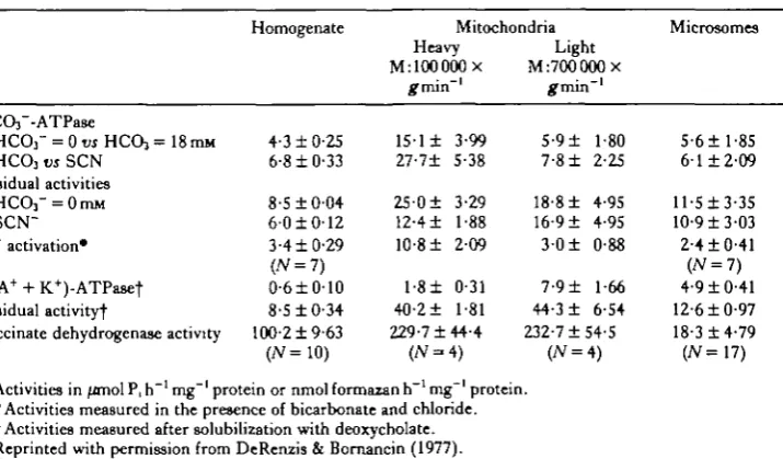
H e results in freshwater eel gill epithelium as did Bornancin et al. (1980) in fresh-water trout gill epithelium. Kinetic studies in these three gill epithelial systems strongly suggested that a (Cl" + HCCVj-stimulated ATPase is involved in the C1~/HCO3~ exchange mechanism and therefore in the acid-base regulation of fresh-water fish. These authors reported a parallelism between the affinities of the ATPase for Cl~ and both the Cl~ affinity for the gill transport mechanism and the Cl~ influx rate. The affinity constants for the CP-stimulated ATPase were 1-0, 5-9 and 23-Omequivl"1 for the goldfish (DeRenzis & Bornancin, 1977), freshwater trout (Bornancin et al. 1980) and freshwater eel (Bornancin et al. 1977) gill epithelium, respectively. The affinity of Cl~ for the transport systems in vivo were 0-07, 0-25 and 1-3 mequivl"1 for the goldfish (DeRenzis & Bornancin, 1977), freshwater trout (Bor-nancin et al. 1980) and freshwater eel (Bor(Bor-nancin et al. 1977) gill epithelium, respec-tively, while the corresponding maximal Cl~ influxes were 55-0, 19-6 and 0-36equiv h"1100g~l. In addition, the finding that Cl~ activation of anion-stimulated ATPase
activity was inhibited by thiocyanate (DeRenzis & Bornancin, 1977) was consistent with transport studies which showed that Cl~ influxes were inhibited by thiocyanate (DeRenzis, 1975). These studies on gill epithelium strongly support the hypothesis that the Cl~-stimulated ATPase is involved in gill anion exchanges that are related to mineral and acid-base homeostasis in freshwater fish.
[image:13.451.53.411.430.639.2]The fiddler crab gill has been shown actively to absorb Cl~ from low salinities (Baldwin & Kirschner, 1976a) and actively to extrude Cl~ in high salinity media (Baldwin & Kirschner, 19766). In concert with these findings DePew & Towle (1979) demonstrated the existence of an anion-stimulated ATPase in the gill cell plasma membrane of fiddler crab and suggested that this enzyme is so situated with its environment that it is highly accessible to Cl~ and HCCh", and thus may play a direct role in active C1~/HCO3~ exchange.
Table 5. Anion-dependent ATPase, (Na + + K+)-ATPase and succinate dehydro-genase activity in the goldfish gills
HCOf-ATPase HCO3" = 0ro HCO3 =
HCO3 vs SCN Residual activities
HCOf = 0mM SCN~ Cl" activation*
(NA+ + K+)-ATPasef Residual activity)-Homogenate 18mM 4-310-25 6-810-33 8-510-04 6-010-12 3-410-29 (JV=7) 0-610-10 8-510-34 Succinate dehydrogenase activity 100-21 9-63
Activities in /molP.h"1
•Activities measured in
(JV=10)
Mitochondria Heavy
M:100000X
1 5 1 1 3-99 27-71 5-38 25-01 3-29 12-41 1-88 10-81 209 1-81 0-31 40-21 1-81 229-7144-4
( N - 4 )
Light M:700000x 5-91 1-80 7-81 2-25 18-81 4-95 16-91 4-95 3 0 1 0-88
7-91 1-66 44-31 6-54 232-7154-5
(JV = 4)
mg~' protein or nmol formazan h"1 mg"1 protein. the presence of bicarbonate and chloride. t Activities measured after solubilization with deoxycholate. Reprinted with permission from DeRenzis & Bornancin (1977).
156 G. A. GERENCSER AND S.-H. LEE
Lee (1982) used an additional approach to the question concerning corresponded* between transport and anion-stimulated ATPase activity. After it was established that anion-stimulated ATPase activity existed in the plasma membrane of blue crab gill epithelium, the animals were adapted to low salinities. This thinking presumed that Cl~/HC03~ exchange should increase under these osmotic stressful conditions, therefore this transport activity should be reflected in an increase in the activity of anion-stimulated ATPase activity. This was indeed the case and Lee (1982) suggested that anion-stimulated ATPase activity appears likely to play an important role in anion transport for osmoregulatory and/or acid-base homeostasis in marine organisms.
Komnick et al. (1980) reported the presence of (CF + HCQr)-stimulated ATPase activity in plasma membranes of larval dragonfly rectum. The Cl~-stimulated ATPase activity was inhibited by thiocyanate as was the Cl~ influx into the rectal epithelia. These results suggested the possible existence of an ATPase-mediated, active C F transport mechanism located in the plasma membrane of larval dragonfly rectal epithelial cells.
In the eel (Anguilla japonica) intestine, relatively recent electrophysiological ex-periments have shown that active transport of CF coupled with water transport markedly increases during seawater adaptation (Ando, Utida & Nagahama, 1975; Ando, 1975). The observed increase in CF absorption raised the question of an associated increase in activity of an enzyme contributing to the transport process. It was demonstrated by Morisawa & Utida (1976) that anion-stimulated ATPase activity existed in an oligomycin-insensitive, thiocyanate-sensitive membrane fraction of eel intestinal enterocytes that was also relatively deficient in cytochrome oxidase activity. Seawater adaptation increased the enzyme activity commensurate with changes in CF and water transport. From these considerations, these authors concluded that the anion-stimulated ATPase played a direct role in CF transport in the eel intestine.
Active CF absorption by the Aplysia californica gut is mediated by a Na+ -independent, electrogenic mechanism (Gerencser, 19836). In an attempt to elucidate the CF transport mechanism, plasma membranes from Aplysia californica enterocytes were isolated by differential centrifugation and sucrose density gradient techniques and assayed for ATP hydrolysing capability (Gerencser & Lee, 1983). Marker enzymes for the plasma membrane fraction included 5'-nucleotidase, glucose-6-phosphatase, alkaline phosphatase and (Na+ + K+)-stimulated ATPase, while suc-cinic dehydrogenase and cytochrome oxidase were used as marker enzymes for the mitochondrial fraction. Both CF and HCCh'-stimulated ATPase activities were found in the plasma membrane fractions, which had virtually no mitochondrial contamination. These anion (CF/HCO3~)-stimulated ATPase activities were in-hibited more than 50% by thiocyanate whereas SITS, amiloride or furosemide had little or no effect on the (Cr/HCC>3~)-stimulated ATPase activity. Additionally, the Na+-independent active C F current traversing the Aplysia californica gut was shown to be 36-6nequivcm~2min~1 (ouabain-insensitive) and more than 50% of this cur-rent was inhibited by thiocyanate (Table 6). These results strongly suggest that the active CF absorptive mechanism in Aplysia californica gut could be a (CF/HCC*3~)-stimulated ATPase found in the enterocyte plasma membrane.
Cl '-stimulated ATPase 157
Table 6. Thiocyanate (SCN~) inhibition of both Aplysia calif ornica enterocyte plasma
membrane Cl '-stimulated ATPase activity and net mucosal to serosal (J%lj Cl''flux across Aplysia calif ornica gut under short-circuited conditions
Cr-ATPasc activity 112 j £ , 36-6 Cl"-ATPase activity + 10mM-SCN" 0-51 J2> + lOmM-SCN" 1-8
CP-ATPase activity is in /jmolPi liberated mg~' protein min"1 while J2, is in nequivcm~2min~'.
active translocation of Cl~ by an enzyme that directly utilizes the energy from ATP hydrolysis is not an unknown phenomenon and has been demonstrated in plants (Hill & Hanke, 1979; Auffret & Hanke, 1981). Indeed the evidence for primary active Cl~ transport in plant cells is almost as convincing .as that for (Na+ + K+)-stimulated ATPase and (Ca2+ + Mg2+)-stimulated ATPase in their respective roles for actively transferring Na+, K+ and Ca2+ across animal plasma membranes. As emphasized by DePont & Bonting (1981) the demonstration that Cl~-stimulated ATPase is involved in primary Cl~ transcellular movement in animal epithelia should satisfy the following criteria: (i) a specific inhibitor for the enzyme should be found or synthesized (e.g., an antibody) and this inhibitor should inhibit the transport process; and (ii) the Cl~-stimulated ATPase should be biochemically isolated and after its incorporation into lipsomes should then be shown to support active Cl~ transport.
This review and some of the work cited herein were supported, in part, by the Whitehall Foundation Grant 78-156 ck 1.
R E F E R E N C E S
AHEARN, G. A., MAGINNIS, L. A., SONG, Y. K. & TORNQUIST, A. (1977). Intestinal water and ion transport
in fresh water malacostracan prawns (Crustacea). In Water Relations in Membrane Transport in Plants and
Animals, (eds A. M. Jungreis, T. K. Hodges, A. Kleineller & S. G. Schultz), pp. 129-142. New York:
Academic Press.
AICKIN, C. C. & THOMAS, R. C. (1977). An investigation of the ionic mechanism of intracellular pH regulation in mouse soleus muscle fibres. J. Physiol., Land. 273, 295-316.
AL-BAZZAZ, F. J. & AL-AWQATI, Q. (1979). Interaction between sodium and chloride transport in canine tracheal mucosa. 7- "PPl- Physiol. 46, 111-119.
ANDO, M. (1975). Intestinal water transport and chloride pump in relation to sea-water adaptation of the eel,
Anguilla japonica. Comp. Biochem. Physiol. S2A, 229—233.
ANDO, M., UTIDA, S. & NAGAHAMA, H. (1975). Active transport of chloride in eel intestine with special reference to sea water adaptation. Comp. Biochem. Physiol. 51A, 27—32.
ARMSTRONG, W. M C D . , SUH, T. K. & GERENCSER, G. A. (1972). Stimulation by anoxia of active chloride transfer in isolated bullfrog intestine. Biochim. biophys.Acta. 225, 647—662.
Au, K. S. (1979). Relationship between rabbit erythrocyte membrane anion-sensitive Mg2+-ATPase and (Ca^+Mg^-ATPase. Int.J. Biochem. 10, 687-689.
AUFFRET, C. A. & HANKE, D. E. (1981). Improved preparation and assay and some characteristics of Cl~-ATPase activity from Limonium vulgare. Biochim. biophys. Acta. 648, 186—191.
BALDWIN, G. F. & KIRSCHNER, L. B. (1976a). Sodium and chloride regulation in Uca adapted to 175% seawater. Physiol. Zool. 49, 158-171.
BALDWIN, G. F. & KIRSCHNER, L. B. (19766). Sodium and chloride regulation in Uca adapted to 10% seawater. Physiol. Zool. 49, 172-180.
BINDER, H. J. & RAWLINS, C. L. (1973). Electrolyte transport across isolated large intestinal mucosa. Am.J.
Physiol. 225, 1232-1239.
BLUM, A. L., SHAH, G., PIERRE, T. S T . , HELANDER, H. F., SUNG, C. P., WIEBLHAUS, V. D. & SACHS, G.
(1971). Properties of soluble ATPase of gastric mucosa. II. Effect of HCQj. Biochim. biophys. Acta. 249,
K
158 G. A. GERENCSER AND S.-H. L E E
BORNANCIN, M., DERENZIS, G. & MAETZ, J. (1977). Branchial Cl transport, anion-stimulated A T P a s e ^ H acid-base balance in Anguilla anguilla adapted to freshwater: Effects of hyperoxia. J. comp. Pkysiol. ^ ^ 313-322.
BORNANCIN, M., DERENZIS, G. & NAON, R. (1980). Cr-HCO3"-ATPase in gills of the rainbow trout:
Evidence for its microsomal localization. Am.J. Pkysiol. 238, R2S1-R2S9.
BOYD, C. A. R. & CHIPPERFIELD, A. R. (1980). Are alkaline phosphatage and bicarbonate-dependent ATPase the same enzyme?7- Physiol., Land. 303, 63P.
CANDIA, O. A. (1972). Ouabain and sodium effects on chloride fluxes across the isolated bullfrog cornea. Am.
J. Physiol. 223, 1053-1057.
CHAN, S. H. P. & BARBOUR, R. L. (1976). Purification and properties of ATPase inhibitor from rat liver mitochondria. Biochim. biophys. Acta. 430, 426-433.
C H I E N . W . & STEVENS, C. E. (1972). Coupled active transport of Na and Cl across forestomach epithelium. Am.
J. Physiol. 223, 997-1003.
COLE, C. H. (1979). Bicarbonate-activated ATPase activity in renal cortex of chronically acidotic rats. Can.jf.
Physiol. Pharmacol. 57, 271-276.
DECNAN, K. J., KARNAKY, K. J. & ZADUNAISKY, J. A. (1977). Active chloride transport in the in vitro opercular skin of a teleost Fundulus heteroclitus, a gill-like epithelium rich in chloride cell. J. Physiol., Land. 271, 155-191.
DEKRUIJFF, B. & DEMEL, R. A. (1974). Polyene antibiotic-sterol interactions in membranes of Acholeplasma
laidlavm cell and lecitin liposomes. I I I . Molecular structure of the polyene antibiotic-cholesterol complexes. Biochim. biophys. Acta. 339, 57-70.
DEPEW, E. F . & TOWLE, D . W. (1979). Bicarbonate stimulated ATPase in plasma membrane fractions of fiddler crab (Uca minax) gill. Mar. Biol. Lett. 1, 59-67.
DEPONT, J. J. H. H. M. & BONTING, S. L. (1981). Anion-sensitive ATPase and ( K++ H+)-ATPase. In
Membrane Transport, (eds S. L. Bonting & J. J. H. H. M. DePont). Elsevier/North Holland Biomedical
Press.
DEPONT, J. J. H. H. M., HANSEN, T . & BONTING, S. L. (1972). An anion sensitive ATPase in lizard gastric mucosa. Biochim. biophys. Acta. 274, 189-200.
DERENZIS, G. (1975). The branchial chloride pump in the goldfish (Carassius auratus): relationship between C P / H C O j " and C1"/C1" exchanges and the effect of thiocyanate. J. exp. Biol. 63, 587-602.
DERENZIS, G. & BORNANCIN, M. (1977). C1~/HCO3~ ATPase in the gills of Carassius auratus: Its inhibition
by thiocyanate. Biochim. biophys. Acta. 467, 192-207.
DERENZIS, G. & MAETZ, J. (1973). Studies on the mechanism of chloride absorption by the goldfish gill: Relation with acid-base regulation. J. exp. Biol. 59, 339-358.
DUBOIS-REYMOND, E. (1848). Untersuchungen uber tierische Elektrizitat. Berlin.
DUFFEY, M. E., THOMPSON, S. M., FRIZZELL, R. A. & SCHULTZ, S. G. (1979). Intracellular chloride
absorp-tion by small intestine of the flounder. Pseudopleuronectes americanus. Fedn Proc. Fedn Am. Socs exp. Biol. 38, 1059.
DUNCAN, C. J. (1975). ATPase in rabbit erythrocytes: Stimulation by HCOj" and by Na+-plus-K+. Life Sci. 16, 955-966.
DURBIN, R. P. & KASBEJKAR, D. K. (1965). Adenosine triphosphate and active transport by the stomach. Fedn
Proc. Fedn Am. Socs exp. Biol. 24, 1377-1381.
EPSTEIN, F. H., MAETZ, J. & DERENZIS, G. (1973). Active transport of chloride by the teleost gill: inhibition by thiocyanate. Am. J. Physiol. 244, 1295-1299.
EJUCSON, A.-C. & SPRING, K. R. (1982). Coupled NaCl entry into Necturus gallbladder epithelial cells. Am.
J. Physiol. 243, C14O-C145.
FARMER, L. L. & EVANS, D. H. (1981). Chloride extrusion in the isolated perfused teleost gill. J. comp. Physiol. 141, 471-476.
FIELD, M., KARNAKY, K. J., SMYTH, P. L., BOLTON, J. E. & KINTER, W. B. (1978). Ion transport across the
isolated intestinal mucosa of the winter flounder, Pseudopleuronectes americanus. I. Functional and struc-tural properties of cellular and paracellular pathways for Na and Cl. J. Membr. Biol. 41, 265—275. FRIZZELL, R. A . , D U G A S , M.& SCHULTZ, S. G. (1975). Sodium chloride transport by rabbit gallbladder: Direct
evidence for a coupled NaCl influx process. J. gen. Physiol. 65, 769—795.
FRIZZELL, R. A., FIELD, M. & SCHULTZ, S. G. (1979). Sodium-coupled chloride transport by epithelial tissues. Am.J. Physiol. 236, F1-F8.
FRIZZELL, R. A., KOCH, M. J. & SCHULTZ, S. G. (1976). Ion transport by rabbit colon. I. Active and passive components. J. Membr. Biol. 27, 297-316.
GALEOTTi, G. (1904). Concerning the E. M. F. which is generated at the surface of animal membranes on contact with different electrolytes. Z.phys. Chem. 49, 542-562.
GASSNER, D . & KOMNICK, H. (1982). The loop diuretic furosemide as a non-competitive inhibitor of C l " / HCOj~-ATPases of vertebrate kidneys and insect rectum. Comp. Biochem. Physiol. 71C, 43-48.
GERENCSER, G. A. (1983a). Invertebrate epithelial transport. An. J . Physiol. 244, R127-R129. GERENCSER, G. A. (19836). Electrophysiology of chloride transport in Aplysia (mollusk) intestine.
Cl '-stimulated ATPase 159
SCSER, G. A. & LEE, S. H. (1983). Inhibition of Cr/HCO3~-stimulated ATPase in Aplysia californica
[it. Proc. int. Union. Physiol. Sri. 15, p. 430.
GRAF, J. & GIEBISCH, G. (1979). Intracellular sodium activity and sodium transport in Necturus gallbladder epithelium. J. Membr. Biol. 47, 327-355.
GRISHAM, C. M. & BARNETT, R. E. (1973). The role of lipid-phase transitions in the regulation of the (sodium and potassium) adenosine tnphosphatase. Biochemistry, N.Y. 12, 2635-2637.
GRISOLIA, S. & MENDELSON, J. (1974). Location of a very active bicarbonate-dependent ATPase in the outer membrane of rat and frog liver mitochondria. Biochem. biophys. Res. Commun. 58, 968-973.
GUNTER-SMITH, P. J. & WHITE, J. F . (1979). Response olAmphiuma small intestine to theophylline: Effect on bicarbonate transport. Am. J. Physiol. 236, E775-E783.
HAOCENBROCK, C. R. (1976). Molecular organization and the fluid nature of the mitochondrial energy trans-ducing membrane. \n Structure of biological Membranes, (eds S. Abrahamsson & I. Pascher). New York and London: Plenum Press.
HANRAHAN, J. & PHILLIPS, J. E. (1983). Mechanism and control of salt absorption in locust rectum. Am. J.
Physiol. 244, R131-R142.
HERRESA, L., LOPES-MORATALLA, N., SANTIAGO, E., PONZ, F. & JORDANA, R. (1978). Effect of bicarbonate
on chloride-dependent transmural potential and ATPase activity in the rectal wall of SMstocerca gregaria.
Revta esp Fisiol. 34, 219—224.
HILL, B. S. & HANKE, D. E. (1979). Properties of the chloride ATPase fromLimomum salt glands: Activation by, and binding to, specific sugars. J . Membr. Biol. 51, 185-194.
Ho, S. & CHAN, D . K. O. (1981). Branchial ATPases and ionic transport in the eel, Anguillajaponica. I I I . HCQf-stimulated SCN" inhibited Mg^-ATPase. Comp. Biochem. Physiol. 68B, 113-117.
HORSTMAN, L. L. & RACKER, E. (1970). Partial resolution of the enzymes catalysing oxidative phosphoryla-tion. XXII. Interaction between mitochondrial adenosine tnphosphatase inhibitor and mitochondrial adenosine triphosphatase. jf. biol. Chem. 245, 1336—1344.
HOUSE, C. R. & GREEN, K. (1965). Ion and water transport in isolated intestine of the marine teleost, Cottus
scorpius.J. exp. Biol. 42, 177-189.
HUBEL, K. A. (1968). The ins and outs of bicarbonate in the alimentary tract. Gastroenterology 56, 647-651.
HUMPHREYS, M. H. JCCHOU, L. Y. N. (1979). Anion-stimulated ATPase activity of brush border from rat small intestine. Am. J. Physiol. 236, E70-E76.
IRITANI, N. & WELLS, W. W. (1976). Properties of a bicarbonate-stimulated ATPase from rat uterus. Biochim.
biophys. Acta. 436, 863-868.
IYENGAR, R., MAILMAN, D . S. & SACHS, G. (1978). Purification of distinct plasma membranes from canine renal medulla. Am. J. Physiol. 234(3), F247-F254.
IZUTSU, J. T . & SIEGEL, I. A. (1972). A microsomal HCOj'-stimulated ATPase from the dog submandibular gland. Biochim. biophys. Acta. 284, 478-484.
IZUTSU, J. T . & SIEGEL, I. A. (1975). Bicarbonate ion-ATPase in rat liver cell fractions. Biochim. biophys. Acta. 382, 193-203.
IZUTSU, K., MADDEN, P. R., WATSON, E. L. & SIEGEL, I. A. (1977). Properties of the HCO3"-stimulated
Mg2+-ATPase activity in red cell membranes. Pflugers Arch, ges. Physiol. 369, 119-124.
KASBEXAR, D . H., DURBIN, R. P. & LINDLEY, D . (1965). An adenosine triphosphatase from frog gastric mucosa. Biochim. biophys. Acta. 105, 472—482.
KERSTETTER, T . & KIRSCHNER, L. B. (1972). Active chloride transport by the gills of rainbow trout (Salmo gairdnetfj.J. exp. Biol. 56, 263-272.
KERSTETTER, T . H. & KIRSCHNER, L. B. (1974). HCOydependent ATPase activity in the gills of rainbow trout
{Salmo gcdrdneri). Comp. Biochem. Physiol. 48B, 581-589.
KIMELBERG, H. K. & BOURKE, R. S. (1973). Properties and localization of bicarbonate-stimulated ATPase activity in rat brain. J. Neurochem. 20, 347-359.
KINNE-SAFFRAN, E. tc KINNE, R. (1974). Presence of bicarbonate-stimulated ATPase in the brush border microvillus membranes of the proximal tubule. Proc. Soc. exp. Biol. Med. 146, 751—753.
KINNE-SAFFRAN, E. & KINNE, R. (1979). Further evidence for the existence of an intrinsic bicarbonate-stimulated Mg2+-ATPase in brush border membranes isolated from rat kidney cortex. J. Membr. Biol. 49, 235-251.
KJJNGENBERG, M. tc PFAFF, E. (1966). Structural and functional compartmentation in mitochondria. In
Regulation of Metabolic Processes in Mitochondria. Vol. 7, (eds J. M. Tager, S. Papa, E. Quagliariello & E.
C. Slater), pp. 180-205. Amsterdam: Elsevier.
KLYCE, S. D. & WONG, R. K. S. (1977). Site and mode of adrenaline action on chloride transport across the rabbit corneal epithelium. J. Physiol., Land. 266, 777—799.
KOMNICK, H., SCHMITZ, M. tc HINSSEN, H. (1980). Biochemischer nachweis von HCQj" und Cl~-abhangigen ATPase-aktivitaten in Rectum von anisopteren Libellenlarven und hemmung der rectalen Chloridaufnahme durch Thiocyanat. Eur.J. Cell Biol. 20, 217-227.
160 G. A. GERENCSER AND S.-H. L E E
LAMBETH, D . O. & HARDY, H. A. (1971). Purification and properties of rat-liver mitochondrial triphosphatase. Eur.J. Biochem. 22, 355-363.
LEAF, A. (1965). Transepithelial transport and its hormonal control in toad bladder. Ergebn Physiol. Chem. exp.
Pharmakol. 56, 216-263.
LEE, S. H. (1982). Salinity adaptation of HCQT-dependent ATPase activity in the gills of blue crab (Callinectes
sapidus). Biochim. biophys. Acta. 689, 143-154.
L E S U E , B. R., SCHWARTZ, J. H. & STEINMETZ, P. R. (1973). Coupling between Cl absorption and HCOj secretion in turtle urinary bladder. Am.J. Physiol. 25, 610—617.
LIANG, C. T . & SACKTOR, B. (1976). Bicarbonate-stimulated ATPase in the renal proximal tubule luminal (brush border) membrane. Archs Biochem. Biophys. 176, 285-297.
MAETZ, J. & GARCIA-ROMEU, F. (1964). The mechanism of sodium and chloride uptake by the gills of a fresh water fish, Carassius auratus. I I . Evidence of NH4"/Na
+
and HCOj~/Cl~ exchanges. J. gen. Physiol. 47, 1209-1227.
MORISAWA, M. & UTIDA, S. (1976). HCO3~-activated adenosine triphosphatase in intestinal mucosa of the eel.
Biochim. biophys. Acta. 445, 458-463.
NAGEL, W., GARCIA-DIAZ, J. F. & ARMSTRONG, W. M C D . (1981). Intracellular ionic activities in frog skin.
J. Membr. Biol. 6, 127-134.
NELLANS, H. N., FRIZZELL, R. A. & SCHULTZ, S. G. (1973). Coupled sodium-chloride influx across the brush border of rabbit ileum. Am.J. Physiol. 225, 467-475.
NELLANS, H. N., FRIZZELL, R. A. & SCHULTZ, S. G. (1974). Brush border processes and transepithelial Na and Cl transport by rabbit ileum. Am. J. Physiol. 226, 1131-1141.
QUAY, J. F. & ARMSTRONG, W. M C D . (1969). Sodium and chloride transport by isolated bullfrog small intestine. Am. J. Physiol. 217, 694-702.
RACKER, E. (1962). ATPase and oxidative phosphorylation. Fedn Proc. Fedn Am. Socs exp. Biol. 21, 54. RENFRO, J. L. (1977). Interdependence of active Na and Cl transport by the isolated urinary bladder of the
teleost, Pseudopleuronectes americanus. J. exp. Zool. 199, 383-390.
ROTHSTEIN, A., CABANTCHIK, Z. I. & KNAUF, P. (1976). Mechanism of anion transport in red blood cells: Role of membrane proteins. Fedn Proc. Fedn Am. Socs exp. Biol. 35, 3-10.
RUSSELL, J. M. & BORON, W. F. (1976). Role of chloride transport in regulation of intracellular pH. Nature,
bond. TM, 73-74.
SACCOMANI, G., SHAH, G., SPENNEY, J. G. & SACHS, G. (1975). Characterization of gastric mucosal
membranes. VIII. The localization of peptides by iodination and phosphorylation. J . Biol. Chem. 250, 4802-4809.
SACHS, G., MITCH, W. E. & HIRSCHOWTTZ, B. I. (1965). Frog gastric mucosal ATPase. Proc. Soc. exp. Biol.
Med. 119, 1023-1027.
SACHS, G., SHAH, G., STRYCH, A., CLJNE, G. & HIRSCHOWTTZ, B. I. (1972). Properties of ATPase of gastric
mucosa. I I I . Distribution of HCOj'-stimulated ATPase in gastric mucosa. Biochim. biophys. Acta. 266, 625-638.
SACHS, G., SPENNEY, J. G. & LEWIN, M. (1978). H+ transport: Regulation and mechanism in gastric mucosa and membrane vesicles. Physiol. Rev. 58, 106—178.
SCHULTZ, S. G. (1979). Chloride transport by gastrointestinal epithelia: An overview. In Mechanisms of
Intestinal Secretion, (ed. H. J. Binder). New York: Alan R. Liss, Inc.
SCHUURMANS STEKHOVEN, F. ic BONTING, S. L. (1981). Transport adenosine triphosphttases: Properties and functions. Physiol. Rev. 61, 1—76.
SENER, A., VALVERDE, I. & MALAISSE, W. J. (1979). Presence of a HCOT-activated ATPase in pancreatic islets. FEBSLett. 105, 40-42.
SETCHELL, B. P., SMITH, M. W. & MUNN, E. A. (1972). The stimulation by bicarbonate of adenosine triphosphatase activity in the seminiferous tubules of rodents and the lack of effect of ouabain.J. Reprod.
Fertil. 28, 413-418.
SILVA, P., STOFF, J., FIELD, M., F I N E , L., FORREST, N. & EPSTEIN, F . H. (1977). Mechanism of active
chloride secretion by shark rectal gland: Role of Na-K-ATPase in chloride transport. Am. J. Physiol. 233, F298-F306.
SIMON, B. (1972). The HCO3~-stimulated ATPase in cat and rabbit pancreatic tissue. In Gastric Secretion, (eds G. Sachs, E. Heinz & K. J. Ullrich), pp. 345-348. New York: Academic Press.
SIMON, B., KINNE, R. & KNAUF, H. (1972a). The presence of a HCQj-ATPase in glandular submandibularis of rabbit. Pflugers Arch. ges. Physiol. 337, 117-184.
SIMON, B., KINNE, R. & SACHS, G. (19726). The presence of a HCOj~-ATPase in pancreatic tissue. Biochim.
biophys. Acta. 282, 293-300.
SIMON, B. & THOMAS, L. (1972). HCOj-stimulated ATPase from mammalian pancreas. Properties and its arrangement with other enzyme activities. Biochim. biophys. Acta. 288, 434—442.
SKADHAUGE, E. (1974). Coupling of transmural flows of NaCl and water in the intestine of the eel (Anguilla
anguilla).J. exp. Biol. 60, 535-546.
SKOU, J. C. (1965). Enzymatic basis for active transport of Na+ and K+ across cell membrane. Physiol. Rev.
C/ '-stimulated ATPase 161
N, R. J., SILVA, P., BEND, J. R. & EPSTEIN, F . H. (1975). Thiocyanate inhibition of ATPase and its relationship to anion transport. Am. J. Physiol. 229, 801—806.
SOUMARMON, A., LEWIN, M., CHERET, A. M. & BONFILS, S. (1974). Gastric HCOvstimulated ATPase:
evidence against its microsomal localization in rat fundus mucosa. Biochim. biophys. Acta. 339, 403-414. SPRING, K. A. & KIMURA, G. (1978). Chloride reabsorption by renal proximal tubules olNecturus.J. Membr.
Biol. 38, 233-254.
STOBBART, R. H. (1967). The effect of some anions and cations upon the fluxes and net uptake of chloride in the larva olAedes aegypti (L) and the nature of the uptake mechanisms for sodium and chloride. J. exp. Biol.
47, 35-57.
TANISAWA, A. S. & FORTE, J. G. (1971). Phosphorylated-intermediate of microsomal ATPase from rabbit gastric mucosa. Archs Biochem. Biophys. 147, 165—175.
THOMAS, R. C. (1977). The role of bicarbonate, chloride and sodium ions in the regulation of intracellular pH in snail neurons. J . Physiol., Land. 273, 317-338.
TURBECK, B. O., NEDERCAARD, S. & KRUSE, H. (1968). An anion-stimulated adenosine triphosphatase from the potassium-transportingmidgut of thelarvtoiHyalophoracecmpia.Biochim. biophys. Acta. 163, 354—361.
TURNBERG, L. A., BISBERDORF, F . A., MORAWAKI, S. G. & FORDTRAN, J. S. (1970). Interrelationships of
chloride, bicarbonate, sodium and hydrogen transport in the human ileum. J. din. Invest. 49, 557—567. USSING, H. H. (1960). The Alkali Metal Ions in Biology. Berlin: Springer-Verlag.
USSING, H. H. & ZERAHN, K. (1951). Active transport of sodium as the source of electric current in the short-circuited isolated frog skin. Acta. physiol. scand. 23, 110-127.
VAN AMELSVOORT, J. M. M., DEPONT, J. J. H. H. M. &BONTING, S. L. (1977a). Is there a plasma membrane-located anion-sensitive ATPase? Biochim. biophys. Acta. 466, 283—301.
VAN AMELSVOORT, J. M. M., D E P O N T , J. J. H. H. M., STOLS, A. L. H. & BONTING, S. L. (19776). Is there
a plasma membrane-located anion-sensitive ATPase? II. Further studies on rabbit kidney. Biochim. biophys.
Acta. 471, 78-91.
VAN AMELSVOORT, J. M. M., JANSEN, J. W. C. M., D E P O N T , J. J. H. H. M. & BONTING, S. L. (1978a). Is
there a plasma membrane-located anion-sensitive ATPase? IV. Distribution of the enzyme in rat pancreas.
Biochim. biophys. Acta. 512, 296-308.
VAN AMELSVOORT, J. M. M., VAN HOOF, P. M. K. B., D E P O N T , J. J. H. H. M. & BONTING, S. L. (19786).
Is there a plasma membrane-located anion-sensitive ATPase? I I I . Identity of the erythrocyte enzyme with (Ca2+ + Mg2*) ATPase. Biochim. biophys. Acta. 507, 83-93.
WAIS, U. & K N A U F , H. (1975). H+ transport and membrane-bound HCC^-ATPase in salivary duct epithelium.
Pftugers Arch. ges. Physiol. 361, 61-64.
WATLJNGTON, C. O., JESSEE, S. D. & BALDWIN, G. (1977). Ouabain, acetazolamide, and Cl" flux in isolated frog skin: Evidence for two distinct active Cl transport mechanisms. Am. J. Physiol. 232, F55O-F558. WHEELER, A. P. & HARRISON, E. W. (1982). Subcellular localization and characterization of HCCV-ATPase
from the mantel of the freshwater clam, Anodonta cataracta. Comp. Biochem. Physiol. 71B, 629-636. WHITE, ] . F. (1980). Bicarbonate-dependent chloride absorption in small intestine: Ion fluxes and intracellular
chloride activities. J. Membr. Biol. 53, 95-107.
WIEBELHAUS, V. D . , SUNG, C. P., HELANDER, H. F . , SHAH, G., BLUM, A. L. & SACHS, G. (1971).
Solubiliza-tion of anion ATPase from Necturus oxyntic cells. Biochim. biophys. Acta. 241, 49-56.
ZADUNAISKY, J. A. (1972). Sodium activation of chloride transport in the frog cornea. Biochim. biophys. Acta.