George Wortzman 1 N. B. Rewcastle2
Received November 4, 1981; accepted after revision February 11, 1982.
Presented in part at the annual meeting of the Association of University Radiologists, New Or -leans, April 1981 .
'Department of Radiology, University of T o-ronto, Mt. Sinai Hospital, 600 University Ave., Toronto, Ontario M5G 1 X5. Address reprint re -quests to G. Wortzman.
2Department of Neuropathology, University of Toronto, Toronto, Ontario. Present address: D e-partment of Pathology, Foothills Hospital, Calgary, Alberta.
AJNR 3:505-512, September/October 1982 0195-6108/82/0305-0505 $00.00 © American Roentgen Ray Society
505
Tomographic Abnormalities
Simulating Pituitary
Microadenomas
False-positive and false-negative interpretations of sellar tomography were found in
about one-fifth of cases in a recent autopsy study correlating the presence of pituitary microadenomas with abnormal sellar tomograms. An analysis of minor variations in the
bony configuration of the sella disclosed variations due to posterior lobe asymmetry,
intercavernous venous channels, bony asymmetry, and an empty sella in 27 of the 120
sellas examined. In some instances, the asymmetry resulted from a combination of
these causes. A further study of 50 pituitary glands in situ showed posterior lobe asymmetry to be a common anomaly (76%) that can produce an obvious disparity between the two halves of the sella. Thus, the minor radiologic criteria of local thinning
of the anterior wall or floor, slant of the floor, or asymmetry of the two halves of the
sella must be interpreted with caution as being indicative of the presence of pituitary microadenoma. In the absence of clinical or biochemical dysfunction, the changes
more likely result from explainable anatomic causes.
A recent autopsy study correlating the presence of pituitary microadenomas and abnormal sellar tomograms urged caution in diagnosing a microadenoma by sellar tomography [1]. False-positive and false-negative interpretation of tomo-graphic findings were found in about one-fifth of 120 cases. The 27 cases of false-positive findings were reexamined as to origin of the minor variations in the bony configuration of the sella. The results of this analysis plus a further study of 50 pituitary glands in situ are the subject of this report.
Materials and Methods
The earlier report [1] contains a detailed description of methods. Briefly, this consisted of an en bloc removal of the pituitary and the sella in an unselected autopsy population only excluding cases with a clinically apparent pituitary tumor. Hypocycloidal tomograms were done on the specimens in anteroposterior (AP) and lateral projections at 2 mm intervals. Interpretation was by three neuroradiologists independently and also together. Criteria for sellar abnormality were the findings of a slanted sellar floor, local cortical thinning of the lamina dura and erosion, or bulging of the anterior wall or floor of the sella. These criteria were based on earlier work by Vezina and Sutton [2] and McLachlan et al. [3]. Care was taken that the normal variations of sphenoid sinus development and septal attachments were not interpreted as abnormal. Cognizance was also taken of the normal variations of the dorsum, which frequently has a concavity centrally with its lateral margins more anteriorly situated.
506 WORTZMAN AND REWCASTLE AJNR:3, September/October 1982
Pathologic study identified 43 adenomas in 32 of the 1 20 pituitaries so examined. These adenomas were mostly under 2 mm in size and distributed chiefly in the periphery of the gland. High false-positive and false-negative interpretation rates of tomographic findings were found in the correlative study then done. Of the 27 cases considered abnormal on tomography, six had a coexisting adenoma, but in no case was the adenoma of such size or position so as to consider it to be the cause of the tomographic abnormality.
The group of 27 cases was reexamined. The microscopic structure of the pituitary gland and the bony sella were correlated with the AP and lateral tomographic studies. The results of this analysis are discussed below.
A further postmortem study of 50 pituitary glands in situ was carried out. This study consisted of careful dissection of the pituitary gland both before and after removal of the dorsum sella to determine the relative positions of the an-terior and posterior lobes of the pituitary in relation to the midline of the sella.
Results
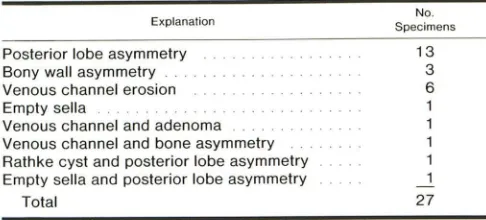
Explanations for sellar variations in the 27 false-positive cases are summarized in table 1. In most cases, one cause for the misinterpretation was found; in four of the cases, more than one factor was instrumental in causing the changes considered compatible with a microadenoma.
Posterior lobe asymmetry was the most common cause of the interpretation of sellar abnormalities and was seen in 15 cases; in 13 of these, it was the only alteration present (figs. 1 and 2). This asymmetry, which consisted of a posterola t-eral rather than a midline relationship of the posterior lobe to the anterior lobe, instigated the second part of this study, verifying this to be a true finding rather than a technical artifact. Intercavernous venous channels were always pres-ent, but, in eight of the cases, the size and position of the channel was considered to be responsible for the abnormal configuration of bony cortex (figs. 3 and 4). In one of these cases, there was a coexisting adenoma; in another, there was bone asymmetry. An asymmetry of the bony walls of the sphenoid sinus that could not be readily attributed to a variation in sinus development or of sinus septal attachment was found in four cases; in three of these, it was the only variation seen. The sellar configuration was considered due to a partially empty sella in two cases (fig. 5); in one, it was the sole explanation. Instances of minor degrees of empty sella in the original study did not cause any significant deformity. Rathke pouch cysts were frequent findings, being seen in 29 of the original 120 subjects. They were present in four of 27 false-positives. In only one case was the presence of the cyst considered to be of significance, but, in that case, there was also a coexisting posterior lobe asymmetry (fig. 6).
The results of the in situ examination of the pituitary gland are summarized in figure 7 with an example of an eccentric posterior pituitary lobe shown in figure 8. The posterior lobe is frequently eccentric in its relation to the anterior lobe and was midline in only 24% of cases. In 14% of cases, the
TABLE 1: Explanations for False-Positive Sellar Abnormalities in Autopsy Specimens
Explanation
Posterior lobe asymmetry Bony wall asymmetry Venous channel erosion Empty sella
Venous channel and adenoma Venous channel and bone asymmetry Rathke cyst and posterior lobe asymmetry Empty sella and posterior lobe asymmetry
Total No. Specimens 13 3 6 27
posterior lobe was situated well laterally. The results of this second study verify that the findings described above in 15 of the 27 cases studied were real rather than due to an artifact or an asymmetry developing in the preparation of the pathology sections.
Discussion
In 1936, Costello [4] found clinically silent pituitary ade-nomas in 225 of 1,000 autopsies. Sections were done in the longest axis of the gland, either sagittally or transversely, cut by hand with thicknesses of 1-1.5 mm. Most adenomas found were 1.5 mm or greater. McCormick and Halmi [5] reported 9.1 % adenomas in 1,600 consecutive autopsies. In our previous report [1], microadenomas were found in 32 (27%) of 120 cases. The adenomas were usually under 2 mm in size. This higher incidence of adenomas is attributed to the greater number of sections taken. Criteria for identi -fication of an adenoma were uniformity of cells, a stromal pattern different from the rest of the gland, and evidence of compression of adjacent pituitary parenchyma. More than one adenoma was found in eight of the 32 cases. Immuno -peroxidase staining identified 16 of 39 microadenomas to be prolactinomas. As in the series of Costello [4], the ade-nomas were usually peripheral in location with some pro-jecting from the surface of the gland under the capsule.
Since the early 1970s, an extensive body of literature pertaining to the sella and pituitary has appeared reflecting advances in the fields of neurosurgery, endocrinology, pa -thology, gynecology, and radiology.
These diagnostic advances include the development of sensitive and specific radioimmunoassays for blood prolac-tin, new staining techniques to identify pituitary cell secre-tory products, refinement of transphenoidal microsurgery, and the medical treatment of the prolactinoma with bromo-criptine [6-16].
[image:2.612.317.560.109.219.2]AJNR:3, September/October 1982 FALSE-POSITIVE SELLAR TOMOGRAPHY
507
Fig. 1 .-Eccentric posterior pituitary lobe. A, Lateral tomographic cut 4 mm to left of midline. Sloped anterior wall. B, Right-sided cut 4 mm from midline. Steeper anterior wall slope, increased AP diameter, and thinned dorsum. C, Histopathology corresponding to A. D, Histopathology corr e-sponding to B. Eccentrically placed posterior lobe of pituitary accounts for marked difference in sel -lar configuration.
A
c
A
c
8
D
appreciated without the aid of pluridirectional tomography. The validity of these criteria has been verified both in ra dio-logic and surgical literature [14, 17-21].
It has long been appreciated that there are, however,
8
D
E
Fig. 2.-Eccentric posterior pituitary lobe. Lat-eral tomographic cuts on left (A) and right (B) of midline. Increased AP diameter evident on right. C and D, Histopathology corresponding to A and B, respectively. As in fig. 1, sellar configuration is determined by eccentric position of posterior lobe of pituitary. E, AP tomographic cut. Sellar floor is slanted down on right.
508
A
B
A
B
c
o
WORTZMAN AND REWCASTLE AJNR:3, September IOctober 1982
Fig. 3.- Venous channels. Left (A) and right (B) tomographic cuts. Deepened floor on B inte r-preted as compatible with microadenoma. C and
D, Corresponding histopathology sections (to A and B, respectively). Large crossing venous sinus beneath pituitary gland causes irregularity of sellar
floor. Low, small posterior lobe on D causes minor erosion of dorsum.
Fig. 4.-Venous channels. Left (A) and right (B) tomographic cuts. Erosion of dorsum and
pos-terior sellar floor on right. C and D, Histopathology sections corresponding to A and B, respectively. Large venous sinus posteroinferior to pituitary
[image:4.617.57.394.88.375.2] [image:4.617.54.389.428.723.2]AJNR:3, September/October 1982
A
/'"
l'c
A
c
FALSE-POSITIVE SELLAR TOMOGRAPHY
B
E
'-D
B
D
509
Fig, 5.- Partially empty sella and eccentric posterior pituitary lobe. Lett (A) and right (B) lo m-ographic culs. Increase in AP diameter of sella on righl with Ihinned dorsum and some undercutling of tuberculum. C and D, Histopalhology sections corresponding 10 A and B, respectively. Parlially empty sella causes undercutling of luberculum and eccenlric posterior pituilary lobe thinning dor -sum. E, AP cuI. Minor depression of sellar floor on righl side partly allributable 10 septal lag.
510 WORTZMAN AND REWCASTLE AJNR:3, September/October 1982
Left
2
19
4% 38%
42%
Middle
12
24%
Right
12
5
24% 10%
34%
Fig. 7.- Posterior pituitary tobe asymmetry. Findings in 50 consecutive
examinations of pituitary glands in situ. Posterior lobe was central in 24%, but eccentric as depicted in the rest. In 14%, the lateral margin of the posterior lobe was flush with the lateral margin of the anterior lobe; the other 62% of less eccentric placed glands had their lateral margins as depicted by
the broken line.
in variations in anatomy of the sphenoid sinus and sellar region with anomalies of the bony walls of the sella being identified [26-28]. Swanson and duBoulay [29] examined 85 cases with no clinical evidence of a pituitary lesion, yet the plain films in 31.7% demonstrated changes that were identical with those described for a small intrasellar lesion. The unanswered question, however, was whether these changes were due to a coexisting but asymptomatic ade-noma. Other authors have pointed out normal variants in sellar configurations secondary to a sloped sellar floor, thinning of the sellar cortex, the pattern of aeration and/or septation of the sphenoid sinus, or thickening of the mucosal membrane of the sinus [30-32].
adjacent sellar floor. The asymmetry creates difficulties when comparing sequential lateral tomographic sections of the sella, as the asymmetry, when due to the eccentric nature of the posterior lobe of the pituitary, is recognized as a unilateral increase in the AP diameter of the sella.
A defect and irregularity of the sellar floor were seen in other cases of this series secondary to crossing venous sinuses. Cysts of the pouch of Rathke proved almost as common as microadenomas in the initial autopsy series, but were not shown to be responsible for any evident bony change. In the case where the partially empty sella was the
Earlier autopsy studies stressed either the incidence of
A
microadenomas or the variations of adjacent structures such as carotid vessels, arachnoid cisterns, cavernous sinuses, and the optic chiasm and nerves [33, 34]. More recent autopsy studies have dealt more with the histopathology of the pituitary gland and its relation to sellar tomographic
abnormalities [1 , 35-37] and have pointed out the nonspe-cificity of the sellar changes previously considered due to a pituitary microadenoma.
The present study carries this a step further and reveals several reasons for apparent abnormalities. The most sig-nificant is an asymmetric position of the posterior lobe of the pituitary gland with its secondary effect on the dorsum sella. This led to the greatest number of errors of interpre -tation despite care being taken during the original study to
avoid errors due to an asymmetry of the dorsum. In a comprehensive study, DiChiro and Nelson [38] pointed out the many irregularities in the appearance of the dorsum and described the depression of bone that accommodates the posterior lobe of the pituitary gland. This depression was
called "fossula hypophyseos" by Karlas, "impressio hypo-physios" by Wegard, and "fossetta pituitaria" by Cardillo and Bossi (all cited in [38]). The resultant thinness of bone in the area of depression varies and the central part may be
absent resulting in a corticated "window" [39]. This depres
-sion of the anterior aspect of the dorsum may involve the
8
Fig. 8.- A, Pituitary gland in situ before removal (from the back and
above). The dorsum has been amputated. The black thread runs in midline.
[image:6.612.84.281.85.219.2] [image:6.612.317.558.227.694.2]AJNR:3. September IOctober 1982 FALSE-POSITIVE SELLAR TOMOGRAPHY 511
sole abnormality, the changes of the sella were considered
to be due to an asymmetric invagination of subarachnoid space.
In addition to the anomalies and changes of the sella described above, there have been reports of other factors
causing changes compatible with an intrasellar lesion. There
is a report of an apparently eroded dorsum that at autopsy proved due to a congenital lack of ossification [40]. Erosion
of the floor of the sella simulating the changes of a pituitary microadenoma has also been reported in two cases of an anomalous arterial communication between the int racavern-ous segments of internal carotid arteries in association with
agenesis of one internal carotid [41].
Pluridirectional tomography is still of aid before
trans-sphenoidal removal of a pituitary microadenoma and usually
reflects the side of the microadenoma and will show the anatomy of sphenoid pneumatization and septation.
How-ever, there are many patients with hormonal dysfunction where an adenoma has been found despite negative
tomo-graphic studies [12, 18]. In a recent radiologic-surgical
correlative study, good correlation existed between the tom-ographic localization and the location of the tumor at surgery
in most cases [42]. In nearly 20% of 89 cases reported, however, the sellar changes suggestive of adenoma on
tomography had no relation to the location of the tumor as seen at surgery. This further indicates the need for caution
in the interpretation of tomographic findings. Protocols for the study of pituitary adenomas have changed radically over
the past 2-3 years as advances in equipment and tech-niques continue, and they are the subject of recent review
articles [43,44].
Interpretation of plain films and tomographic studies of
the sella turcica must be undertaken with caution. In addition
to variations of sphenoid pneumatization and septation, one can be misled by a change in the AP diameter of the sella secondary to eccentric position of the posterior lobe of the pituitary. An anatomic review shows the posterior lobe to be more frequently eccentric in its relation to the anterior lobe than to being a truly midline attachment. In addition, bone defects simulating erosion of the sellar floor may be
sec-ondary to intercavernous venous channels, bony wall
asym-metry, or to anomalies of sphenoid sinus development and other explainable anatomic variations.
ACKNOWLEDGMENT
We thank Christine Bobkowski for help in manuscript preparation.
REFERENCES
1. Burrow GN. Wortzman G, Rewcastle NB, Holgate RC, Kovacs
K. Microadenomas of the pituitary and abnormal sellar tom o-grams in an unselected autopsy series. N Engl J Med 1981 ;304: 156-158
2. Vezina JL, Sutton T J. Prolactin-secreting pituitary micr oaden-omas: roentgenologic diagnosis. AJR 1974; 120 :46-54 3. McLachlan MSF, Wright AD, Doyle FH. Plain film and t
omo-graphic assessment of the pituitary fossa in 140 acromegalic patients. Br J Radiol 1970;43: 360-369
4. Costello RT. Subclinical adenoma of the pituitary gland. Am J Patho/1936; 12: 205-215
5. McCormick WF, Halmi NS. Absence of chromophobe ade no-mas from a large series of pituitary tumors. Arch Patho! Lab Med 1971;92:231-238
6. Rawe SE, Williamson HO. Levine JH, Phansey SA. Hungerford
D, Adkins WY. Prolactinomas: surgical therapy. indications and
results. Surg Neuro/1980;14: 161 -167
7. Hwang P, Guyda H, Friesen H. A radioimmunoassay for human prolactin. Proc Natl Acad Sci USA 1971 ;68: 1902-1906 8. Kleinberg DL, Noel GL, Frantz AG. Galactorrhea: a study of
235 cases. including 48 with pituitary tumors. N Eng! J Med
1977; 296: 589-600
9. McCarty KS Jr. Bredesen DE. Vogel FS. Neoplasms of the anterior pituitary. Neurosurgery 1978;3: 96-1 04
10. Sirek AMT. Corenblum B. Horvath E. Rewcastle B. Ezrin C. Kovacs K. A new look at pituitary adenomas: structure eluc i-dating function. Can Med Assoc J 1976; 114: 225-229 11. Corenblum T. Sirek AMT. Horvath E. Kovacs K. Ezrin C. Human
mixed somatotrophic and lactotrophic pituitary adenomas. J Clin Endocrinol Metab 1976;42: 857 -863
12. Kovacs K. Horvath E. Bayley TA. Hassaram ST. Ezrin C. Silent corticotroph cell adenoma with lysosomal accumulation and crinophagy: a distinct clinicopathologic entity. Am J Med 1978;64: 492-499
13. Wilson CB. Dempsey LC. Transsphenoidal microsurgical
re-moval of 250 pituitary adenomas. J Neurosurg 1978;48: 13-22
14. Hardy J. Transsphenoidal surgery of hypersecreting pituitary tumors. In: Kohler PO, Ross GT. eds. Diagnosis and treatment of pituitary tumors. International Congress Series No. 303.
Excerpta Med 1973; 179: 94
15. Hardy J. Microsurgery of pituitary disorders. Ann R Coli Surg
Eng/1980;13:294-298
16. Reichlin S. The prolactinoma problem. N Eng! J Med
1979;300:313-315
17. Robertson WD, Newton TH. Radiologic assessment of pituitary microadenomas. AJR 1978; 131 : 489-492
18. Richmond IL. Newton TH. Wilson CB. Prolactin-secreting pi -tuitary adenomas: correlation of radiographic and surgical findings. AJNR 1980; 1 : 1 3-1 6
19. Chang RJ. Keye WR Jr. Young JR. Wilson CB. Jaffe RB.
Detection. evaluation. and treatment of pituitary micro adeno-mas in patients with galactorrhea and amenorrhea. Am J Obstel Gyneco/1977;128:356-363
20. Geehr RB. Allen WE III. Rothman SLG. Spencer DD. Pluridirec-tional tomography in the evaluation of pituitary tumors. AJR 1978; 130: 1 05-1 09
21. Antunes JL. Housepian EM. Frantz AG. et al. Prolactin-secre
t-ing pituitary tumors. Ann Neurol 1977;2: 148-153
22. Cope VZ. The internal structure of the sphenoidal sinus. J Anal 1917;51 :127-136
23. Van Alyea OE. Sphenoid sinus. Anatomic study. with consi d-eration of the clinical significance of the structural
character-istics of the sphenoid sinus. Arch Otolaryngol 1941;
34:225-253
24. Peele JC. Unusual anatomical variations of the sphenoid
si-nuses. Laryngoscope 1957;67: 208-237
25. Hammer G. Radberg C. The sphenoidal sinus. An anatomical and roentgenologic study with reference to transsphenoid hy-pophysectomy. Acta Radiol (Stockh) 1961 ;56: 401 -422 26. Renn WHo Rhoton AL Jr. Microsurgical anatomy of the sellar
region. J Neurosurg 1975;43: 288-298
27. Rhoton AL Jr. Hardy DG. Chambers SM. Microsurgical
anat-omy and dissection of the sphenoid bone. cavernous sinus and
512 WORTZMAN AND REWCASTLE AJNR:3, September jOctober 1982
28. Fujii K, Chambers SM, Rhoton AL Jr. Neurovascular relati on-ships of the sphenoid sinus. J Neurosurg 1979;50: 31-39
29. Swanson HA, duBoulay G. Borderline variants of the normal
pituitary fossa. Br J Radio/1975;48: 366-369
30. Dubois PJ, Orr DP, Hoy RJ, Herbert DL, Heinz ER. Normal
sellar variations in frontal tomograms. Radiology
1979;131: 1 05-11 0
31. Bruneton IN, Drouillard JP, Sabatier JC, Elie GP, Tavernier JF.
Normal variants of the sella turcica. Radiology
1979;131 :99-104
32. Tenner MS, Weitzner I Jr. Pitfalls in the diagnosis of erosive changes in expanding lesions of the pituitary fossa. Radiology
1980; 137: 393-396
33. Bergland RM, Ray BS, Torack RM. Anatomical variations in the pituitary gland and adjacent structures in 225 human
autopsy cases. J Neurosurg 1968;28: 93-99
34. McLachlan MSF, Williams ED, Doyle FH. Applied anatomy of
the pituitary gland and fossa. A radiological and histopathol
og-ical study based on 50 necropsies. Br J Radiol 1968;41 :782-788
35. Wortzman G, Holgate RC, Rewcastle NB, Burrow GN. Abnor -mal sellas and pituitary adenomas in 120 post mortem sphen -oid specimens (abstr). AJNR 1980; 1 : 365
36. Turski PA, Newton TH, Horton B. Anatomic correlation with
complex motion tomography in 100 sphenoid specimens
(abstr). AJNR 1980; 1 : 365
37. Muhr C, Bergstrom K, Grimelius L, Larsson S-G. A parallel
study of the roentgen anatomy of the sella turcica and the
histopathology of the pituitary gland in 205 autopsy specimens.
Neuroradiology 1981 ;21 : 55-65
38. Di Chiro G, Nelson KB. The volume of the sella turcica. AJR 1962;87: 989-1 008
39. Berger PE, Harwood-Nash DC, Fitz CR. The dorsum sellae in infancy and childhood. Pediatr Radio/1976;4: 212-220 40. Penkrot RJ, Bures C. The "apparently" eroded dorsum sella:
a new anomaly. AJR 1979;132:1005-1006
41. Kishore PRS, Kaufman AB, Melichar FA. Intrasellar carotid anastomosis simulating pituitary microadenoma. Radiology
1979;132: 381-383
42. Raji MR, Kishore PRS, Becker DP. Pituitary microadenoma: a
radiological-surgical correlative study. Radiology 1981;
139:95-99
43. Kricheff II. The radiologic diagnosis of pituitary adenoma. An
overview. Radiology 1979;131 :263-265
44. Robertson HJ, Rose A, Ehmi B, England G, Meriweather R.