(
E
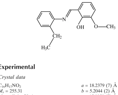
)-2-[(2-Ethylphenyl)iminomethyl]-6-methoxyphenol
Serap Yazıcı,a* C¸ig˘dem Albayrak,bI:smail Gu¨mru¨kc¸u¨og˘lu,c I:smet S¸enelaand Orhan Bu¨yu¨kgu¨ngo¨ra
aDepartment of Physics, Faculty of Arts and Sciences, Ondokuz Mayıs University, TR-55139 Kurupelit–Samsun, Turkey,bSinop Faculty of Education, Sinop University, TR-57000 Sinop, Turkey, andcDepartment of Chemistry, Ondokuz Mayıs University, TR-55139 Kurupelit–Samsun, Turkey
Correspondence e-mail: yserap@omu.edu.tr
Received 26 December 2009; accepted 28 December 2009
Key indicators: single-crystal X-ray study;T= 150 K; mean(C–C) = 0.002 A˚; disorder in main residue;Rfactor = 0.044;wRfactor = 0.115; data-to-parameter ratio = 15.4.
The molecule of the title compound, C16H17NO2, adopts the phenol–imine tautomeric form with a strong intramolecular O—H N hydrogen bond and an E conformation with respect to the azomethine C N bond. The dihedral angle between the aromatic rings is 21.23 (9). The ethyl group is disordered over two orientations with occupancies of 0.598 (6) and 0.402 (6). In the crystal, the molecules are linked into chains along thebaxis by C—H interactions.
Related literature
For general background to o-hydroxy Schiff bases, see: Stewart & Lingafelter (1959); Calligariset al.(1972); Maslen & Waters (1975). For the photochromic and thermochromic characteristics of Schiff base compounds, see: Cohen et al.
(1964); Moustakali-Mavridis et al. (1980); Hadjoudis et al.
(1987); Xuet al.(1994). For a related structure, see: Yu¨ceet al.
(2004).
Experimental
Crystal data
C16H17NO2
Mr= 255.31 Monoclinic,P21=c
a= 18.2379 (7) A˚ b= 5.2044 (2) A˚ c= 15.0950 (7) A˚
= 113.788 (3)
V= 1311.05 (9) A˚3
Z= 4
MoKradiation
= 0.09 mm1
T= 150 K
0.580.390.08 mm
Data collection
Stoe IPDS II diffractometer Absorption correction: integration
(X-RED32; Stoe & Cie, 2002) Tmin= 0.961,Tmax= 0.993
18335 measured reflections 3014 independent reflections 2353 reflections withI> 2(I) Rint= 0.072
Refinement
R[F2> 2(F2)] = 0.044
wR(F2) = 0.115
S= 1.03 3014 reflections 196 parameters
H atoms treated by a mixture of independent and constrained refinement
max= 0.26 e A˚
3
min=0.32 e A˚
[image:1.610.48.239.571.725.2]3
Table 1
Hydrogen-bond geometry (A˚ ,).
Cg1 is the centroid of the C8–C13 ring.
D—H A D—H H A D A D—H A
O1—H1 N1 0.98 (2) 1.68 (2) 2.6023 (15) 156 (2) C14—H14c Cg1i
0.96 2.83 3.6241 (18) 141
Symmetry code: (i)xþ1;yþ1 2;zþ
3 2.
Data collection:X-AREA(Stoe & Cie, 2002); cell refinement: X-AREA; data reduction: X-RED32 (Stoe & Cie, 2002); program(s) used to solve structure: SHELXS97(Sheldrick, 2008); program(s) used to refine structure: SHELXL97 (Sheldrick, 2008); molecular graphics:ORTEP-3 for Windows(Farrugia, 1997); software used to prepare material for publication:WinGX(Farrugia, 1999).
The authors acknowledge the Faculty of Arts and Sciences, Ondokuz Mayıs University, Turkey, for the use of the Stoe IPDS II diffractometer (purchased under grant No. F279 of the University Research Fund).
Supplementary data and figures for this paper are available from the IUCr electronic archives (Reference: CI5005).
References
Calligaris, M., Nardin, G. & Randaccio, L. (1972).Coord. Chem. Rev.7, 385– 403.
Cohen, M. D., Schmidt, G. M. J. & Flavian, S. (1964).J. Chem. Soc.pp. 2041– 2051.
Farrugia, L. J. (1997).J. Appl. Cryst.30, 565. Farrugia, L. J. (1999).J. Appl. Cryst.32, 837–838.
Hadjoudis, E., Vitterakis, M. & Maviridis, I. M. (1987).Tetrahedron,43, 1345– 1360.
Maslen, H. S. & Waters, T. N. (1975).Coord. Chem. Rev.17, 137–176. Moustakali-Mavridis, I., Hadjoudis, B. & Mavridis, A. (1980).Acta Cryst.B36,
1126–1130.
Sheldrick, G. M. (2008).Acta Cryst.A64, 112–122.
Stewart, J. M. & Lingafelter, E. C. (1959).Acta Cryst.12, 842–845.
Stoe & Cie (2002).X-AREAandX-RED. Stoe & Cie, Darmstadt, Germany. Xu, X.-X., You, X.-Z., Sun, Z.-F., Wang, X. & Liu, H.-X. (1994).Acta Cryst.
C50, 1169–1171.
Yu¨ce, S., O¨ zek, A., Albayrak, C¸., Odabas¸og˘lu, M. & Bu¨yu¨kgu¨ngo¨r, O. (2004). Acta Cryst.E60, o718–o719.
Acta Crystallographica Section E
Structure Reports
Online
supporting information
Acta Cryst. (2010). E66, o287 [https://doi.org/10.1107/S1600536809055573]
(
E
)-2-[(2-Ethylphenyl)iminomethyl]-6-methoxyphenol
Serap Yaz
ı
c
ı
,
Ç
i
ğ
dem Albayrak,
İ
smail G
ü
mr
ü
k
çü
o
ğ
lu,
İ
smet
Ş
enel and Orhan B
ü
y
ü
kg
ü
ng
ö
r
S1. Comment
o-Hydroxy Schiff bases derived from the reaction of o-hydroxyaldehydes with aniline have been examined extensively
(Steward & Lingafelter, 1959; Calligaris et al., 1972; Maslen & Waters, 1975). Schiff base compounds display interesting
photochromic and thermochromic features and can be classified in terms of these (Cohen et al., 1964;
Moustakali-Mavridis et al., 1980; Hadjoudis et al., 1987). Photo- and thermochromism arise via H atom transfer from the hydroxy O
atom to the N atom (Hadjoudis et al., 1987; Xu et al., 1994).
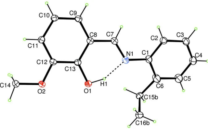
The molecule of the title compound (Fig. 1) exists in the phenol-imine form which is confirmed by C13—O1 and C7—
N1 bond distances. These distances agree with the corresponding distances in
1-{4-[(2-hydroxybenzylidene)amino]-phenyl}ethanone, a related structure [C—O = 1.3500 (17) and C—N = 1.2772 (16) Å; Yüce et al., 2004].
The title molecule is not planar; the dihedral angle between the two benzene rings is 21.23 (9)°. An intramolecular O1—
H1···N1 hydrogen bond (Fig. 1) generates an S(6) ring-motif.
The crystal structure is stabilized by weak C—H···π interactions involving H14C and C8–C13 ring (Fig. 2).
S2. Experimental
A solution of 3-methoxysalicylaldehyde (0.5 g 3.3 mmol) in ethanol (20 ml) was added to a solution of 2-ethylaniline (0.4
g 3.3 mmol) in ethanol (20 ml). The reaction mixture was stirred for 1 h under reflux. Single crystals of the title
compound were obtained by slow evaporation of an ethanol solution (yield 72%, m.p. 339-340 K).
S3. Refinement
The ethyl group is disordered over two orientations with occupancies of 0.598 (6) and 0.402 (6). Atom H1 was located in
a difference map and refined freely. The remaining H atoms were placed in calculated positions and constrained to ride
Figure 1
The molecular structure of the title compound, with the atomic numbering scheme. Displacement ellipsoids are drawn at
the 30% probability level. The dashed line indicates a hydrogen bond. Only the major disorder component is shown.
Figure 2
[image:3.610.137.481.339.639.2](E)-2-[(2-Ethylphenyl)iminomethyl]-6-methoxyphenol
Crystal data
C16H17NO2
Mr = 255.31 Monoclinic, P21/c
Hall symbol: -P 2ybc a = 18.2379 (7) Å b = 5.2044 (2) Å c = 15.0950 (7) Å β = 113.788 (3)° V = 1311.05 (9) Å3
Z = 4
F(000) = 544 Dx = 1.293 Mg m−3
Mo Kα radiation, λ = 0.71073 Å Cell parameters from 2353 reflections θ = 1.5–28.0°
µ = 0.09 mm−1
T = 150 K Plate, brown
0.58 × 0.39 × 0.08 mm
Data collection
Stoe IPDS II diffractometer
Radiation source: fine-focus sealed tube Graphite monochromator
Detector resolution: 6.67 pixels mm-1
ω scan
Absorption correction: integration (X-RED32; Stoe & Cie, 2002) Tmin = 0.961, Tmax = 0.993
18335 measured reflections 3014 independent reflections 2353 reflections with I > 2σ(I) Rint = 0.072
θmax = 27.6°, θmin = 2.4°
h = −23→23 k = −6→6 l = −19→19
Refinement
Refinement on F2
Least-squares matrix: full R[F2 > 2σ(F2)] = 0.044
wR(F2) = 0.115
S = 1.03 3014 reflections 196 parameters 0 restraints
Primary atom site location: structure-invariant direct methods
Secondary atom site location: difference Fourier map
Hydrogen site location: inferred from neighbouring sites
H atoms treated by a mixture of independent and constrained refinement
w = 1/[σ2(F
o2) + (0.052P)2 + 0.279P]
where P = (Fo2 + 2Fc2)/3
(Δ/σ)max = 0.001
Δρmax = 0.26 e Å−3
Δρmin = −0.32 e Å−3
Extinction correction: SHELXL97 (Sheldrick, 2008), Fc*=kFc[1+0.001xFc2λ3/sin(2θ)]-1/4
Extinction coefficient: 0.024 (3)
Special details
Experimental. 320 frames, detector distance = 100 mm
Geometry. All e.s.d.'s (except the e.s.d. in the dihedral angle between two l.s. planes) are estimated using the full covariance matrix. The cell e.s.d.'s are taken into account individually in the estimation of e.s.d.'s in distances, angles and torsion angles; correlations between e.s.d.'s in cell parameters are only used when they are defined by crystal symmetry. An approximate (isotropic) treatment of cell e.s.d.'s is used for estimating e.s.d.'s involving l.s. planes.
Refinement. Refinement of F2 against ALL reflections. The weighted R-factor wR and goodness of fit S are based on F2,
conventional R-factors R are based on F, with F set to zero for negative F2. The threshold expression of F2 > σ(F2) is used
only for calculating R-factors(gt) etc. and is not relevant to the choice of reflections for refinement. R-factors based on F2
are statistically about twice as large as those based on F, and R- factors based on ALL data will be even larger.
Fractional atomic coordinates and isotropic or equivalent isotropic displacement parameters (Å2)
x y z Uiso*/Ueq Occ. (<1)
H16A 0.0628 0.3425 0.5702 0.080* 0.402 (6)
H16B 0.0188 0.3027 0.4581 0.080* 0.402 (6)
H16C 0.0970 0.4691 0.5006 0.080* 0.402 (6)
C15A 0.1206 (3) 0.0813 (10) 0.5183 (4) 0.0411 (12) 0.402 (6)
H15A 0.0944 −0.0727 0.5274 0.049* 0.402 (6)
H15B 0.1734 0.0953 0.5705 0.049* 0.402 (6)
C15B 0.09657 (17) 0.2040 (7) 0.4866 (2) 0.0380 (9) 0.598 (6)
H15C 0.1114 0.3839 0.4967 0.046* 0.598 (6)
H15D 0.0388 0.1903 0.4639 0.046* 0.598 (6)
C16B 0.13777 (19) 0.0531 (6) 0.5796 (3) 0.0470 (9) 0.598 (6)
H16D 0.1220 0.1202 0.6285 0.070* 0.598 (6)
H16E 0.1948 0.0681 0.6009 0.070* 0.598 (6)
H16F 0.1226 −0.1244 0.5682 0.070* 0.598 (6)
H1 0.2630 (14) 0.462 (4) 0.5862 (17) 0.077 (7)*
O1 0.29046 (6) 0.5645 (2) 0.64484 (7) 0.0387 (3)
N1 0.23740 (7) 0.3745 (3) 0.47070 (8) 0.0376 (3)
C12 0.38182 (8) 0.9118 (3) 0.68144 (9) 0.0314 (3)
O2 0.37858 (6) 0.9107 (2) 0.77070 (7) 0.0386 (3)
C9 0.38032 (8) 0.8881 (3) 0.49637 (10) 0.0344 (3)
H9 0.3798 0.8812 0.4345 0.041*
C13 0.33418 (7) 0.7266 (3) 0.61589 (9) 0.0306 (3)
C8 0.33213 (7) 0.7187 (3) 0.52183 (9) 0.0315 (3)
C14 0.42206 (9) 1.1110 (3) 0.83509 (10) 0.0406 (3)
H14A 0.4162 1.0939 0.8952 0.061*
H14B 0.4015 1.2747 0.8066 0.061*
H14C 0.4777 1.0989 0.8468 0.061*
C11 0.42846 (8) 1.0776 (3) 0.65443 (10) 0.0338 (3)
H11 0.4602 1.1993 0.6982 0.041*
C7 0.27988 (8) 0.5403 (3) 0.45018 (10) 0.0346 (3)
H7 0.2772 0.5468 0.3874 0.042*
C2 0.20659 (9) 0.1179 (3) 0.32266 (10) 0.0407 (3)
H2 0.2493 0.1925 0.3133 0.049*
C10 0.42817 (8) 1.0634 (3) 0.56190 (10) 0.0347 (3)
H10 0.4605 1.1732 0.5447 0.042*
C3 0.16088 (10) −0.0684 (3) 0.25904 (11) 0.0474 (4)
H3 0.1729 −0.1186 0.2073 0.057*
C1 0.18945 (8) 0.1950 (3) 0.40048 (10) 0.0378 (3)
C4 0.09745 (10) −0.1796 (3) 0.27231 (11) 0.0479 (4)
H4 0.0666 −0.3053 0.2298 0.057*
C6 0.12496 (10) 0.0848 (4) 0.41384 (13) 0.0594 (5)
C5 0.08014 (10) −0.1034 (4) 0.34882 (13) 0.0568 (5)
H5 0.0373 −0.1794 0.3575 0.068*
Atomic displacement parameters (Å2)
U11 U22 U33 U12 U13 U23
C16A 0.049 (2) 0.056 (3) 0.061 (3) 0.001 (2) 0.028 (2) −0.013 (2)
C15B 0.0310 (13) 0.0355 (19) 0.0482 (16) 0.0008 (13) 0.0168 (12) 0.0063 (14) C16B 0.0583 (17) 0.0449 (16) 0.042 (2) −0.0002 (13) 0.0248 (15) 0.0062 (13)
O1 0.0388 (5) 0.0449 (6) 0.0382 (5) −0.0100 (5) 0.0216 (4) −0.0055 (5)
N1 0.0279 (5) 0.0472 (7) 0.0369 (6) −0.0015 (5) 0.0124 (5) −0.0066 (5)
C12 0.0324 (6) 0.0346 (7) 0.0296 (6) 0.0033 (6) 0.0151 (5) 0.0015 (5)
O2 0.0447 (5) 0.0440 (6) 0.0327 (5) −0.0101 (5) 0.0214 (4) −0.0068 (4)
C9 0.0384 (7) 0.0358 (7) 0.0318 (6) 0.0057 (6) 0.0169 (6) 0.0043 (5)
C13 0.0271 (6) 0.0332 (7) 0.0338 (6) 0.0025 (5) 0.0146 (5) 0.0020 (5)
C8 0.0290 (6) 0.0337 (7) 0.0315 (6) 0.0053 (5) 0.0117 (5) 0.0016 (5)
C14 0.0491 (8) 0.0413 (9) 0.0362 (7) −0.0070 (7) 0.0220 (6) −0.0083 (6)
C11 0.0358 (7) 0.0316 (7) 0.0345 (7) −0.0002 (6) 0.0148 (6) 0.0000 (6)
C7 0.0307 (6) 0.0404 (8) 0.0318 (6) 0.0055 (6) 0.0116 (5) −0.0002 (6)
C2 0.0402 (7) 0.0450 (9) 0.0350 (7) −0.0007 (6) 0.0132 (6) −0.0004 (6)
C10 0.0381 (7) 0.0329 (7) 0.0372 (7) 0.0013 (6) 0.0193 (6) 0.0050 (6)
C3 0.0531 (9) 0.0507 (10) 0.0350 (7) −0.0020 (8) 0.0143 (7) −0.0054 (7)
C1 0.0299 (6) 0.0454 (9) 0.0340 (7) −0.0001 (6) 0.0088 (5) −0.0047 (6)
C4 0.0426 (8) 0.0488 (9) 0.0396 (8) −0.0027 (7) 0.0035 (6) −0.0058 (7)
C6 0.0407 (8) 0.0860 (14) 0.0566 (10) −0.0213 (9) 0.0248 (8) −0.0273 (10)
C5 0.0376 (8) 0.0732 (13) 0.0584 (10) −0.0174 (8) 0.0180 (8) −0.0171 (9)
Geometric parameters (Å, º)
C16A—C15A 1.529 (7) C9—C10 1.370 (2)
C16A—H16A 0.96 C9—C8 1.4035 (19)
C16A—H16B 0.96 C9—H9 0.93
C16A—H16C 0.96 C13—C8 1.4058 (17)
C15A—C6 1.611 (5) C8—C7 1.4521 (19)
C15A—H15A 0.97 C14—H14A 0.96
C15A—H15B 0.97 C14—H14B 0.96
C15B—C16B 1.516 (5) C14—H14C 0.96
C15B—C6 1.523 (3) C11—C10 1.3966 (18)
C15B—H15C 0.97 C11—H11 0.93
C15B—H15D 0.97 C7—H7 0.93
C16B—H16D 0.96 C2—C3 1.382 (2)
C16B—H16E 0.96 C2—C1 1.391 (2)
C16B—H16F 0.96 C2—H2 0.93
O1—C13 1.3490 (16) C10—H10 0.93
O1—H1 0.98 (2) C3—C4 1.378 (2)
N1—C7 1.2781 (19) C3—H3 0.93
N1—C1 1.4193 (18) C1—C6 1.394 (2)
C12—O2 1.3722 (15) C4—C5 1.373 (2)
C12—C11 1.3834 (19) C4—H4 0.93
C12—C13 1.4034 (19) C6—C5 1.394 (2)
O2—C14 1.4274 (17) C5—H5 0.93
C15A—C16A—H16A 109.5 C9—C8—C7 119.54 (12)
C15A—C16A—H16B 109.5 C13—C8—C7 120.87 (12)
C15A—C16A—H16C 109.5 O2—C14—H14B 109.5
H16A—C16A—H16C 109.5 H14A—C14—H14B 109.5
H16B—C16A—H16C 109.5 O2—C14—H14C 109.5
C16A—C15A—C6 100.8 (4) H14A—C14—H14C 109.5
C16A—C15A—H15A 111.6 H14B—C14—H14C 109.5
C6—C15A—H15A 111.6 C12—C11—C10 120.47 (13)
C16A—C15A—H15B 111.6 C12—C11—H11 119.8
C6—C15A—H15B 111.6 C10—C11—H11 119.8
H15A—C15A—H15B 109.4 N1—C7—C8 122.06 (12)
C16B—C15B—C6 105.7 (3) N1—C7—H7 119.0
C16B—C15B—H15C 110.6 C8—C7—H7 119.0
C6—C15B—H15C 110.6 C3—C2—C1 120.71 (14)
C16B—C15B—H15D 110.6 C3—C2—H2 119.6
C6—C15B—H15D 110.6 C1—C2—H2 119.6
H15C—C15B—H15D 108.7 C9—C10—C11 120.11 (13)
C15B—C16B—H16D 109.5 C9—C10—H10 119.9
C15B—C16B—H16E 109.5 C11—C10—H10 119.9
H16D—C16B—H16E 109.5 C4—C3—C2 120.00 (14)
C15B—C16B—H16F 109.5 C4—C3—H3 120.0
H16D—C16B—H16F 109.5 C2—C3—H3 120.0
H16E—C16B—H16F 109.5 C2—C1—C6 119.58 (14)
C13—O1—H1 101.6 (13) C2—C1—N1 122.67 (13)
C7—N1—C1 120.97 (12) C6—C1—N1 117.68 (13)
O2—C12—C11 124.55 (12) C5—C4—C3 119.48 (15)
O2—C12—C13 115.46 (11) C5—C4—H4 120.3
C11—C12—C13 119.99 (12) C3—C4—H4 120.3
C12—O2—C14 115.69 (10) C5—C6—C1 118.45 (15)
C10—C9—C8 120.50 (12) C5—C6—C15B 121.37 (16)
C10—C9—H9 119.7 C1—C6—C15B 119.27 (17)
C8—C9—H9 119.7 C5—C6—C15A 115.8 (2)
O1—C13—C12 118.68 (11) C1—C6—C15A 121.60 (19)
O1—C13—C8 122.03 (12) C4—C5—C6 121.78 (16)
C12—C13—C8 119.27 (12) C4—C5—H5 119.1
C9—C8—C13 119.58 (12) C6—C5—H5 119.1
C11—C12—O2—C14 5.35 (19) C3—C2—C1—N1 176.31 (14)
C13—C12—O2—C14 −175.41 (12) C7—N1—C1—C2 25.3 (2)
O2—C12—C13—O1 −0.49 (18) C7—N1—C1—C6 −157.73 (16)
C11—C12—C13—O1 178.79 (12) C2—C3—C4—C5 0.2 (3)
O2—C12—C13—C8 178.24 (11) C2—C1—C6—C5 0.9 (3)
C11—C12—C13—C8 −2.48 (19) N1—C1—C6—C5 −176.19 (17)
C10—C9—C8—C13 −1.1 (2) C2—C1—C6—C15B −168.4 (2)
C10—C9—C8—C7 177.95 (13) N1—C1—C6—C15B 14.5 (3)
O1—C13—C8—C9 −178.51 (12) C2—C1—C6—C15A 156.9 (3)
C12—C13—C8—C9 2.80 (19) N1—C1—C6—C15A −20.1 (4)
O1—C13—C8—C7 2.45 (19) C16B—C15B—C6—C5 95.4 (3)
C12—C13—C8—C7 −176.23 (12) C16B—C15B—C6—C1 −95.6 (3)
C13—C12—C11—C10 0.4 (2) C16A—C15A—C6—C5 −108.3 (3)
C1—N1—C7—C8 −177.25 (12) C16A—C15A—C6—C1 95.1 (3)
C9—C8—C7—N1 176.91 (13) C16A—C15A—C6—C15B 0.4 (3)
C13—C8—C7—N1 −4.1 (2) C3—C4—C5—C6 0.1 (3)
C8—C9—C10—C11 −1.0 (2) C1—C6—C5—C4 −0.7 (3)
C12—C11—C10—C9 1.3 (2) C15B—C6—C5—C4 168.4 (2)
C1—C2—C3—C4 0.1 (3) C15A—C6—C5—C4 −158.1 (3)
C3—C2—C1—C6 −0.6 (3)
Hydrogen-bond geometry (Å, º)
Cg1 is the centroid of the C8–C13 ring.
D—H···A D—H H···A D···A D—H···A
O1—H1···N1 0.98 (2) 1.68 (2) 2.6023 (15) 156 (2)
C14—H14c···Cg1i 0.96 2.83 3.6241 (18) 141