3-Nitro-1
H
-1,2,4-triazole
Madhukar Hemamalini and Hoong-Kun Fun*‡
X-ray Crystallography Unit, School of Physics, Universiti Sains Malaysia, 11800 USM, Penang, Malaysia
Correspondence e-mail: [email protected]
Received 22 November 2010; accepted 25 November 2010
Key indicators: single-crystal X-ray study;T= 100 K; mean(N–C) = 0.001 A˚;
Rfactor = 0.035;wRfactor = 0.092; data-to-parameter ratio = 20.1.
The asymmetric unit of the title compound, C2H2N4O2, contains two crystallographically independent molecules in which the triazole rings are essentially planar, with maximum deviations of 0.003 (1) A˚ in both molecules. The dihedral angle between the two 1H-1,2,4-triazole rings is 56.58 (5). In
the crystal, molecules are linkedviaintermolecular N—H N and C—H O hydrogen bonds, forming a supramolecular chain along thebaxis.
Related literature
For details and applications of 1H-1,2,4-triazole derivatives, see: Desenko (1995); Voset al.(1983); van Albadaet al.(1984); Al-Kharafiet al.(1986); Gupta & Bhargava (1978); Joneset al.
(1965); Bennuret al.(1976). For the stability of the tempera-ture controller used in the data collection, see: Cosier & Glazer (1986).
Experimental
Crystal data
C2H2N4O2
Mr= 114.08 Monoclinic,P21=c
a= 8.7818 (1) A˚
b= 10.0726 (2) A˚
c= 9.9703 (1) A˚
= 107.081 (1)
V= 843.03 (2) A˚3
Z= 8
MoKradiation
= 0.16 mm1
T= 100 K
0.480.330.30 mm
Data collection
Bruker SMART APEXII CCD area-detector diffractometer Absorption correction: multi-scan
(SADABS; Bruker, 2009)
Tmin= 0.928,Tmax= 0.954
11450 measured reflections 3081 independent reflections 2768 reflections withI> 2(I)
Rint= 0.022
Refinement
R[F2> 2(F2)] = 0.035
wR(F2) = 0.092
S= 1.05 3081 reflections 153 parameters
H atoms treated by a mixture of independent and constrained refinement
max= 0.50 e A˚
3
min=0.40 e A˚
3
Table 1
Hydrogen-bond geometry (A˚ ,).
D—H A D—H H A D A D—H A
N2A—H1N1 N1Ai
0.885 (15) 1.995 (15) 2.8540 (9) 163.4 (15) N2B—H1N2 N1Bii 0.857 (16) 2.057 (16) 2.9128 (10) 176.0 (16) C1A—H1AA O2Aiii
0.93 2.50 3.1129 (10) 124
C1B—H1BA O2Bii
0.93 2.51 3.0451 (11) 117
Symmetry codes: (i) x;yþ1 2;z
1
2; (ii) xþ1;y 1 2;zþ
3 2; (iii)
xþ2;yþ1 2;zþ
3 2.
Data collection:APEX2(Bruker, 2009); cell refinement:SAINT (Bruker, 2009); data reduction:SAINT; program(s) used to solve structure: SHELXTL (Sheldrick, 2008); program(s) used to refine structure:SHELXTL; molecular graphics:SHELXTL; software used to prepare material for publication:SHELXTLandPLATON(Spek, 2009).
MH and HKF thank the Malaysian Government and Universiti Sains Malaysia for the Research University grant No. 1001/PFIZIK/811160. MH also thanks Universiti Sains Malaysia for a post-doctoral research fellowship.
Supplementary data and figures for this paper are available from the IUCr electronic archives (Reference: IS2634).
References
Albada, G. A. van, de Graaff, R. A. G., Haasnoot, J. G. & Reedijk, J. (1984).
Inorg. Chem.23, 1404–1408.
Al-Kharafi, F. M., Al-Hajjar, F. H. & Katrib, A. (1986).Corros. Sci.26, 257– 264.
Bennur, S. C., Jigajinni, V. B. & Badiger, V. V. (1976).Rev. Roum. Chim.21, 757–762.
Bruker (2009).APEX2,SAINTandSADABS. Bruker AXS Inc., Madison, Wisconsin, USA.
Cosier, J. & Glazer, A. M. (1986).J. Appl. Cryst.19, 105–107. Desenko, S. M. (1995).Khim. Geterotsikl. Soedin.pp. 2–24. Gupta, A. K. & Bhargava, K. P. (1978).Pharmazie,33, 430–431.
Jones, D. H., Slack, R., Squires, S. & Wooldridge, K. R. H. (1965).J. Med. Chem.8, 676–680.
Sheldrick, G. M. (2008).Acta Cryst.A64, 112–122. Spek, A. L. (2009).Acta Cryst.D65, 148–155.
Vos, G., le Febre, R. A., de Graaff, R. A. G., Haasnoot, J. G. & Reedijk, J. (1983).J. Am. Chem. Soc.105, 1682–1683.
Acta Crystallographica Section E Structure Reports
Online
supporting information
Acta Cryst. (2011). E67, o15 [https://doi.org/10.1107/S1600536810049287]
3-Nitro-1
H
-1,2,4-triazole
Madhukar Hemamalini and Hoong-Kun Fun
S1. Comment
1H-1,2,4-Triazole ring systems are typical planar six-π-electron partially aromatic systems, and are used, along with their
derivatives, as starting materials for the synthesis of many heterocycles (Desenko, 1995). Substituted 1H-1,2,4-triazoles
have also been actively studied as bridging ligands coordinating through their vicinal N atoms and some have special
structures with interesting magnetic properties (Vos et al., 1983; van Albada et al., 1984). Studies also indicate that the
1H-1,2,4-triazole system is associated with anticorrosion (Al-Kharafi et al., 1986) and anti-inflammatory action (Gupta &
Bhargava, 1978) and other pharmacological activities by exhibiting antiviral, anti-asthmatic, diuretic, analgesic,
antimicrobial, antidepressant and antifungal effects (Jones et al., 1965; Bennur et al., 1976).
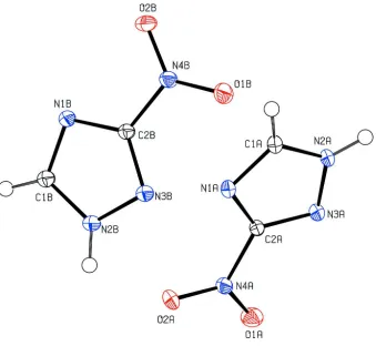
The asymmetric unit of the title compound consists of two crystallographically independent 3-nitro-1H-1,2,4-triazole
molecules (A & B) with very similar geometry (Fig. 1). The 1H-1,2,4-triazole units are essentially planar with maximum
deviations of 0.003 (1) Å for atom N1A (molecule A) and 0.003 (1) Å for atom C2B (molecule B). The dihedral angle
between the two 1H-1,2,4-triazole (N1A—N3A/ C1A–C2A) and (N1B—N3B/C1B–C2B) rings is 56.58 (5)°.
In the crystal structure (Fig. 2), molecules are connected via N2A—H1N1···N1A, N2B—H1N2···N1B, C1A—
H1AA···O2A and C1B—H1BA···O2B (Table 1) hydrogen bonds to form a one-dimensional supramolecular chain along
the b-axis.
S2. Experimental
Hot methanol solution (20 ml) of 3-nitro-1H-1,2,4-triazole (57 mg, Aldrich) was warmed over a heating magnetic stirrer
for 5 minutes. The resulting solution was allowed to cool slowly at room temperature. Crystals of the title compound
appeared from the mother liquor after a few days.
S3. Refinement
Atoms H1N1 and H1N2 were located from a difference Fourier map and refined freely [refined N—H distances
0.857 (16) and 0.885 (15) Å]. The remaining H atoms were positioned geometrically [C—H = 0.93 Å] and were refined
Figure 1
The asymmetric unit of the title compound. Displacement ellipsoids are drawn at the 50% probability level.
Figure 2
The crystal packing of the title compound, showing a hydrogen-bonded (dashed lines) molecular chain.
3-Nitro-1H-1,2,4-triazole
Crystal data
C2H2N4O2 Mr = 114.08
Monoclinic, P21/c
Hall symbol: -P 2ybc a = 8.7818 (1) Å b = 10.0726 (2) Å c = 9.9703 (1) Å
V = 843.03 (2) Å3 Z = 8
F(000) = 464 Dx = 1.798 Mg m−3
[image:3.610.127.477.417.548.2]T = 100 K Block, colourless
0.48 × 0.33 × 0.30 mm
Data collection
Bruker SMART APEXII CCD area-detector diffractometer
Radiation source: fine-focus sealed tube Graphite monochromator
φ and ω scans
Absorption correction: multi-scan (SADABS; Bruker, 2009) Tmin = 0.928, Tmax = 0.954
11450 measured reflections 3081 independent reflections 2768 reflections with I > 2σ(I) Rint = 0.022
θmax = 32.7°, θmin = 2.9° h = −13→11
k = −15→13 l = −15→15
Refinement
Refinement on F2
Least-squares matrix: full R[F2 > 2σ(F2)] = 0.035 wR(F2) = 0.092 S = 1.05 3081 reflections 153 parameters 0 restraints
Primary atom site location: structure-invariant direct methods
Secondary atom site location: difference Fourier map
Hydrogen site location: inferred from neighbouring sites
H atoms treated by a mixture of independent and constrained refinement
w = 1/[σ2(F
o2) + (0.0495P)2 + 0.2412P]
where P = (Fo2 + 2Fc2)/3
(Δ/σ)max = 0.001
Δρmax = 0.50 e Å−3
Δρmin = −0.40 e Å−3
Special details
Experimental. The crystal was placed in the cold stream of an Oxford Cryosystems Cobra open-flow nitrogen cryostat (Cosier & Glazer, 1986) operating at 100.0 (1) K.
Geometry. All s.u.'s (except the s.u. in the dihedral angle between two l.s. planes) are estimated using the full covariance matrix. The cell s.u.'s are taken into account individually in the estimation of s.u.'s in distances, angles and torsion angles; correlations between s.u.'s in cell parameters are only used when they are defined by crystal symmetry. An approximate (isotropic) treatment of cell s.u.'s is used for estimating s.u.'s involving l.s. planes.
Refinement. Refinement of F2 against ALL reflections. The weighted R-factor wR and goodness of fit S are based on F2,
conventional R-factors R are based on F, with F set to zero for negative F2. The threshold expression of F2 > 2σ(F2) is
used only for calculating R-factors(gt) etc. and is not relevant to the choice of reflections for refinement. R-factors based on F2 are statistically about twice as large as those based on F, and R- factors based on ALL data will be even larger.
Fractional atomic coordinates and isotropic or equivalent isotropic displacement parameters (Å2)
x y z Uiso*/Ueq
O1A 0.73082 (8) −0.04055 (7) 0.51717 (7) 0.01914 (13)
O2A 0.85716 (9) 0.01680 (7) 0.73151 (6) 0.01888 (14)
N1A 1.01266 (8) 0.21389 (7) 0.64087 (7) 0.01307 (13)
N2A 0.99857 (9) 0.24376 (7) 0.42001 (7) 0.01273 (13)
N3A 0.89747 (8) 0.14183 (7) 0.41773 (7) 0.01286 (13)
N4A 0.82618 (9) 0.02813 (7) 0.60358 (7) 0.01348 (13)
C1A 1.06543 (10) 0.28585 (8) 0.55150 (8) 0.01356 (14)
H1AA 1.1381 0.3552 0.5768 0.016*
C2A 0.91199 (9) 0.13008 (8) 0.55271 (8) 0.01167 (14)
O1B 0.75840 (8) 0.41600 (7) 0.50676 (7) 0.02046 (14)
N1B 0.51771 (8) 0.42579 (7) 0.73353 (7) 0.01312 (13)
N2B 0.51813 (9) 0.20833 (7) 0.72132 (7) 0.01419 (13)
N3B 0.60998 (9) 0.24714 (7) 0.64058 (7) 0.01375 (13)
N4B 0.68913 (8) 0.46461 (7) 0.58504 (7) 0.01361 (13)
C1B 0.46484 (10) 0.31423 (8) 0.77581 (8) 0.01436 (15)
H1BA 0.4002 0.3102 0.8347 0.017*
C2B 0.60451 (9) 0.37710 (8) 0.65365 (8) 0.01189 (14)
H1N1 1.0120 (19) 0.2722 (16) 0.3403 (16) 0.034 (4)*
H1N2 0.5028 (18) 0.1259 (16) 0.7343 (15) 0.030 (4)*
Atomic displacement parameters (Å2)
U11 U22 U33 U12 U13 U23
O1A 0.0195 (3) 0.0170 (3) 0.0203 (3) −0.0044 (2) 0.0049 (2) −0.0008 (2) O2A 0.0278 (3) 0.0179 (3) 0.0136 (3) 0.0007 (2) 0.0102 (2) 0.0037 (2) N1A 0.0164 (3) 0.0135 (3) 0.0103 (3) −0.0005 (2) 0.0055 (2) −0.0003 (2) N2A 0.0164 (3) 0.0133 (3) 0.0099 (3) 0.0001 (2) 0.0060 (2) 0.0011 (2) N3A 0.0156 (3) 0.0128 (3) 0.0107 (3) 0.0004 (2) 0.0048 (2) 0.0009 (2) N4A 0.0161 (3) 0.0118 (3) 0.0144 (3) 0.0020 (2) 0.0072 (2) 0.0018 (2) C1A 0.0163 (3) 0.0142 (3) 0.0115 (3) −0.0007 (3) 0.0060 (3) −0.0005 (2) C2A 0.0146 (3) 0.0109 (3) 0.0107 (3) 0.0013 (2) 0.0056 (2) 0.0010 (2) O1B 0.0240 (3) 0.0185 (3) 0.0245 (3) 0.0046 (2) 0.0158 (3) 0.0025 (2) O2B 0.0264 (3) 0.0106 (3) 0.0260 (3) −0.0006 (2) 0.0131 (3) 0.0004 (2) N1B 0.0151 (3) 0.0111 (3) 0.0143 (3) 0.0010 (2) 0.0062 (2) 0.0004 (2) N2B 0.0178 (3) 0.0101 (3) 0.0161 (3) −0.0002 (2) 0.0072 (2) 0.0006 (2) N3B 0.0163 (3) 0.0111 (3) 0.0149 (3) 0.0009 (2) 0.0062 (2) 0.0007 (2) N4B 0.0144 (3) 0.0119 (3) 0.0153 (3) 0.0015 (2) 0.0055 (2) 0.0018 (2) C1B 0.0166 (3) 0.0119 (3) 0.0160 (3) 0.0006 (3) 0.0070 (3) 0.0005 (2) C2B 0.0128 (3) 0.0103 (3) 0.0127 (3) 0.0007 (2) 0.0041 (2) 0.0007 (2)
Geometric parameters (Å, º)
O1A—N4A 1.2241 (10) O1B—N4B 1.2239 (9)
O2A—N4A 1.2289 (9) O2B—N4B 1.2329 (9)
N1A—C1A 1.3329 (10) N1B—C1B 1.3307 (10)
N1A—C2A 1.3455 (10) N1B—C2B 1.3462 (10)
N2A—C1A 1.3383 (10) N2B—C1B 1.3421 (10)
N2A—N3A 1.3531 (10) N2B—N3B 1.3539 (9)
N2A—H1N1 0.885 (15) N2B—H1N2 0.857 (16)
N3A—C2A 1.3194 (9) N3B—C2B 1.3178 (10)
N4A—C2A 1.4506 (10) N4B—C2B 1.4476 (10)
C1A—H1AA 0.9300 C1B—H1BA 0.9300
C1A—N1A—C2A 101.26 (6) C1B—N1B—C2B 100.99 (7)
C1A—N2A—N3A 110.72 (6) C1B—N2B—N3B 110.52 (7)
C1A—N2A—H1N1 129.9 (10) C1B—N2B—H1N2 128.3 (10)
N3A—N2A—H1N1 119.4 (10) N3B—N2B—H1N2 121.2 (10)
O1A—N4A—O2A 125.11 (7) O1B—N4B—O2B 124.56 (7)
O1A—N4A—C2A 118.18 (6) O1B—N4B—C2B 118.56 (7)
O2A—N4A—C2A 116.70 (7) O2B—N4B—C2B 116.86 (6)
N1A—C1A—N2A 110.12 (7) N1B—C1B—N2B 110.33 (7)
N1A—C1A—H1AA 124.9 N1B—C1B—H1BA 124.8
N2A—C1A—H1AA 124.9 N2B—C1B—H1BA 124.8
N3A—C2A—N1A 117.27 (7) N3B—C2B—N1B 117.63 (7)
N3A—C2A—N4A 121.04 (7) N3B—C2B—N4B 121.29 (7)
N1A—C2A—N4A 121.66 (6) N1B—C2B—N4B 121.08 (7)
C1A—N2A—N3A—C2A −0.05 (8) C1B—N2B—N3B—C2B 0.10 (9)
C2A—N1A—C1A—N2A −0.45 (9) C2B—N1B—C1B—N2B −0.54 (9)
N3A—N2A—C1A—N1A 0.33 (10) N3B—N2B—C1B—N1B 0.30 (10)
N2A—N3A—C2A—N1A −0.27 (9) N2B—N3B—C2B—N1B −0.49 (9)
N2A—N3A—C2A—N4A −178.46 (7) N2B—N3B—C2B—N4B 179.31 (7)
C1A—N1A—C2A—N3A 0.46 (9) C1B—N1B—C2B—N3B 0.66 (9)
C1A—N1A—C2A—N4A 178.64 (7) C1B—N1B—C2B—N4B −179.14 (7)
O1A—N4A—C2A—N3A −5.31 (11) O1B—N4B—C2B—N3B 4.58 (12)
O2A—N4A—C2A—N3A 173.84 (7) O2B—N4B—C2B—N3B −176.50 (8)
O1A—N4A—C2A—N1A 176.57 (7) O1B—N4B—C2B—N1B −175.62 (8)
O2A—N4A—C2A—N1A −4.27 (11) O2B—N4B—C2B—N1B 3.29 (11)
Hydrogen-bond geometry (Å, º)
D—H···A D—H H···A D···A D—H···A
N2A—H1N1···N1Ai 0.885 (15) 1.995 (15) 2.8540 (9) 163.4 (15)
N2B—H1N2···N1Bii 0.857 (16) 2.057 (16) 2.9128 (10) 176.0 (16)
C1A—H1AA···O2Aiii 0.93 2.50 3.1129 (10) 124
C1B—H1BA···O2Bii 0.93 2.51 3.0451 (11) 117