Silver indium diphosphate, AgInP
2O
7Hafid Zouihri,a*‡ Mohamed Saadi,aBoujemaa Jaberband Lehcen El Ammaria
a
Laboratoire de Chimie du Solide Applique´e, Faculte´ des Sciences, Universite´ Mohammed V-Agdal, Avenue Ibn Batouta, BP 1014, Rabat, Morocco, andbCentre National pour la Recherche Scientifique et Technique, Division UATRS, Angle Allal AlFassi et Avenue des FAR, Hay Ryad, BP 8027, Rabat, Morocco
Correspondence e-mail: [email protected]
Received 9 December 2010; accepted 14 December 2010
Key indicators: single-crystal X-ray study;T= 296 K,P= 0.0 kPa; mean(P–O) = 0.002 A˚;Rfactor = 0.021;wRfactor = 0.048; data-to-parameter ratio = 36.9.
Polycrystalline material of the title compound, AgInP2O7, was synthesized by traditional high-temperature solid-state methods and single crystals were grown from the melt of a mixture of AgInP2O7and B2O3as flux in a platinium crucible. The structure consists of InO6octahedra, which are corner-shared to PO4 tetrahedra into a three-dimensional network with hexagonal channels running parallel to the c axis. The silver cation, located in the channel, is bonded to seven O atoms of the [InP2O7] framework with Ag–O distances ranging from 2.370 (2) to 3.015 (2) A˚ . The P2O7diphosphate anion is characterized by a P—O—P angle of 137.27 (9) and a nearly eclipsed conformation. AgInP2O7 is isotypic with the
MIFeP2O7(MI= Na, K, Rb, Cs and Ag) diphosphate family.
Related literature
For properties of MIFeP2O7 (M I
= Na, K, Rb, Cs and Ag) diphosphates, see: Terebilenko et al. (2010); Hizhnyi et al.
(2008); Whangboet al.(2004); Vitinset al.(2000). For isotypic structures, see: Belkouchet al.(1995); Gabelica-Robertet al.
(1982); Moya-Pizarroet al.(1984); Mercaderet al.(1990).
Experimental
Crystal data
AgInP2O7
Mr= 396.63 Monoclinic,P21=c
a= 7.4867 (3) A˚
b= 8.2620 (3) A˚
c= 9.8383 (5) A˚
= 112.038 (2)
V= 564.09 (4) A˚3
Z= 4
MoKradiation
= 8.11 mm 1
T= 296 K
0.080.060.05 mm
Data collection
Bruker X8 APEXII CCD area-detector diffractometer Absorption correction: multi-scan
(SADABS; Sheldrick, 1999)
Tmin= 0.563,Tmax= 0.667
21692 measured reflections 3730 independent reflections 3245 reflections withI> 2(I)
Rint= 0.035
Refinement
R[F2> 2(F2)] = 0.021
wR(F2) = 0.048
S= 1.03 3730 reflections
101 parameters
max= 1.62 e A˚ 3
min= 2.04 e A˚ 3
Data collection:APEX2(Bruker, 2005); cell refinement:SAINT
(Bruker, 2005); data reduction:SAINT; program(s) used to solve structure:SHELXS97(Sheldrick, 2008); program(s) used to refine structure: SHELXL97 (Sheldrick, 2008); molecular graphics:
ORTEP-3 for Windows(Farrugia, 1997); software used to prepare material for publication:WinGX(Farrugia, 1999).
The authors thank the Unit of Support for Technical and Scientific Research (UATRS, CNRST) for the X-ray data collection.
Supplementary data and figures for this paper are available from the IUCr electronic archives (Reference: BR2154).
References
Belkouch, J., Monceaux, L. & Courtine, P. (1995).Mater. Res. Bull.30, 149– 160.
Bruker (2005).APEX2andSAINT. Bruker AXS Inc., Madison, Wisconsin, USA.
Farrugia, L. J. (1997).J. Appl. Cryst.30, 565. Farrugia, L. J. (1999).J. Appl. Cryst.32, 837–838.
Gabelica-Robert, M., Goreaud, M., Labbe, Ph. & Raveau, B. (1982).J. Solid State Chem.45, 389–395.
Hizhnyi, Y. A., Oliynyk, A., Gomenyuk, O., Nedilko, S. G., Nagornyi, P., Bojko, R. & Bojko, V. (2008).Opt. Mater.30, 687–689.
Mercader, R. C., Terminiello, L., Long, G. J., Reichel, D. G., Dickhaus, K., Zysler, R., Sanchez, R. & Tovar, M. (1990).Phys. Rev. B Condens. Matter, 42, 25–32.
Moya-Pizarro, T., Salmon, R., Fournes, S. L., Le Flem, G., Wanklyn, B. S. & Hagenmuller, P. (1984).J. Solid State Chem.53, 387–397.
Sheldrick, G. M. (1999).SADABS. University of Go¨ttingen, Germany. Sheldrick, G. M. (2008).Acta Cryst.A64, 112–122.
Terebilenko, K. V., Kirichok, A. A., Baumer, V. N., Sereduk, M., Slobodyanik, N. S. & Gu¨tlich, P. (2010).J. Solid State Chem.183, 1473–1476.
Vitins, G., Kanepe, Z., Vitins, A., Ronis, J., Dindune, A. & Lusis, A. (2000).J. Solid State Electochem.4, 146–152.
Whangbo, M.-H., Dai, D. & Koo, H.-J. (2004).Dalton Trans.pp. 3019–3025. Acta Crystallographica Section E
Structure Reports
Online
ISSN 1600-5368
supporting information
Acta Cryst. (2011). E67, i2 [https://doi.org/10.1107/S1600536810052451]
Silver indium diphosphate, AgInP
2O
7Hafid Zouihri, Mohamed Saadi, Boujemaa Jaber and Lehcen El Ammari
S1. Comment
The diphosphates AIMIIIP
2O7 (AI = Li, Na, K, Rb, Cs and Ag; MIII = Al, Ga, Cr, Fe, In, Y) are extensively studied for their
electrical and optical properties and for its perspective application as magnetic materials (Terebilenko et al. (2010);
Hizhnyi et al. (2008); Whangbo et al. (2004); Vitins et al. (2000)). The crystal structures of most of these compounds are
known except a few cases in which the crystal growth is difficult. In this context, the present paper reports on the
determination of AgInP2O7 crystal structure from X-ray diffraction single-crystal data.
The structure of this phosphate is characterized by a three-dimensional network built up from indum octahedra linked to
diphosphate groups by a corner-sharing. Each InO6 octahedra is surrounded by six PO4 tetrahedra belonging to five
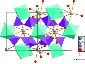
different P2O7 groups (see Fig.1 and Fig.2). As a result of these blocks, assemblage three-dimensional-framework is
formed with hexagonal channels, where silver cations reside. Although, the coordination sphere of Ag+ cations is
composed of seven O2- anions in an irregular geometry, located at Ag–O distances between 2.370 (2) and 3.015 (2) Å (see
Fig.2). Furthermore, the diphosphate group contains two distorted PO4 tetrahedra sharing one corner and display a nearly
eclipsed conformation. The P–O bond-lengths range between 1.492 (2) Å for terminal P1–O1 and 1.606 (2) Å for the
bridging P2–O7 bond. Therefore, a P1–O7–P2 angle of 137.27 (9) ° is wider than 133.6 (3)° and 132.9 (3) ° reported for
both AgFeP2O7 and NaFeP2O7 respectively (Belkouch et al. (1995); Gabelica-Robert et al. (1982); Moya-Pizarro et al.
(1984); Mercader et al. (1990)).
Silver indium diphosphate (pyrophosphate) is isostructural to AIFeP
2O7 (AI = Na, K, Rb, Cs and Ag) diphosphates
family and is categorized as a dichromate type.
S2. Experimental
AgInP2O7 in the form of single crystals was prepared by stoichiometric reaction of AgNO3, (NH4)2HPO4 and In2O3 in
B2O3 flux. The mixture was heated at 773 K under ambiante atmosphere for 6 h and 973 K for 2 h with intermediate
grindings to ensure complete reaction. Subsequent melting at 1323 K followed by slow cooling to room temperature at a
rate of 12°K h-1 resulted in colourless crystals of the title compound.
S3. Refinement
The highest and deepest hole residual peak in the final difference Fourier map are located at 0.49 Å and 0.58 Å,
Figure 1
Partial plot of AgInP2O7 crystal structure shawing plyhedra linkage. Displacement ellipsoids are drawn at the 50%
probability level. Symmetry codes: (i) -x + 1, y - 1/2, -z + 1/2; (ii) -x, y - 1/2, -z + 1/2; (iii) -x + 1, -y + 1, -z + 1; (iv) x, -y
+ 3/2, z - 1/2; (v) x - 1, y, z; (vi) x - 1, -y + 3/2, z - 1/2; (vii) -x, -y + 1, -z + 1.
Figure 2
[image:3.610.137.475.382.637.2]Silver indium diphosphate
Crystal data
AgInP2O7
Mr = 396.63
Monoclinic, P21/c
Hall symbol: -P 2ybc
a = 7.4867 (3) Å
b = 8.2620 (3) Å
c = 9.8383 (5) Å
β = 112.038 (2)°
V = 564.09 (4) Å3
Z = 4
F(000) = 728
Dx = 4.670 Mg m−3
Mo Kα radiation, λ = 0.71073 Å Cell parameters from 317 reflections
θ = 2.5–30.2°
µ = 8.11 mm−1
T = 296 K Block, colourless 0.08 × 0.06 × 0.05 mm
Data collection
Bruker X8 APEXII CCD area-detector diffractometer
Radiation source: fine-focus sealed tube Graphite monochromator
ω and φ scans
Absorption correction: multi-scan (SADABS; Sheldrick, 1999)
Tmin = 0.563, Tmax = 0.667
21692 measured reflections 3730 independent reflections 3245 reflections with I > 2σ(I)
Rint = 0.035
θmax = 41.0°, θmin = 2.9°
h = −13→13
k = −15→15
l = −18→18
Refinement
Refinement on F2
Least-squares matrix: full
R[F2 > 2σ(F2)] = 0.021
wR(F2) = 0.048
S = 1.03 3730 reflections 101 parameters 0 restraints
Primary atom site location: structure-invariant direct methods
Secondary atom site location: difference Fourier map
w = 1/[σ2(F
o2) + (0.017P)2 + 0.9979P]
where P = (Fo2 + 2Fc2)/3
(Δ/σ)max = 0.001
Δρmax = 1.62 e Å−3
Δρmin = −2.04 e Å−3
Extinction correction: SHELXL97 (Sheldrick, 2008), Fc*=kFc[1+0.001xFc2λ3/sin(2θ)]-1/4
Extinction coefficient: 0.0171 (4)
Special details
Geometry. All s.u.'s (except the s.u. in the dihedral angle between two l.s. planes) are estimated using the full covariance matrix. The cell s.u.'s are taken into account individually in the estimation of s.u.'s in distances, angles and torsion angles; correlations between s.u.'s in cell parameters are only used when they are defined by crystal symmetry. An approximate (isotropic) treatment of cell s.u.'s is used for estimating s.u.'s involving l.s. planes.
Refinement. Refinement of F2 against ALL reflections. The weighted R-factor wR and goodness of fit S are based on F2,
conventional R-factors R are based on F, with F set to zero for negative F2. The threshold expression of F2 > σ(F2) is used
only for calculating R-factors(gt) etc. and is not relevant to the choice of reflections for refinement. R-factors based on F2
are statistically about twice as large as those based on F, and R- factors based on ALL data will be even larger.
Fractional atomic coordinates and isotropic or equivalent isotropic displacement parameters (Å2)
x y z Uiso*/Ueq
In1 0.242354 (15) 0.495357 (12) 0.247622 (11) 0.00618 (3) Ag1 −0.20911 (3) 0.52697 (2) 0.30478 (2) 0.02442 (4)
P1 0.57689 (6) 0.74758 (5) 0.46083 (4) 0.00600 (6)
O1 0.6810 (2) 0.86792 (17) 0.40464 (15) 0.0141 (2)
O2 0.6836 (2) 0.71622 (16) 0.62241 (14) 0.0136 (2)
O3 0.52473 (17) 0.59259 (15) 0.36935 (14) 0.01027 (19) O4 0.04427 (18) 0.91166 (17) 0.35059 (15) 0.0123 (2)
O5 0.1917 (2) 0.79561 (16) 0.60976 (14) 0.0126 (2)
O6 0.13231 (18) 0.61348 (15) 0.39564 (14) 0.01054 (19) O7 0.37868 (18) 0.83601 (16) 0.44239 (16) 0.0128 (2)
Atomic displacement parameters (Å2)
U11 U22 U33 U12 U13 U23
In1 0.00656 (4) 0.00614 (4) 0.00593 (4) −0.00024 (3) 0.00244 (3) −0.00045 (3) Ag1 0.01913 (7) 0.02720 (8) 0.03341 (9) −0.00235 (6) 0.01725 (7) −0.01071 (7) P1 0.00574 (14) 0.00643 (14) 0.00582 (14) −0.00025 (11) 0.00217 (11) 0.00044 (11) P2 0.00588 (14) 0.00706 (14) 0.00644 (14) 0.00073 (11) 0.00195 (11) −0.00090 (12) O1 0.0185 (6) 0.0141 (5) 0.0133 (5) −0.0053 (4) 0.0104 (5) 0.0012 (4) O2 0.0195 (6) 0.0098 (5) 0.0070 (4) −0.0006 (4) −0.0001 (4) 0.0018 (4) O3 0.0072 (4) 0.0099 (5) 0.0130 (5) −0.0012 (3) 0.0030 (4) −0.0040 (4) O4 0.0084 (5) 0.0135 (5) 0.0135 (5) 0.0034 (4) 0.0023 (4) 0.0041 (4) O5 0.0211 (6) 0.0099 (5) 0.0072 (4) −0.0009 (4) 0.0058 (4) −0.0023 (4) O6 0.0116 (5) 0.0097 (5) 0.0123 (5) −0.0019 (4) 0.0067 (4) −0.0039 (4) O7 0.0078 (5) 0.0100 (5) 0.0217 (6) 0.0010 (4) 0.0069 (4) −0.0020 (4)
Geometric parameters (Å, º)
In1—O1i 2.0799 (13) Ag1—O5vii 2.7829 (14)
In1—O4ii 2.1120 (12) Ag1—O6vii 3.0153 (14)
In1—O2iii 2.1133 (13) P1—O1 1.4919 (13)
In1—O5iv 2.1401 (13) P1—O2 1.5097 (13)
In1—O3 2.1562 (12) P1—O3 1.5292 (13)
In1—O6 2.1569 (12) P1—O7 1.6021 (13)
Ag1—O3v 2.3703 (12) P2—O4 1.5101 (13)
Ag1—O6 2.4757 (13) P2—O5 1.5158 (13)
Ag1—O4ii 2.4865 (14) P2—O6 1.5295 (13)
Ag1—O2vi 2.6991 (14) P2—O7 1.6062 (13)
Ag1—O7ii 2.7744 (15)
O1i—In1—O4ii 90.70 (6) O3v—Ag1—O5vii 94.99 (4)
O1i—In1—O2iii 86.35 (6) O6—Ag1—O5vii 104.03 (4)
O4ii—In1—O2iii 89.82 (6) O4ii—Ag1—O5vii 80.85 (4)
O1i—In1—O5iv 89.04 (5) O2vi—Ag1—O5vii 157.25 (4)
O4ii—In1—O5iv 93.79 (5) O7ii—Ag1—O5vii 71.00 (4)
O2iii—In1—O5iv 174.18 (6) O3v—Ag1—O6vii 72.37 (4)
O1i—In1—O3 96.36 (5) O6—Ag1—O6vii 88.04 (4)
O4ii—In1—O3 172.86 (5) O4ii—Ag1—O6vii 119.79 (4)
O2iii—In1—O3 89.55 (5) O2vi—Ag1—O6vii 148.46 (4)
O5iv—In1—O3 87.43 (5) O7ii—Ag1—O6vii 119.64 (4)
O4ii—In1—O6 82.94 (5) O1—P1—O2 111.17 (8)
O2iii—In1—O6 92.62 (5) O1—P1—O3 113.17 (8)
O5iv—In1—O6 92.35 (5) O2—P1—O3 113.16 (8)
O3—In1—O6 89.98 (5) O1—P1—O7 104.17 (8)
O3v—Ag1—O6 134.04 (4) O2—P1—O7 107.40 (8)
O3v—Ag1—O4ii 155.99 (4) O3—P1—O7 107.10 (7)
O6—Ag1—O4ii 69.47 (4) O4—P2—O5 115.20 (8)
O3v—Ag1—O2vi 85.86 (5) O4—P2—O6 113.76 (8)
O6—Ag1—O2vi 91.29 (4) O5—P2—O6 109.65 (8)
O4ii—Ag1—O2vi 89.15 (5) O4—P2—O7 100.90 (8)
O3v—Ag1—O7ii 102.17 (4) O5—P2—O7 109.63 (8)
O6—Ag1—O7ii 123.47 (4) O6—P2—O7 107.01 (7)
O4ii—Ag1—O7ii 54.04 (4) P1—O7—P2 137.27 (9)
O2vi—Ag1—O7ii 86.57 (4)