Three-dimensional computed tomographic volume
rendering imaging as a teaching tool in veterinary
radiology instruction
H. Lee
1, J. Kim
1, Y. Cho
2, M. Kim
1, N. Kim
1, K. Lee
11College of Veterinary Medicine, Chonbuk National University, Jeonju, Republic of Korea 2Radiologic Technology, Daegu Health College, Daegu, Republic of Korea
ABSTRACT: The educational value of three-dimensional computed tomography (3D CT) volume rendering imaging was compared to conventional plain radiographic instruction in a veterinary radiology class. Veterinary radiology is an important subject in veterinary medicine and has been well-recognized as a primary diagnostic method. Many junior and senior students have difficulty interpreting two dimensional radiographs that depict three-dimensional organs. A total of 158 junior veterinary students with knowledge of anatomy, pathology, physiol-ogy, and other basic subjects were divided into two groups; Group 1 (n = 45) received conventional radiographic instruction using normal and representative abnormal canine thoracic and abdominal radiographs followed by repetition of the same one week later, while Group 2 (n = 113) received plain radiograph instruction as in Group 1 followed by volume-rendered 3D CT images from the same canine patient one week later. The evaluations were performed at the end of each instruction. In Group 1, the majority did not understand the radiographic signs and no significant improvement was observed. In Group 2, 13% and 20% of the students learned only from radiographs, and understood the thoracic and abdominal radiographic alterations, respectively. After studying the 3D CT images, more than 94% of the students deduced the reasons for the radiographic alterations on the radiographs (P
< 0.001). These results strongly suggest that 3D CT imaging is an effective tool for teaching radiographic anatomy to veterinary medical students.
Keywords: three-dimensional CT; teaching; veterinary radiology; dog
Supported by the Korea Research Foundation Grant funded by the Korean Government (MOEHRD, Basic Research Promotion Fund) (Grant No. KRF-2008-313-E00650).
Because traditional radiology provides only flat images, it is often difficult to arrive at the three-dimensional configuration based on them. This requires integrating knowledge of anatomy, physi-ology, and physics of imaging. It was reported that a lack of radiographic anatomy instruction can lead to errors in interpretation (Lamb et al., 2007). This implies that sufficient knowledge of anatomy is an important element in the interpretation of radio-graphs. Various teaching methods for radiographic anatomy and gross anatomy have been developed (Reidy et al., 1978; Erkonen et al., 1990; Scrivani et al., 2000; Croy and Dobson, 2003; Vandeweerd et al., 2007; Jacobson et al., 2009).In particular, teach-ing both anatomy and radiology simultaneously has
MATERIAL AND METHODS
Experimental design is described in Table 1.
Participated students
Three classes with a total of 158 junior veterinary medical students participated in this study. The stu-dents were divided into two groups; Group 1: 113 students who received repeated explanation of plain radiographic signs and Group 2: 45 students who first received instruction with plain radiographic signs, which was then followed by 3D CT imaging education (Table 1). The students have completed a basic curriculum including classes in anatomy, physiology, and pathology. They had not previously undergone instruction in radiographic anatomy.
Group 1
Instruction with Plain radiograph. Initially,
plain thoracic and abdominal radiographs of a
normal dog and corresponding radiographs of a canine patient with a heartworm infection showing an enlarged pulmonary trunk and a canine patient with splenomegaly were presented to the students. Mainly, the radiographic findings showed normal and abnormal cardiovascular systems in the tho-racic part and a summation effect of the spleen in the abdomen.
1st evaluation. Right after the instruction,
evalu-ation was performed to analyze how the students understood the normal radiographic anatomy, as well as the representative radiographic findings of an enlarged pulmonary trunk on the thoracic radiograph and summated splenic image as well as radiographic signs of splenomegaly on the ab-dominal radiograph.
[image:2.595.72.526.395.711.2]The anonymous evaluation sheet consisted of a multiple-choice questionnaire to determine how the individual student understanded the normal and abnormal radiographic findings. The sheets were collected immediately and then analyzed. The possible answers to the questions were as follows;
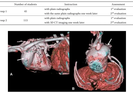
Table 1. Experimental design
Number of students Instruction Assessment
Group 1 45 with plain radiographs 1st evaluation
with the same plain radiographs one week later 2nd evaluation
Group 2 113 with plain radiographs 1
st evaluation
with 3D CT imaging one week later 2nd evaluation
1 = did not understand, 2 = somewhat, but not fully understood, 3 = full recognition.
Repeated instruction with Plain radiograph.
Plain radiograph explanation was made repeatedly.
2nd evaluation. Identical assessment was
per-formed as in the 1st evaluation.
Group 2
Instruction with Plain radiograph. Same
in-struction as was given to Group 1.
1st evaluation. Identical assessment was
[image:3.595.107.500.85.320.2]per-formed as in the 1st evaluation for Group 1. Figure 2. Threshold and opacity-adjusted 3D volume-rendering image mimicking the thoracic plain radiograph of the same dog in Figure 1. A = left lateral view shows enlarged main pulmonary artery (arrow), faintly observed on the lateral thoracic radiograph. B = cranial view displays the enlarged pulmonary trunk at one to two o’clock (arrow), corresponding to the bulge presented in the ventrodorsal radiograph
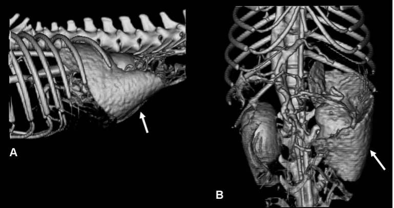
[image:3.595.112.497.500.703.2]Instruction with 3D CT image. Right after the
first instruction and evaluation, 3D CT images from the same dogs were shown to the students. The CT data was reformatted using commercial software (Rapida, Infinity, Korea). The principal re-constructed 3D images were produced using a vol-ume rendering technique used in student teaching. The 3D CT images displaying an apparent enlarged pulmonary trunk and splenomegaly was given to the students (Figures 1 and 3). Additionally, the summation effect observable on the plain radio-graphs was reemphasized by showing the thresh-old and opacity-adjusted volume rendering images (Figures 2 and 4).
2nd evaluation. Identical assessment was
per-formed as in the 1st evaluation.
Statistical analysis
A one-way ANOVA test was used with SPSS 12.0 software. A P < 0.05 was considered to be statisti-cally significant.
RESULTS
Group 1
Instruction with Plain radiograph. Most of
[image:4.595.129.501.85.280.2]the students did not fully understand the radio-graphic findings in regard to the enlarged pul-monary artery and spleen at the first instruction. Only 13% and 20% of the students completely Figure 4. Threshold and opacity-adjusted 3D volume-rendered image mimicking the abdominal plain radiograph of the same dog from Figure 1. A = left lateral view shows accentuated triangular splenic shape (arrow), compatible with the summation effect shown on the lateral abdominal radiograph. B = front view displays the triangular spleen on the lateral side of the abdominal wall (arrow), corresponding to the summation effect presented in the ventrodorsal radiograph
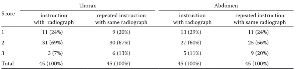
Table 2. Cognitive rate comparison between plain radiographs and 3D CT images of canine thorax and abdomen in Group 1
Score
Thorax Abdomen
instruction
with radiograph with same radiographrepeated instruction with radiographinstruction with same radiographrepeated instruction
1 11 (24%) 9 (20%) 13 (29%) 11 (24%)
2 31 (69%) 30 (67%) 27 (60%) 25 (56%)
3 3 (7%) 6 (13%) 5 (11%) 9 (20%)
Total 45 (100%) 45 (100%) 45 (100%) 45 (100%)
[image:4.595.62.542.627.738.2]comprehended the changes in the thoracic and abdominal regions, respectively, after repeated instruction. A majority of students, 67% and 56%, partially understood the changes in the thoracic and abdominal regions, respectively. And 20% and 24% showed no understanding of the respec-tive changes (Table 2).
Repeated instruction with Plain radiograph. The
results of repeating the instruction with plain radio-graphs were not significantly different compared to the previous instruction. Most of the students were not helped to fully understand the radiographic signs on the plain radiographs after repeated instruction. Thus, repeating the instruction with the same plain radiographs did not have a positive effect on student learning in this study (Table 2).
Group 2
Instruction with Plain radiograph. Most of the
students did not fully understand the radiographic findings in regard to the enlarged pulmonary artery and spleen. Only 13% and 20% of the students com-pletely comprehended the changes in the thoracic and abdominal regions, respectively. A majority of students, 65% and 52%, partially understood the changes in the thoracic and abdominal regions, re-spectively. 22% and 28% showed no understanding of the respective changes (Table 3).
Instruction with 3D CT image. The students
who did not fully understand the radiographic find-ings on the plain radiographs were able to see the pathologic change after instruction with the 3D CT images. More than 94% of the students complete-ly understood the radiographic alterations on the X-ray films (Table 3). Only a small number of stu-dents, 6% and 3%, displayed a partial
understand-ing. None of the students demonstrated a complete lack of understanding after being shown the 3D CT images (Table 3).
DISCUSSION
Three-dimensional CT images were shown and explained to veterinary medical students in a radi-ology class in order to improve their understanding of radiographic anatomy. Interpretation remains an important faculty for veterinary medical stu-dents. Plain radiography is easily accessible and cost effective. However, an understanding of other advanced imaging modalities, such as computed to-mography, magnetic resonance imaging, and even nuclear medicine, is becoming more important to veterinary students. Because radiology is based on a sound knowledge of anatomy, and a good under-standing of anatomy is required in order to properly interpret any imaging modality. Individual or small group studies of images would be an ideal method for radiology instruction. Also, having instructors present is helpful even if the students are working at their own pace. Realistically, this method is not al-ways possible due to demands on both the student’s and faculty’s time. With these limitations in mind, the effectiveness of radiology instruction has been found to be greatest when computer-assisted teach-ing programs are available in addition to traditional teaching methods (Goldberg et al., 1990; Scrivani et al., 2000; Croy and Dobson, 2003; Mastrangelo et al., 2003; Vandeweerd et al, 2007).
Although an effective educational environment provides students with knowledge of radiology and gross anatomy simultaneously, many students will not entirely understand the radiographic anatomy. Therefore, it is often not easy for students to
com-Table 3. Cognitive rate comparison between plain radiographs and 3D CT images of canine thorax and abdomen in Group 2
Score Thorax Abdomen
radiograph 3D CT image radiograph 3D CT image
1 25 (22%) 0 32 (28%) 0
2 73 (65%) 7 (6%)* 59 (52%) 3 (3%)*
3 15 (13%) 106 (94%)* 22 (20%) 110 (97%)*
Total 113 (100%) 113 (100%) 113 (100%) 113 (100%)
[image:5.595.67.535.624.728.2]prehend complex anatomy based on a traditional teaching system with film-based radiographic im-ages.
Though learning depends on a student’s ability to absorb the knowledge presented in a complicated medical course, improved teaching techniques can certainly aid in the learning process. Instruction using the 3D CT volume-rendered images as an adjunct to traditional radiology teaching showed dramatic results in a radiology class.
The 3D CT technique is used for the preopera-tive planning of complex diseases, such as pelvic and acetabular fractures, craniofacial anomalies, and laparoscopic surgery (Dahlen and Zwipp, 2001; Mastrangelo et al., 2002; Gaia et al., 2005; Cimerman and Kristan, 2007).
Initially, most of the students had great difficultly understanding how organs, such as the heart, great vessels, and spleen, are displayed on the X-ray films. A great amount of effort must be devoted to under-standing three-dimensional organs presented on two dimensional radiographs. Few students were able to understand why the pulmonary trunk was shown in the one to two o’clock position on the ventrodorsal thoracic radiograph. Consequently, it is easy to see how an enlarged main pulmonary artery, which can indicate a heartworm infection or right-sided heart problem, cannot be easily detected. Similarly, understanding the triangular shape of the spleen on the X-ray films was difficult for the students. Although the summation effect, which shows increased radiopacity, can be under-stood by the students, the summated triangular splenic silhouette could not easily be explained to the students. Most of the students who under-stood the simple anatomy of the spleen were not able to understand the configuration of the spleen on the radiograph. Although the same instruction was repeated one week later, a majority of students still failed to fully understand. While repeating the conventional instruction with plain radiographs did not improve the degree of student comprehen-sion significantly, most of the students were able to completely understand the normal radiographic anatomy after instruction using 3D CT volume-rendered images of the thorax and abdomen. In particular, threshold and opacity-adjusted 3D CT abdominal images showing the entire spleen clearly are very helpful for students to understand the ex-act radiographic anatomy as well as gross anatomy. Moreover, adjusted 3D CT images which mimic plain radiographs could facilitate understanding as
to why the spleen has triangular soft tissue opac-ity on abdominal radiographs. An explanation of why the spleen appears much larger would be even easier (Love and Berry, 2002). In conclusion, this easily understandable teaching material, 3D CT volume-rendered imaging, substantially supports students in learning and comprehending completely radiographic anatomy and abnormal radiographic findings as well as gross anatomy. Therefore, 3D CT volume-rendered images might be very useful and helpful for both students and teachers in veterinary radiology classes.
Acknowledgement
The authors would like to thank Dr. HS Kwak (Radiologist, Chonbuk National University Hospital)) and Dr. HW Kang (College of Veterinary Medicine of Chonbuk National University). Supported by the Korea Research Foundation Grant funded by the Korean Government (MOEHRD, Basic Research Promotion Fund) (Grant No. KRF-2008-313-E00650).
REfERENCES
Brown GA, Firoozbakhsh K, Gehlert RJ (2001): Three-dimensional CT modeling versus traditional radiology techniques in treatment of acetabular fractures. Iowa Orthopaedic Journal 21, 20–24.
Cimerman M, Kristan A (2007): Preoperative planning in pelvic and acetabular surgery: the value of advanced computerised planning modules. Injury 38, 442– 449.
Croy BA, Dobson H (2003): Radiology as a tool for teach-ing veterinary anatomy. Journal of Veterinary Medical Education 30, 264–269.
Dahlen C, Zwipp H (2001): Computer-assisted surgical planning. 3-D software for the PC. Unfallchirurg 104, 466–479.
Edelson G, Saffuri H, Obid E, Vigder F (2009): The three-dimensional anatomy of proximal humeral fractures.
Journal of Shoulder and Elbow Surgery 18, 535–544. Erkonen WE, Albanese MA, Smith WL, Pantazis NJ
(1990): Gross anatomy instruction with diagnostic im-ages. Investigative Radiology 25, 292–294.
ning for laparoscopic surgery. Studies in Health Technology and Informatics 85, 274–279.
Mastrangelo MJ Jr, Adrales G, McKinlay R, George I, Witzke W, Plymale M, Witzke D, Donnelly M, Stich J, Nichols M, Park AE (2003): Inclusion of 3-D computed tomography rendering and immersive VR in a third year medical student surgery curriculum. Studies in Health Technology and Informatics 94, 199–203. Reidy J, Williams J, Dilly N, Fraher J (1978): The learning
of radiological anatomy by medical students. Clinical Radiology 29, 591–592.
Scrivani PV, Dykes NL, Lonsdale RA, Wilson KM, Ed-mondson KM (2000): An approach to student learning in clinical radiology. Veterinary Radiology & Ultra-sound 41, 392–395.
Vandeweerd JM, Davies JC, Pinchbeck GL, Cotton JC (2007): Teaching veterinary radiography by e-learning versus structured tutorial: a randomized, single-blinded controlled trial. Journal of Veterinary Medical Education 34, 160–167.
Received: 2010–09–29 Accepted after corrections: 2010–11–28
Goldberg HI, Fell S, Myers HJ, Taylor RC (1990): A computer-assisted, interactive radiology learning pro-gram. Investigative Radiology 25, 947–951.
Jacobson S, Epstein SK, Albright S, Ochieng J, Griffiths J, Coppersmith V, Polak JF (2009): Creation of virtual patients from CT images of cadavers to enhance inte-gration of clinical and basic science student learning in anatomy. Medical Teacher 31, 749–751.
Lamb CR, Pfeiffer DU, Mantis P (2007): Errors in radio-graphic interpretation made by veterinary students.
Journal of Veterinary Medical Education 34, 157–159. Linton A, Schoenfeld-Tacher R, Whalen LR (2005): De-veloping and implementing an assessment method to evaluate a virtual canine anatomy program. Journal of Veterinary Medical Education 32, 249–254.
Love NE, Berry CR (2002): Interpretation paradigms for the abdomen-canine and feline. In: Thrall DE (ed.): Textbook of Veterinary Diagnostic Radiology. 4th ed.
WB Saunders, Philadelphia. 483–492.
Mastrangelo MJ Jr, Stich J, Hoskins JD, Witzke W, George I, Garrison J, Nichols M, Park AE (2002): Advance-ments in immersive VR as a tool for preoperative
plan-Corresponding Author:
Kichang Lee, Chonbuk National University, College of Veterinary Medicine, Department of Veterinary Surgery and Radiology, 664-14, 1 ga, DuckJin-dong, Jeonju 561-756, Republic of Korea