1
Emotion recognition problems after brain injury:
Development of the Brief Emotion Recognition Test (BERT)
Joanne Carole Howe
A thesis submitted in partial fulfillment of the requirements of the University of the West of England, Bristol, for the degree of
Professional Doctorate in Counselling Psychology
Faculty of Health and Social Sciences University of the West of England, Bristol
September 2018
2 ABSTRACT
Difficulty recognising emotion can have a major impact on psychosocial outcome
following acquired brain injury. Current measures are time consuming and outdated.
The need to have an easily administered screening test which enables clinicians to
quickly assess this ability has been identified. In this thesis, the development of the
Brief Emotion Recognition Test (BERT) is described. The test consists of 14 short
video clips of actors portraying positive, negative and neutral emotions. After watching
each video clip viewers are asked to choose which emotion is being portrayed from a
list of six emotions (happy, sad, surprise, anger, fear, disgust) and neutral. Half of the clips include facial expressions only (“no phrase”) and the other half include facial expressions and congruent vocal cues in the form of neutral carrier phrases (“with phrase”). The performance of 92 neurologically healthy adults was compared with
that of 20 adults who had sustained moderate-to-severe acquired brain injury (ABI).
Performance on the BERT was found to be correlated with an existing measure of
emotion recognition (Emotion Evaluation Test - EET). Correlations were higher in the
ABI group than the neurologically healthy group. Test retest reliability in the ABI group was good, and moderate in the neurologically healthy group. Overall the ABI group’s
performance on the BERT was impaired relative to the neurologically healthy group. Both groups performed worse on the “with phrase” BERT, with the ABI participants
finding this part of the test particularly difficult. In the neurologically healthy group,
intelligence was not found to be associated with performance on the BERT. However, it was found to be associated with the ABI group’s performance on the “with phrase"
BERT. The groups differed in education and intelligence. Education was not found to
be a significant predictor of group differences, intelligence was. The neurologically healthy group were more accurate regarding five clips in the ‘no phrase’ condition; two of the seven in the ‘with phrase’ trial; and in the total overall score. Overall, findings
for this pilot study suggest the BERT provides a useful means of rapidly screening for
emotion recognition difficulties after brain injury. Further research is needed establish the new test’s psychometric properties. Evaluation of the findings, relevance to
counselling psychology, implications for practice and areas for further investigations
3 Acknowledgements
Enormous thanks to my supervisory team. Doctor Tony Ward has been a fantastic director of studies providing expert knowledge, encouragement and support throughout. Professor Nick Alderman has shared his knowledge, experience and expertise and offered support. Thank you to Doctor Nancy Snook who joined the supervisory team recently and has provided feedback on my thesis and help and guidance with the analysis. I have learnt a lot from my supervisory team for which I am grateful.
Thank you to all the participants who took part in this research study, without whom this work would not have been possible. I would also like to thank the people at Headway Bedford, Headway Somerset and Headway Swindon for supporting this research and facilitating recruitment.
I would like to thank Keith Mitchell for his help and technical expertise in facilitating the recoding of the actors in the video clips and other assistance he has provided during the research. I really appreciate his help. I would also like to thank Paul White for his help with the statistical analysis. Thank you also to Geraldine Truffil, Eamon Fulcher and their team for their help in putting the new test in an on-line format.
Finally, special thanks to my husband Geoff Howe for his unfailing support and faith in me. I am grateful to him for his encouragement and patience which have undoubtedly helped me to persevere with this work. Thank you also to our two children Alex and Georgia. Without my family’s love, encouragement and support I would have found it far more difficult to complete this research.
Jo Howe
4
Table of Contents
Page
Abstract 2
Acknowledgements 3
Table of Contents 4-6
List of figures 7
List of tables 8
Chapter 1 Literature review
1.1 Acquired brain injury 9-14
1.2 Emotion recognition problems after ABI 14-30
1.3 A theory of emotion recognition 30-35
1.4 Empathy and emotion recognition 35-39
1.5 Existing emotion recognition tests 39-42
1.6 Rationale and aims of research 42-48
Chapter 2 Methodology
2.1 Introduction 49
2.2 Aims of Methodology 49
2.3 Recruitment 49-54
2.4 Ethical Practice 54-56
2.5 Procedures 57
2.5.1 Development of the BERT 57-62
2.5.2 Other materials 62-65
2.5.3 Administration of new test and other measures to control group
65-66
2.5.4 Collection of validity data from the ABI group 66-68
2.6 Data analysis 68-70
2.7 Summary of methodology 71
Chapter 3 Results and Analysis of Results
3.1 Introduction 72
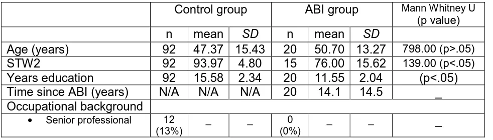
3.2 Group characteristics 72-73
3.3 Reliability – Test retest 73
3.3.1 Test retest reliability - control group 73-75
3.3.2 Test retest reliability - ABI group 75-76
3.3.3 Summary of reliability 76
3.4 Concurrent validity 76
3.4.1 Concurrent validity – control group 76-78
3.4.2 Concurrent validity – ABI group 78-79
3.4.3 Summary of concurrent validity 80
3.5 Discriminant validity 80
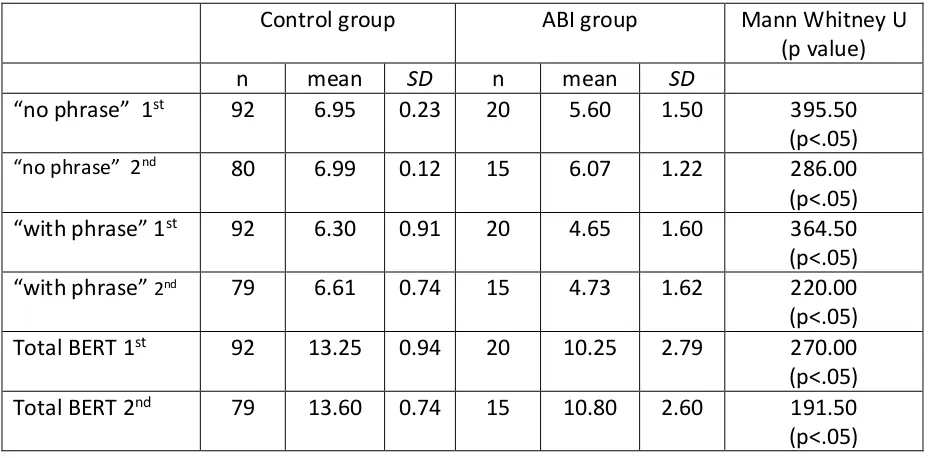
3.5.1 Between group differences on the BERT 80-81
3.5.2 Comparison of performance on the “no phrase” and “with phrase” BERT
81-82
3.5.3 Multiple regression analysis on education and STW2 82-84 3.5.4 Between group differences on the other measures 84-85 3.5.5 Performance of the control and ABI groups on each item
of the BERT
85-86
5
3.5.7 The groups’ performance on the “with phrase” BERT 87-88 3.5.8 Between group differences on the other measures 88-89
3.5.9 Summary of discriminant validity 89
3.6 Clinical cut off scores 89-91
3.6.1 Summary of clinical cut off scores 91
Chapter 4 Discussion
4.1 Introduction 92
4.2 Were the aims of the study achieved? 92
4.2.1 Aim 1 – To develop a short, simple, valid and reliable screening instrument to use in clinical, research and
rehabilitation contexts for detection of emotion recognition problems using contemporary stimulus items
92
4.2.2 Aim 2 – The new screening measure would consist of actors portraying the six basic emotions and neutral. There would be two parts to the test. One part would screen for deficits recognising facial expressions (no vocal cues), and the second part would screen for deficits recognising emotions expressed facially with prosody congruent to the facial expression
92-93
4.2.3 Aim 3 – To generate normative data from an age stratified sample of neurologically healthy participants and compare the performance of the neurologically healthy participants with a sample of participants with ABI
93
4.2.4 Aim 4 – To explore whether performance on the BERT had any association with general intelligence, gender,
education or age.
93-97
4.3 The main findings and whether these support the hypotheses 97 4.3.1 Hypothesis 1 – The new test would be reliable. There
would be test retest reliability
97-99
4.3.2 Hypothesis 2 – There would be evidence of validity.
Performance on the BERT would be correlated with performance on the EET, an existing emotion recognition scale which would confirm criterion validity
99
4.3.3 Hypothesis 3 – Performance on the BERT would be correlated with the ability to empathise as measured by scores on two existing empathy rating scales (BEES and IRI)
99-103
4.3.4 Hypothesis 4 – Performance of the ABI group would be worse than the neurologically healthy participants on the BERT and EET
103-104
4.3.5 Hypothesis 5 – Performance would be improved in both groups in the second part of the new test (“with phrase” BERT) which provides more cues (facial expression and vocal cues) and there would be smaller group differences for the “with phrase” BERT than the “no phrase” BERT
104-109
4.3.6 Hypothesis 6 – It would be possible to calculate clinical cut off scores
109-110
4.4 How to use the BERT – What are the different parts of the BERT screening for?
110-111
6
4.4.2 The “with phrase” BERT 111-112
4.4.3 Administering both the “no phrase” and “with phrase” BERT
112
4.5 Findings on valence 112-116
4.6 Contributions made by the current study 116
4.6.1 Development of a screening tool that can rapidly detect emotion recognition difficulties after ABI
116-117
4.6.2 Screening for emotion recognition problems shortly after ABI
117
4.6.3 Informing rehabilitation 117-119
4.6.4 Support for previous research findings 119
4.6.5 The finding that neutral verbal content appears to make emotion recognition more difficult
119-120
4.6.6 The BERT could add to the debate on theories of emotion recognition
121
4.7 Relevance of current study to Counselling Psychology 121-124
4.8 Limitations of the current study 124
4.8.1 Floor or ceiling effects 125
4.8.2 Is the BERT ecological? 125
4.8.3 Participant sample 126
4.8.4 Ethnicity and age of the actors used in the video clips 127
4.8.5 Are other cognitive processes involved? 127-128
4.8.6 Forced rather than open responses 128
4.8.7 Self-report questionnaires 129
4.8.8 The use of STW2 129-130
4.8.9 Limitations of a computerised screening test 130-131
4.8.10 Not varying order of clips or counterbalancing 131-132
4.9 How work done in current study could be developed 132
4.9.1 Larger and different ABI samples 132
4.9.2 Influence of age on emotion recognition 132-133
4.9.3 Influence of pre-morbid characteristics 133
4.9.4 Influence of affective state: anxiety and depression 133-134
4.9.5 The role of alexithymia 134-135
4.9.6 The characteristics and impairments of individuals who Score below the BERT cut off scores
135-136
4.9.7 Understanding the role of emotion recognition in social cognition
137-138
4.9.8 Rehabilitation training 138-139
4.9.9 Other clinical populations with emotion recognition problems
139
4.10 Conclusion 140-143
References 144-164
Appendices 165-209
7
[image:7.595.75.525.116.243.2]List of Figures
Figure No. Description Page
Figure 1 Example of BERT: Screen shot of practice item 1 of the
“no phrase” BERT 62
Figure 2 Means and SDs of the control and ABI groups’ scores on
the BERT
82
Figure 3 Percentage of control and ABI participants who got each item of the “no phrase” BERT correct
87
Figure 4 Percentage of control and ABI participants who got each
8
List of Tables
Table No. Description Page
Table 1 Levels of ABI severity (Department of Defense and Department of Veterans Affairs, 2008)
12
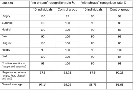
Table 2 Average recognition rates of the individuals who generated the final pool of clips and the control group
60
Table 3 Summary of the groups’ characteristics 73
Table 4 Spearman’s Rho correlations for the control and ABI groups’ performance on first and second administrations of the BERT
73
Table 5 Percentage agreement across two administrations of the BERT for the control and ABI groups
74
Table 6 Spearman’s Rho correlation figures and p<.05 significance values for the control group on the BERT and other measures
77
Table 7 Spearman’s Rho correlation figures and p<.05 significance values for the ABI group on the BERT and other measures
79
Table 8 Performance of the groups on the BERT 81
Table 9 Results of multiple regression analysis for prediction of performance on the BERT
84
Table 10 Groups performance on the EET, BEES and IRI 84
Table 11 Fisher’s Exact Test results for control and ABI groups performance on each item of the BERT
86
Table 12 Clinical cut off scores for the BERT and percentage of participants below cut off scores
90
Table 13 Means, SDs and p values for ABI participants’ below and
above the cut off scores on the EET, BEES, IRI and STW2
9 Chapter 1: Literature Review
1.1 Acquired brain injury
Acquired brain injury (ABI) is brain damage that occurs after birth. There are many
types of ABI including those resulting from external traumatic events such as accidents
and physical assaults and those resulting from internal events such as strokes, brain
tumours and brain infections.
Incidence
ABI is a major health issue affecting approximately 10 million people worldwide
(Langlois, Rutland-Brown & Wald, 2006). It will be one of the major causes of death
and disability by the year 2020 (Hyder, Wunderlich, Puvanachandra, Gururaj &
Kobusingye, 2007). Brain injury is the most common cause of death and disability in
people aged 1–40 years in the UK and each year 1.4 million people attend emergency
departments in England and Wales with a recent head injury (NICE 2014; Clinical
Guidance 176).
Personality and behavioural changes after ABI
Cognitive and neuro-physical impairments are common after ABI. Depending on
factors such as the location and extent of damage; age (brain plasticity); premorbid
functional and intellectual level; medical and emotional health; and the support system
that exists, physical and cognitive recovery can often occur within the first few months
after injury (Zillmer, Spiers & Culbertson, 2008). As well as cognitive and physical
impairments, individuals can suffer a range of personality and behavioural changes
such as becoming self-centred, insensitive to others needs, slowness, poor memory
10
evident shortly after injury and long-term (Langois et al., 2006; Milders, Ietswaart,
Crawford & Currie, 2006; Oddy, Coughlan, Tylerman & Jenkins, 1985; Rosenberg,
McDonald, Rosenberg & Westbrook, 2016). The research suggests that these
changes in personality and behaviour do not dissipate over time (Marsh, Knight &
Godfrey, 1990; McKinlay, Brooks, Bond, Martinage & Marshall, 1981; Oddy,
Humphrey & Uttley, 1978) without intervention. Most people rely on their partners or
families to support them after an ABI (Wood, Liossi & Wood, 2005). The consequence
of this is that the impact of ABI is not just on those who have suffered the injury but
also on their partners and families (Anderson, Parmenter & Mok, 2002; Endberg &
Teasdale, 2004; Hoofian, Gilboa, Vakil & Donovick, 2001; Harris, Godfrey, Partridge
& Knight, 2001; Kreutzer, Gervasio & Camplar, 1994). Changes in family and social
relationships and overall quality of life are often reported (Teasdale & Endberg, 2005).
Early studies identified that a lot of families complain about personality, behavioural
and emotional changes in the person who has experienced a brain injury. For
example, Thomsen (1974), in a study of 40 severely brain injured individuals and their
families found that 84% of families complained of changes which included behaving in
socially inappropriate ways, egocentricity, self-centeredness, being argumentative,
disinterested and insensitivity to others. In a follow up study, Thomsen (1984) found
that the psychosocial consequences of brain injury, namely, emotional problems and
personality change were more debilitating than physical disabilities and increased the
risk of social isolation, caregiver distress and unemployment. Thomsen’s work
highlighted how psychosocial changes after ABI have a significant impact on
individuals and the importance of addressing these in order to improve outcome.
Brooks et al. (1986) carried out a one year and five year follow up of a group of 55
11
42 of the original participants and assessed that these were a good representation of
the original sample. Using structured interviews, a close relative of each brain injured
patient was asked about the patient’s physical and mental state, behaviour, self-care
abilities and personality. Relatives were asked to report any changes in the patient
which had emerged after the injury and were still present. Relatives were also asked
to report any strain or distress experienced as a result of the changes. This was
measured using a seven point self-report scale ranging from the low point “I feel no
strain as a result of changes in my spouse/relative”, to the maximum of “I feel severe
strain…” Seventy four percent of relatives reported personality change at the five year
follow up (up from 60% at one year follow up) and personality change was found to be
related to high levels of distress in the relatives.
Research shows ABI has a significant impact on family functioning and levels of
psychological distress (for example: Hoofian et al., 2001; Kreutzer et al., 1994;
Kreutzer, Marwitz, Hsu, Williams & Riddick 2007; Livingston, Brooks & Bond, 1985).
After ABI there is a high likelihood of depression, family burden and loneliness and
this is associated with difficulties the ABI person has in social situations (Hoofian et
al., 2001; Testa, Malec, Moessner & Brown, 2006). Families are confronted with
dealing with many changes including cognitive, behavioural and neuro-physical
changes. Anxiety and depression is evident in 25-30% of relatives and 60-80% report
some emotional distress (Kreutzer et al., 1994; Livingston et al., 1985).
ABI severity
ABI severity is usually defined as ‘mild’, ‘moderate’ or ‘severe’ according to the amount
of altered consciousness experienced after the injury (Saatman & Duhaime, 2008).
12
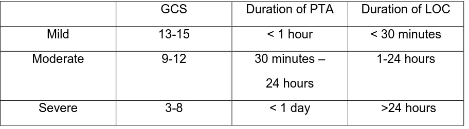
levels of ABI severity by three factors: the Glasgow Coma Scale (GCS; Teasdale &
Jennet, 1974); duration of post trauma amnesia (PTA) and the duration of loss of
consciousness (LOC) after the injury. The GCS is used to assess the central nervous
system status of a patient. It has three elements; eye response, verbal response and
motor response. These elements are scored out of five and summed up. The
maximum score is 15 which indicates a fully awake patient and the minimum score is
three which indicates deep coma or a brain-dead state (Jennet & Bond, 1975). Table
1 illustrates how levels of ABI severity are defined by the Department of Defense and
Department of Veterans Affairs (2008). The current study has used this table to
[image:12.595.72.528.378.504.2]categorise ABI severity.
Table 1. Levels of ABI severity (Department of Defense and Department of Veterans Affairs, 2008)
GCS Duration of PTA Duration of LOC
Mild 13-15 < 1 hour < 30 minutes
Moderate 9-12 30 minutes –
24 hours
1-24 hours
Severe 3-8 < 1 day ˃24 hours
Reduced social contact and loneliness
Research has established that the number of relationships and frequency of social
contact decreases after ABI and there is a tendency to rely on family for social contact
(Elsass & Kinsella, 1987; Thomsen, 1974). People with ABI have reported loneliness
as being a main problem for them (Oddy et al., 1985). Difficulty maintaining
relationships including established relationships built up over many years occurs and
there is evidence that even when the number of acquaintances before and after injury
remains constant the number of close friends decreases (Endberg & Teasdale, 2004).
13
the number of friends that the person with ABI has maintained contact with (Weddell,
Oddy & Jenkins, 1980). It has been suggested that reduced social contact and high
levels of loneliness may be as a result of the difficulties people with ABI have in social
situations (Hoofian et al., 2001).
Therefore, after ABI people often have reduced social contact. The social contact that
does occur is different to what it was before the ABI in terms of frequency and type
and loneliness and social isolation are problems for people after ABI.
Breakdown in marital relationships
Divorce and separation rates are higher than average after ABI. Marital breakdown
after a “severe” outcome been assessed as up to 78% (Thomsen, 1984) and 42% in
those with “good” outcomes (Tate, Lulham & Broe, 1989; Wood & Yurdakul, 1997).
Deterioration in marriages following ABI has been found to be directly associated with
loneliness and altered interpersonal skills of the ABI individual (Wedcliffe & Ross,
2001).
Gosling and Oddy (1999) reported poor marital dissatisfaction and lack of emotional
responsiveness and expressed affection in couples 1-7 years following ABI. They also
found that partners were more dissatisfied with the relationship than the person who
had had the ABI. This may suggest that sometimes after ABI individuals are not aware
of the impact their behaviour is having on relationships (Koskinen, 1998; Wood, 2001).
Research shows that difficulties engaging and interacting socially is a particular
problem after brain injury (Greenwood, 1999). There is evidence that after brain injury,
behaviour can appear to be lacking in empathy, be inappropriate and self-centred and
14
Wood & Williams, 2008). Despite these difficulties being well documented, there is a
lack of literature on how neuro-behavioural problems affect the relationships that
people with ABI have (Wood et al., 2005).
1.2 Emotion recognition problems after ABI
Psychosocial functioning problems often pose greater problems to adjustment after
injury than cognitive or physical functioning (Gratton & Ghahramanlou, 2002; Yates,
2003). The impairments that contribute to psychosocial problems are not well
understood (MIlders, Fuchs & Crawford, 2003). They are likely to be multifactorial and
include cognitive, physical and emotional factors as well as external factors such as
reduced social and financial opportunities (Bornhofen & McDonald, 2008a). The
occurrence of emotion recognition problems after ABI has been identified. Research
has shown that a substantial number of people are impaired in their ability to recognize
emotional facial expressions after ABI (McDonald & Flanagan, 2004; McDonald &
Saunders, 2005; Radice-Neumann, Zupan, Babbage & Willer, 2007; Rosenberg et al.,
2016).
Studies investigating the proportion of people who have emotion recognition problems
after ABI have reported various figures, some as high as 51% (Biszak & Babbage,
2014). In a meta-analysis of 13 studies involving 296 adults with moderate-to-severe
ABI and 296 matched controls (Babbage et al., 2011) it was reported that there was a
relatively large effect size (1.1 SD) differentiating people with ABI from matched controls. This meta-analysis estimated that up to 39% of people with ABI experience
problems recognising facial emotions from static photographs.
Brain injuries, especially those that result from traumatic impact such as in motor
15
prefrontal, temporal and parietal lobes, the amygdala and other structures which have
connections within and to the limbic system (Radice-Neumann et al., 2007). These
areas are associated with emotion and so it is perhaps not surprising that problems
with emotion recognition often occur after ABI.
Emotions are communicated largely through facial expressions (Jackson & Moffat,
1987). Difficulty recognising facial emotion can interfere with a person’s ability to
interpret how others are feeling and is likely to be related to the communication
problems and difficulties in social relationships that are often reported after ABI
(Radice-Neumann et al., 2007). Negative consequences of not being able to interpret
facial emotional expressions may include not responding appropriately to others; not
being able to gauge the appropriateness of their own behaviour; and not fully
understanding the communication of others (McDonald, 2003). Despite the fact that
emotion recognition is a critical aspect in the development and maintenance of social
relationships (Radice-Neumann at al., 2007) and problems in relationships after ABI
have been recognised for some time, it is only relatively recently that research into
impairments in emotion recognition following ABI has been carried out (McDonald,
2013).
In a recent study, May et al. (2017), found there was an association between poor
post-injury behaviour (including community integration) and emotion recognition after
ABI. They also found that this association could not be explained fully by injury
severity, time since injury or education. This finding supports the suggestion that
emotion recognition is an important factor in aiding and maintaining good social
functioning. We know that post injury social behaviour is related to social outcome
(Struchan, Pappadis, Sander, Burrows & Myszka, 2011), and that changes in social
16
Emotion recognition problems present shortly after injury
Research has investigated whether emotion recognition problems are present shortly
after injury in order to try to establish whether the deficits are caused by the injury itself
or secondary factors. Research comparing emotion recognition problems in
participants who were an average of just 2.6 months post injury (Green et al., 2004)
and during first 60 days of injury (Borgaro, Prigatano, Kwasnica, Alcott & Cutter, 2004)
with matched controls found that the ABI participants were significantly less accurate
than controls identifying emotions. In other words, the findings of these studies
suggest that deficits in emotion recognition exist shortly after injury and are therefore
caused by the injury itself rather than environmental changes or from secondary
factors such as depression or anxiety which often occur after ABI. As well as there
being evidence that emotion recognition deficits exist shortly after ABI, longitudinal
research has demonstrated that without intervention emotion recognition impairments
in facial and vocal expressions do not change over time (Ietswaart, Milders, Crawford,
Currie & Scott, 2008; Milders et al., 2003).
As a significant proportion of individuals with ABI have impairments in emotion
recognition and research shows these problems are as a result of the injury rather
than secondary factors such as anxiety or depression, it makes sense for these
impairments to be screened for shortly after injury. Indeed, Borgaro et al. (2004) have
highlighted the importance of screening for emotion recognition deficits during
rehabilitation and have suggested that the availability of a brief screening measure
would be particularly useful, especially in an acute setting where patients’ ability to
17
Emotion recognition research using photographs
One of the first studies to report emotion recognition problems after ABI was that of
Prigatano and Pribram (1982). They used the Ekman and Friesen (1971) black and
white photographs of facial expressions. These photographs portray the six basic
emotions (happy, sad, fear, anger, surprise, disgust) and neutral that have been
demonstrated by Ekman and Friesen (1971) to be universal across cultures. A
substantial number of subsequent studies using photographs have reported findings
of emotion recognition impairments after ABI (for example, Borgaro et al., 2004;
Green, Turner & Thompson, 2004; Jackson & Moffatt, 1987). In addition these studies
have established that emotion recognition problems after ABI are common and have
drawn attention to how emotion recognition problems are likely to adversely affect
social and emotional behaviour.
Vocal emotion recognition
Emotion in voices is portrayed in the meaning of the vocal content (the what) and also in prosody (the how) (Dimoska, McDonald, Pell, Tate & James, 2010). The term prosody includes the intonation pattern (pitch contour) of speech, word stress (a
complex subjective variable based on timing, pitch, and loudness) and pauses that
sometimes occur at the ends sentences (Wingfield, Lahar & Stine, 1989).
Research has established that a significant number of individuals with ABI have
difficulties recognising emotion from vocal expressions (Braun, Baribeau, Ethier,
Daigneault & Proulx, 1989; Hornak, Rolls & Wade, 1996; Marquardt, Rios-Brown,
Richburg, Seibert & Cannito, 2001; McDonald & Flanagan, 2004; McDonald,
Flanagan, Martin & Saunders, 2004; Milders et al., 2003; Spell & Frank, 2000; Zupan,
18
vocal and facial emotion recognition (Knox & Douglas 2009; McDonald & Saunders
2005; Milders et al., 2003; Spell & Frank 2000; Zupan, Babbage, Neumann & Willer,
2014) and have found that individuals with ABI perform less well on both types of tasks
compared to matched controls without brain injury, though impairment has been found
to be greater for vocal emotion than facial emotion recognition (Spell & Frank, 2000).
Whilst deficits in interpreting facial emotion recognition has been widely explored,
difficulties interpreting vocal cues of emotion has received much less attention (Zupan
et al., 2009). Accurate interpretation of vocal emotion cues is important, particularly
when facial cues are absent or ambiguous because these cues contribute to more
accurate identification of emotion (Zupan et al., 2009). One of the first studies to
compare face and vocal emotion recognition (Braun et al., 1989) reported that ABI
participants were impaired in facial emotion recognition but were not impaired
identifying emotions from verbal narratives with the exception of narratives portraying
anger where there was significant impairment. However, subsequent research has
established that difficulties recognising vocal emotion is not limited to the emotion
anger and that individuals with ABI have difficulty recognising vocal expressions of all
emotions (for example; Ietswaart et al., 2008; McDonald & Saunders, 2005; Milders et
al., 2003).
Taken together the studies have established that as well as there being facial emotion
recognition problems after ABI there are also vocal emotion recognition problems. The
studies have also highlighted how vocal emotion recognition deficits after ABI
contribute to social communication problems (Spell & Frank, 2000; Zupan, et al., 2009)
and these problems are likely to adversely affect social integration and social
functioning (McDonald & Flanagan, 2004; McDonald & Saunders, 2005; Orbelo, Testa
19 Dynamic multi-modal displays of emotion
Static displays of emotion in photographs are simple to use and allow the viewer time
to view a fixed emotional expression. However, in everyday communication we use
verbal cues, facial expressions, context, past experience and other non-verbal
information cues to make sense of social interactions (Bornhofen & McDonald, 2008a).
In other words recognising emotions displayed in photographs or audio recordings
(unimodal displays) is arguably different to recognising emotions in real life social
interactions as they fail to provide the viewer with all the cues and information which
are present in most normal interpersonal exchanges (Bornhofen & McDonald, 2008a).
In the last decade or so, progress has been made in developing tests of emotion
recognition that are dynamic (moving) and multimodal making them more ecological.
The Awareness of Social Inference Test (TASIT; McDonald, Flanagan, Rollings &
Kinch, 2003) is one test that simulates real life settings in the form of videoed vignettes.
The TASIT has three parts. The first part is the Emotion Evaluation Test (EET). This
consists of 28 video vignettes of professional actors portraying one of the six standard
emotions and neutral. All the scripts are ambiguous monologues or dialogues devoid
of specific emotional content. The video vignettes are used to emulate real emotional
expressions and viewers have to identify the emotional expression being portrayed by
the designated actor in the vignette by choosing an emotion from a list of the six
standard emotions and neutral. Responses are recorded on a sheet and scored
manually.
McDonald and Saunders (2005) used the EET in a study involving 34 adults with
severe ABI and 28 adults without brain injuries. Four different media were used: still
20
found that individuals with ABI performed more poorly than individuals in the control
group in all of the four different media. McDonald and Saunders (2005) discovered
that the ABI group were significantly impaired recognising emotions in both the audio
and audio-visual displays and eight of the thirty four were significantly impaired
recognising the emotions in the photographs whilst only one of the thirty four was
impaired in the recognition of the moving visual displays with no sound. The ABI group
was most impaired in the dynamic audio visual task. The findings led the authors to
suggest that recognition of emotion in visual moving displays involves different brain
systems (for example the parietal cortices), to recognising emotions in still displays.
Knox and Douglas (2009) compared the performance of 13 individuals with ABI and
13 matched controls on emotion recognition of static stimuli (Ekman photos) and
dynamic displays (18 items from the EET presented without no sound). The study
found that the ABI group were impaired interpreting facial expressions in both static
and dynamic displays, though performed significantly worse on the dynamic display
task. The control group’s performance was similar in both the tasks. The finding in
this study that dynamic displays of emotion were more difficult for individuals with ABI
to identify than static photographs is different to the findings of McDonald and
Saunders (2005).
Knox and Douglas (2009) suggest the reason why individuals in the ABI group
performed worse on the dynamic stimuli task may be that the dynamic displays
demanded more executive functioning than the static stimuli (photographs). They
suggest that when viewing dynamic displays the viewer has to focus on a greater
number of cues and information and the performance of individuals with ABI is affected
21
working memory deficits. Further, the authors point out that static images allow the
viewer to look at a single image for a longer time and do not require the viewer to rely
as much on their working memory, therefore they minimise the impact of slowed
information processing or attention deficit.
Damage after ABI is diffuse so deficits in facial emotion recognition with vocal content
may be due to difficulty processing cues in the visual and/or auditory modality or how
the cues in the two modalities are integrated (Madigan, DeLuca, Diamond,
Tramontano, & Averill, 2000). Zupan et al. (2009) argue that information from the
visual and auditory channels are automatically integrated during the interpretation of
emotion. They suggest that when both facial and vocal cues are available, an emotion
can be more confidently identified if what is seen and heard are perceived to be
consistent with one another (e.g both sad). They argue that perception of emotion is
a bimodal process and that social interactions rely on the interpretation and integration
of facial and vocal emotion cues. This explains why congruous facial and vocal
expressions are easier to understand than incongruent ones (Marquardt et al., 2001).
It has been suggested that emotional processing is dependent on emotional control
(Pessoa et al., 2002) and that problems in emotional recognition may be due to
insufficient resources such as attentional control (Ridout et al., 2007) being available
to process all the information.
Williams and Wood (2010a) replicated the study of McDonald and Saunders (2005) to
investigate if different media presentation affected emotion recognition. They
expected individuals with ABI to find dynamic displays more difficult than static
displays, on the basis that audio visual media (dynamic displays) requires the
individual to process conversational content as well as emotion and prosody and this
22
2010a). Williams and Williams (2010a) asked 64 participants with ABI and matched
controls to complete an audio visual test (the EET) and a static photograph test (the
Ekman 60 faces test). The study found that the ABI group was significantly impaired
on both the audio visual test and the static photographs test compared to the control
group. Moreover, it was found that both groups were more accurate in recognising
emotions displayed in audio visual format, though the difference was more marked in
the ABI group. The authors suggest that this finding may be explained by the fact that
the audio-visual test provided more clues than static photographs and these cues help
us to recognise emotions. They also point out that the static photographs used in the
study were black and white and visually outdated.
Williams and Wood (2010a) concluded from their study that mode of stimulus is an
important factor influencing emotion recognition abilities. They suggested that their
findings provide evidence that emotion recognition is affected by type of media
presentation and that their findings may give support to Adolphs (2002), and Adolphs,
Tranel and Damasio (2003) suggestion that different neural pathways process
different types of emotional stimuli. Adolphs et al. (2003) suggest that dynamic
emotional media is processed by the parietal cortical systems, whereas still emotional
media is processed by other neural pathways including the limbic system, bilateral
inferior and anterior temporal lobe and medial frontal cortices.
Subsequent research by Zupan and Neumann (2014) supports Williams and Wood’s
(2010a) finding that individuals with ABI find it easier to recognise emotions from
context rich dynamic stimuli than static photographs. The reason may be as Williams
and Wood (2010a) suggest, namely that there are more cues available in dynamic
23
emotion (de Gelder & Vroomen, 2000; Zupan et al., 2009) then the more information
and cues available the easier it is to recognise the emotion (Williams & Wood, 2010a).
However, Zupan et al. (2009) acknowledge that more research is needed on the
integration of facial and vocal emotion cues of emotion in static versus dynamic stimuli.
Therefore, the majority of the research comparing emotion recognition of static stimuli
and dynamic audio visual stimuli has found that individuals with ABI find dynamic audio
visual displays easier.
Valence - Negative and positive emotions
Research suggests that some emotions are generally easier to recognise than others.
Happy facial expressions have been found to be identified more accurately, earlier
and faster than other facial expressions (Calvo & Lundqvist, 2008). Identification of
negative emotions (i.e. sadness, anger, disgust and fear) has been found to be more
difficult and slower to recognise after ABI (Adolphs et al., 1999; Calvo & Lundqvist,
2008; Croker & McDonald, 2005; McDonald et al., 2003; Jackson & Moffat, 1987;
Williams & Wood, 2010a). Also, research has shown that those with ABI are poorer
at judging neutral expressions than matched controls (McDonald et al., 2003).
However, whilst there appears to be increased difficulty in identifying neutral items,
neutral items are not mistakenly identified as any particular emotion by individuals with
ABI (Williams & Wood, 2010a).
Are particular neural structures involved in recognising different emotions and different media?
The fact there is a valence difference has led some to propose that this is because
there is independent cognitive processing of specific emotions (Parry, Young, Saul &
24
in processing different emotions (Adolphs, 2002; Wang et al., 2002). There is
evidence that the amygdala is involved in recognising fear (Adolphs, Baron-Cohen &
Tranel, 2002; Calder, Young, Rowland, Perrett, Hodges & Etcoff, 1996; Adolphs,
Tranel, Damasio & Damasio, 1994). The amygdala has also been associated with
recognising sad expressions (Adolphs & Tranel, 2004). It has been estimated that
approximately 50% of patients with amygdala damage have impaired ability to
recognise sad facial expressions (Fine & Blair, 2000).
There is also research suggesting that the ventral striatum is involved in recognising
anger (Calder, Keane, Lawrence & Manes, 2004). Further, it has been reported that
functional neuroimaging demonstrates that the facial expressions associated with
disgust engage different regions of the brain (insula and putamen) than other facial
expressions (Calder, Keane, Manes, Antoun & Young, 2000; Sprengelmeyer, Young,
Calder, Kamat, Lange & Homberg, 1996).
The temporal structures of the brain have been identified as being important in emotion
recognition by Rankin, Kramer and Miller, (2005). Facial emotion recognition has been
associated with the right superior temporal sulcus and the amygdala and orbitofrontal
cortex (Iidaka et al., 2001; Narumoto et al., 2001; Rosen et al., 2002; and Winston et
al., 2002). Voice prosody has been associated with the right superior temporal sulcus
and the amygdala (Adolphs, 1999; Scott, Morris, Scott & Dolan, 1999, Young & Calder,
1997).
The literature demonstrates that different modes of stimuli affects emotion recognition
performance in individuals with ABI (McDonald & Saunders, 2005; Wood & Williams,
2010a, Zupan et al., 2009). Evidence that different neural pathways may be involved
25
involving individuals with brain lesions (Adolphs et al., 2003; Tolmeo et al., 2016; and
Wang et al., 2002)
Adolphs et al. (2003) found that a patient who had extensive bilateral brain lesions
could only recognise happiness from static images or single verbal labels but could
recognise all basic emotions except disgust from dynamic displays and stories. The
authors suggest dynamic emotional media may be processed via the parietal cortical
systems whereas the limbic system, bilateral inferior and anterior temporal lobe and
medial frontal cortices may be involved in processing still emotional media.
Wang et al. (2002) in their study of a schizophrenic patient with bilateral anterior
cingulate gyrus lesions and a lesion in right amygdala found that this patient was
significantly worse at recognising fear compared to three groups of controls (normal
control group; brain injured group with lesions that did not include the amygdala,
hippocampus or cingulate gyrus and a schizophrenic group). Recognition of the other
five basic emotions was not significantly different from that of the controls. The authors
concluded that their finding that this patient was worse at identifying fear but not the
other basic emotions supports the view that the brain has separable networks for
processing different emotions and that the cingulate gyrus and amygdala are involved
in the recognition of fear. Moreover, Ridout et al. (2007) reported that patients with
lesions to the anterior cingulate cortex (ACC) demonstrated impaired facial emotion
recognition performance (as measured by the EET) than those without ACC lesions.
Further evidence comes from the study of Tolomeo et al. (2016). They compared
emotion recognition in patients who had treatment resistant depression and who had
undergone bilateral anterior cingulotomy, with patients who had treatment resistant
26
patients who had the surgery were worse at recognising the negative emotions of fear,
disgust and anger and had no impairment in recognising the facial expressions of
surprise, happy or sadness. They also found that larger volume lesions predicted
more impairment in identifying fear, anger and disgust but did not predict identification
of happy or surprise. The research findings of Tolomeo et al. (2016) provides some
evidence that the anterior mid-cingulate cortex is part of a network associated with the
experience of negative emotion and pain and engages cognitive control processes for
optimising behaviour in the presence of negative emotion and pain. This finding
supports the existing evidence that the anterior cingulate has a causal role in
recognising negative emotions.
Taken together these studies have implications for individuals with ABI. They provide
evidence that different neural systems are involved in the recognition of different
emotions and different types of media. The implication is that in the future it may be
possible to identify the type of emotion recognition deficits an individual has from the
location or type of injury sustained or the location or type of injury from the emotion
recognition deficit.
Subjective emotional experience
Reduced emotional experience has been reported after ABI. Hornak, et al. (1996)
obtained self-reports of participants’ ability to experience the emotions of sadness,
enjoyment, anger, fear and disgust since sustaining their injury. They found that
several of the participants with ABI reported blunted affect, particularly for negative
emotions such as sadness. In another study investigating subjective emotional
experience, Croker and McDonald (2005) found that the majority of participants with
27
although the pattern of changes differed greatly between individuals. They found that
reduced subjective experience, especially of sadness and fear was associated with
poor emotion matching but not emotion labelling. Other research has reported that
damage to the prefrontal cortex results in a decreased ability to experience one’s own
emotions and this may make it difficult to perceive emotion in others (Hornak et al,
2003).
Alexithymia
Alexithymia is difficulty differentiating feelings and body sensations and difficulty
identifying and describing one’s own emotions (Bird & Cook, 2013). Those who have
alexithymia usually have externally orientated and concrete style of thinking (Taylor,
Bagby & Parker 1997) and tend to reflect less on their emotions as well as others’
emotions than those without this trait (Lane et al., 1996).
The incidence of alexithymia in the general population is estimated to be between 10
and 14.8% (Berthoz, Pouga & Wessa, 2010; Koponen, et al., 2005; Wood & Williams,
2010b). The majority of research investigating the relationship between alexithymia
and deficits in emotion recognition in neurologically healthy populations has found that
there is an association (Lane et al., 1996; Parker, Taylor, & Bagby, 1993; Parker,
Prkachin & Prkachin, 2005; Prkachin, Casey & Prkachin, 2009). Alexithymia appears
to be a contributor in behavioural and psychiatric disorders including disordered
eating, somatoform disorders and depression and anxiety (Connelly & Denney, 2007;
Pedrosa-Gil et al., 2008; Ridout, Thom & Wallis, 2010). It has been linked to poor
self-awareness (Allerdings & Alfano, 2001), anxiety and depression (Wood, Williams &
28
The Toronto Alexithymia Scale (TAS-20; Parker, Taylor & Bagby, 1993) has been
used in the ABI literature to measure alexithymia. Using this measure it has been
found that there are high levels of acquired alexithymia following ABI (Henry, Phillips,
Crawford, Theorodou & Summers, 2006; Koponen et al., 2005; Williams & Wood,
2010b). For example, Williams and Wood (2010b) found that 60.9% of individuals in
their ABI group had alexithymia compared to 10.9% in the control group. It has been
suggested that what appears to be poor emotion recognition (or empathy) may be due
to alexithymia (Bird & Cook, 2013).
There is a paucity of literature looking at the link between alexithymia and emotion
recognition problems after ABI (McDonald, Rosenfeld, Henry, Togher & Bornhofen
2011). McDonald et al. (2011) investigated levels of alexithymia (using the TAS-20)
and emotion recognition performance on two tasks (matching and labelling photos of
the 6 basic emotions) in 20 individuals with ABI and 20 adults without brain injuries.
The study found that the association between alexithymia symptoms and emotion
perception deficits was generally limited. The only correlation they found between
alexithymia and emotion perception in the ABI group was between the labelling of
positive emotions and the TAS-20 (measure of alexithymia) subscale “Externally
Orientated Thinking”. The authors reported that this was surprising because most of
the participants including the ABI participants in the study found the positive emotions
easy. Consistent with existing research (such as Henry et al., 2006; Kopenen et al.,
2005; Williams & Wood, 2010b), the study found that the ABI participants had a higher
incidence of alexithymia and were more impaired in the recognition of negative
emotions than the neurologically healthy controls, but that these scores were not
29
deficits in emotion recognition in ABI are not part of a broader disorder of acquired
alexithymia as measured by the TAS-20.
One of the most recent studies to look at the relationship between alexithymia and
emotion recognition problems in people with ABI was conducted byNeumann, Zupan,
Malec and Hammond (2014). In this study 60 individuals with ABI and 60 age and
gender matched controls were evaluated for alexithymia (using the TAS-20); facial and
vocal emotion recognition using Diagnostic Assessment of Non-verbal Accuracy 2-
Adult Faces and Voices (DANVA2-Faces; Norwicki & Duke, 1994); and empathy using
the Interpersonal Reactivity Index (IRI; Davis1980). The study found that the
participants with ABI had significantly higher alexithymia; poorer facial and vocal
emotion recognition; and lower empathy scores. For the ABI participants, facial and
vocal emotion recognition variances were significantly explained by alexithymia (12%
and 8%, respectively); however, the majority of the variances were accounted for by
one of the subscales of the TAS-20, externally-oriented thinking. The authors
concluded that people who have a tendency to avoid thinking about emotions
(externally-oriented thinking) are more likely to have problems recognizing others’
emotions and assuming others’ points of view.
Other studies using the TAS-20 with ABI populations (Henry et al., 2006; Wood &
Williams 2007; Wood, Williams & Kalyani, 2009) suggest that alexithymia and emotion
recognition are associated. It has been suggested that acquired alexithymia in ABI is
a generalised deficit in processing emotional stimuli which includes emotion
recognition problems (Wood & Williams, 2007). Further, there is evidence that
acquired alexithymia in individuals with ABI may be linked to the development of
somatoform disorders and poor self-awareness (Allerdings & Alfano, 2001; Wood,
30
However, the use of the TAS-20 with its three subscales has not been properly
investigated in ABI samples (Wood et al., 2009) and the reliability of the TAS-20
subscales in the ABI population has been questioned by some including Henry et al.,
(2006); and McDonald et al. (2011). Poor verbal skills, and memory problems have
been found to contribute to being assessed as having alexithymia as measured by the
TAS-20 (Wood & Williams, 2007). Further, the TAS-20 is a self-report measure and
is therefore vulnerable to the influence of other variables such as subjective ratings of
emotional experience, self-perception, affective disorders and lack of self-awareness
(Testa et al., 2006; Wood et al., 2009) which are known to be present after ABI (Croker
& McDonald, 2005; Hornak et al., 1996).
Alexithymia and its possible role in emotion recognition problems is mentioned in this
chapter for completeness. Alexithymia was not measured in the current study for a
number of reasons. Firstly, there are questions about the use of the TAS-20 in ABI
samples (Henry et al., 2006; McDonald et al., 2011. Wood et al., 2009). Further
investigations are needed to ascertain its appropriateness in measuring alexithymia in
ABI population. Further, the main aims of the current study were to develop a
screening test of emotion recognition, generate normative data from an age stratified
sample and compare the performance of neurologically healthy participants with a
sample of individuals with ABI. It is acknowledged that in any research project there
are limitations on what can be investigated and the author recognises that further
research investigating the role of alexithymia (and other factors such as executive
function and affect) in emotion recognition would be beneficial. This is discussed in
more detail in the discussion chapter of this thesis.
31
Research has therefore established that emotion recognition difficulties after ABI often
exist and negative emotions like fear, anger, disgust and sadness are more difficult to
recognise than positive emotions such as happiness. Moreover, research findings
report that emotion recognition is variable according to the location of the ABI (Calder
et al., 1996; Hornak et al., 2003; Wang et al., 2002) and the media being used (Knox
& Douglas, 2009; McDonald & Saunders, 2005; Wood & Williams, 2010a).
How do we recognise emotions? One theory is that we use mirror neurons. A mirror
neuron is a neuron that fires both when an animal acts and when the animal observes
the same action performed by another (Rizzolatti & Craighero, 2004). Brain imaging
research has demonstrated that neurons show activity during the execution and also
the observation of an action (Rizzolatti, Fogassi & Gallesse, 2001). In humans, brain
activity consistent with that of mirror neurons has been found in the premotor cortex,
the supplementary motor area, the primary somatosensory cortex and the inferior
parietal cortex (Molenberghs, Cunnington & Mattingley, 2009). Levenson and Ruef
(1992), have called this “shared physiology”. Psychologists sometimes refer to this as
“emotional contagion” (Hatfield, Cacioppo & Rapson, 1994), “affective empathy”
(Zahn-Waxler, Robinson & Emde, 1992) and “automatic emotional empathy” (Hodges
& Wegner, 1997).
Gallesse (2001) in his “shared manifold” hypothesis of emotion recognition suggests
that recognising emotions is not exclusively dependent upon visual abilities or the
capacity to represent the mental states of self and others which is often referred to as
the ability to mentalize (Frith & Frith, 2003) or “theory of mind” (Baron-Cohen, 1995).
According to this theory the same neural structures that are involved in processing
and controlling actions, felt sensations and emotions are also active when the actions,
32
observe a particular emotional expression our motor system becomes active and
“resonates” with that emotion as if we were feeling the same emotion. Gallesse (2003)
suggests that it is through this “shared manifold” that intersubjective communication
and ability to understand others is possible. According to Gallesse (2001), action
observation implies action stimulation. He points to the everyday occurrences of
observing others yawning or laughing which result in us performing the same action
as evidence of this.
According to Gallesse (2001), our felt capacity to have relationships with others and
how we “mirror” ourselves in the behaviour of others and recognise them as similar to
us are rooted in empathy and it is empathy that enables us to generate a link between
ourselves and others and have meaningful relationships. According to this theory
emotions experienced by others become meaningful to us because we can share them
with them. We understand the emotions of others through a mirror matching
mechanism.
Radice-Neumann at al. (2007) rely on similar presumptions in their two stage theory
of facial emotion recognition. They suggest that the first stage involves an individual
correctly recognising the relevant facial features that depict the emotion (Tarr &
Gauthier, 2000; Haxby, Hoffman & Gobbini, 2000). The second stage requires the
individual to correctly interpret his or her own emotional state (Adolphs, Damasio,
Tranel, Cooper & Damasio, 2000; Hornak et al., 2003). Thus, the second part of facial
emotion recognition is self-emotion processing which involves the interpretation of
one’s own emotional state to facilitate the identification of the feelings of others
(Adolphs, 2002; Adolphs Damasio, Tranel, Cooper & Damasio, 2000). Damasio
(1999) suggests that one of the mechanisms which enables us to feel emotion is the
33
The idea that the perception of someone’s emotional state activates the observer’s
corresponding representations which in turn activate somatic and autonomic
responses (Blair, 2005) has some evidence to support it. It has been reported that
when someone is impaired recognising a particular emotion they are also impaired in
subjectively experiencing that emotion (Calder et al., 1996). Further, there are studies
that have reported evidence supporting “mirroring” of facial emotions and how this
ability is sometimes not seen in those with ABI (Knox and Douglas, 2009; McDonald
et al., 2011).
However, some neuroscientists, for example Hickok (2008) argue that whilst mirror
neuron theories are interesting and on the face of it are a reasonable idea, there is a
lack of empirical evidence to support them. Hickok (2008) points out that the studies
by Rizzolati et al. (2001; 2004) were based on motor responses in monkeys and that
to extend this work to emotions in humans is overgeneralising. What is clear is that
more research is needed to better understand mirror neurons and the role they play in
humans and emotion recognition.
The new screening measure of emotion recognition deficits developed in the current
study will add to the debate about how we recognise emotions. It could be used to
screen for emotion recognition problems whilst investigating through measurement of
physiological responding and neuroimaging (for example using Magnetic Resonance
Imaging (MRI) and functional Magnetic Resonance Imaging (fMRI)), whether the same
neural structures that are involved in controlling actions, sensations and emotions are
also active when the actions sensations and emotions are detected in others as
suggested by Gallesse (2003). In other words, the new screening measure could be
useful in investigating evidence of associations between emotional recognition
34
responding to emotional stimuli. The current research findings on physiological
responding to emotional stimuli is now discussed.
Physiological responses to emotional stimuli
Physiological emotional under-arousal has been reported in individuals with mild head
injury (Baker & Good, 2014). Hopkins, Dywan and Segalowitz (2002) measured the
electro dermal activity (EDA) of a group of participants with ABI and matched controls
when presented with faces that varied in emotional expression found that the ABI
group failed to increase EDA in response to negative facial expressions and had a
reduced ability to identify negative emotions particularly fear compared to the matched
controls. De Sousa et al. (2011) measured physiological responding using facial
electromyography (EMG) and skin conductance using finger electrodes. They
reported that there was reduced physiological responses to the emotional expression
of anger in their ABI injury group compared to the control group.
In a study investigating startle response, Williams and Wood (2012) measured EMG
activity and the eye blink component of startle reflex recorded electromyographically
from the orbicularis oculi muscle beneath the left eye. Eye blink amplitude and latency
were computer scored using a commercial startle system (SR-HLAB San Diego
Instruments, CA). The performance of individuals with ABI and matched controls, was
compared. It was found that the ABI group produced the usual attenuation responses
of startle response to pleasant pictures but a significantly lower startle response to
unpleasant pictures and also rated the unpleasant pictures as less arousing that the
control group. Measures of attention and information processing speed were also
taken, though some of this data was incomplete. The authors concluded that
35
ABI and suggest that ABI may disrupt the neural pathways and structures which are
involved in the aversive-defensive motivational system. They also argue that their
findings support the conclusion offered by Saunders, McDonald and Richardson
(2006) that there is a reduction in emotional responsiveness following ABI and not
attention or information processing.
Together, the research on physiological responding provides evidence that
physiological responding to emotional stimuli is affected by ABI and negative emotions
seem to be particularly affected. The evidence suggests that some people with ABI
may process negative emotions differently to people who do not have an ABI. The
current study will contribute to the literature in this area by providing a quick and easy
to administer stimuli that will enable deficits in emotion recognition whilst
simultaneously measuring physiological responses.
1.4 Empathy and emotion recognition
According to Grattan and Eslinger (1989) empathy “refers to the capacity to apprehend
another person’s situation or state of mind in such a way that there is a potential for
sharing and increased understanding through an interpersonal relationship” (p.176).
The ability to recognise and understand emotions is part of empathy, and difficulties
recognising emotions are likely to interfere with a person’s ability to interpret how
others are feeling, in other words their ability to empathise (Grattan & Eslinger, 1989).
Empathy is commonly understood to have two components (Baron-Cohen, 1995;
Davis, 1980). One of the components is understanding the feelings of others. This is
commonly known as cognitive empathy and is understood to denote the ability to
recognise the mental state of another person and the ability to take the mental
36
emotional states i.e. understand other people. Cognitive empathy is thought to involve
perspective taking and theory of mind (Eslinger, 1998).
The second component of empathy is sharing the feelings of others which is commonly
known as affective or emotional empathy (Baron-Cohen, 1995). Emotional empathy
is the capacity to experience affective reactions to the observed experiences of others,
it is our emotional reactions to people (Baron-Cohen, 1995, Davis, 1980,
Shamay-Tsoory, 2011). It is the ability to emotionally resonate with others’ feelings while
understanding that they are distinct from one’s own (Baron-Cohen & Wheelwright,
2004). Emotional empathy is generally thought to be an unconscious process
involving the sharing of emotions, affective responsiveness and emotional contagion.
The key difference between cognitive and emotional empathy is that the former
involves understanding the other person’s point of view, whereas emotional empathy
includes sharing or experiencing the other person’s feelings (Mehrabian & Epstein,
1972). The ability to recognise emotions is often seen as a precursor for empathy
(Shamay-Tsoory, Tomer, Berger & Aharon-Peretz, 2003). Evidence of a relationship
between emotion recognition and empathy in neurologically healthy individuals exists
(Lawrence et al., 2006).
Theories of empathy have many similarities to the theories of emotion recognition
mentioned above. Preston and de Waal’s (2002) theory of empathy is based on a
perception-action model. According to this model, perceiving another in a given
situation automatically results in matching the other’s neural state because perception
and action rely on the same neural circuits. The perception of an object’s state
activates the subject’s corresponding representations which in turn activate somatic