Ethyl 4-chloro-3-nitrobenzoate
Hao-Yuan Li,aBo-Nian Liu,aShi-Gui Tang,bYe-Ming Xua and Cheng Guoa*
a
College of Science, Nanjing University of Technology, Xinmofan Road No. 5 Nanjing, Nanjing 210009, People’s Republic of China, andbCollege of Life Sciences and Pharmaceutical Engineering, Nanjing University of Technology, Nanjing 210009, People’s Republic of China
Correspondence e-mail: [email protected]
Received 28 November 2007; accepted 24 December 2007
Key indicators: single-crystal X-ray study;T= 298 K; mean(C–C) = 0.003 A˚; Rfactor = 0.046;wRfactor = 0.130; data-to-parameter ratio = 14.6.
In the molecule of the title compound, C9H8ClNO4, an
intramolecular C—H O hydrogen bond results in the formation of a planar five-membered ring, which is nearly coplanar with the adjacent six-membered ring, the rings being oriented at a dihedral angle of 4.40 (3). In the crystal structure, intermolecular C—H O hydrogen bonds link the molecules.
Related literature
For related literature, see: Jo¨nsson et al. (2004). For bond-length data, see: Allenet al.(1987).
Experimental
Crystal data
C9H8ClNO4 Mr= 229.61
Monoclinic,C2=c a= 12.930 (3) A˚
b= 7.4820 (15) A˚ c= 20.945 (4) A˚ = 92.11 (3)
V= 2024.9 (7) A˚3 Z= 8
MoKradiation = 0.37 mm1 T= 298 (2) K 0.400.300.10 mm
Data collection
Enraf–Nonius CAD-4 diffractometer
Absorption correction: scan (Northet al., 1968) Tmin= 0.866,Tmax= 0.964 1984 measured reflections
1984 independent reflections 1449 reflections withI> 2(I) 3 standard reflections
frequency: 120 min intensity decay: none
Refinement
R[F2> 2(F2)] = 0.045 wR(F2) = 0.130 S= 1.06 1984 reflections
136 parameters
H-atom parameters constrained max= 0.16 e A˚3
min=0.21 e A˚3
Table 1
Hydrogen-bond geometry (A˚ ,).
D—H A D—H H A D A D—H A
C2—H2B O2 0.97 2.29 2.706 (3) 104
C8—H8A O2i
0.93 2.53 3.357 (3) 148
Symmetry code: (i)xþ1 2;yþ
1 2;z.
Data collection: CAD-4 Software (Enraf–Nonius, 1989); cell refinement: CAD-4 Software; data reduction: XCAD4 (Harms & Wocadlo, 1995); program(s) used to solve structure: SHELXS97
(Sheldrick, 2008); program(s) used to refine structure:SHELXL97
(Sheldrick, 2008); molecular graphics:SHELXTL (Siemens, 1996); software used to prepare material for publication:SHELXTL.
The authors thank Dr Shan Liu. Nanjing University of Technology. for useful discussion and the Center of Testing and Analysis, Nanjing University. for support.
Supplementary data and figures for this paper are available from the IUCr electronic archives (Reference: HK2403).
References
Allen, F. H., Kennard, O., Watson, D. G., Brammer, L., Orpen, A. G. & Taylor, R. (1987).J. Chem. Soc. Perkin Trans. 2, pp. S1–19.
Enraf–Nonius (1989).CAD-4 Software. Version 5.0. Enraf–Nonius, Delft, The Netherlands.
Harms, K. & Wocadlo, S. (1995).XCAD4. University of Marburg, Germany. Jo¨nsson, D., Warrington, B. H. & Ladlow, M. (2004).J. Comb. Chem.6, 584–
595.
North, A. C. T., Phillips, D. C. & Mathews, F. S. (1968).Acta Cryst.A24, 351– 359.
Sheldrick, G. M. (2008).Acta Cryst.A64, 112–122.
Siemens (1996). SHELXTL. Version 5.06. Siemens Analytical X-ray Instruments Inc., Madison, Wisconsin, USA.
Acta Crystallographica Section E
Structure Reports
Online
supporting information
Acta Cryst. (2008). E64, o523 [doi:10.1107/S1600536807068304]
Ethyl 4-chloro-3-nitrobenzoate
Hao-Yuan Li, Bo-Nian Liu, Shi-Gui Tang, Ye-Ming Xu and Cheng Guo
S1. Comment
Some derivatives of benzoic acid are important chemical materials. As part of our ongoing studies, we synthesized the
title compound, (I), and report herein its crystal structure.
In the molecule of (I), (Fig. 1) the bond lengths and angles are within normal ranges (Allen et al., 1987). The
intramolecular C—H···O hydrogen bond (Table 1) results in the formation of a planar five-membered ring B
(C2/H2B/C3/O1/O2). Ring A (C4—C9) is, of course, planar and the dihedral angle between them is A/B = 4.40 (3)°. So,
rings A and B are also nearly co-planar.
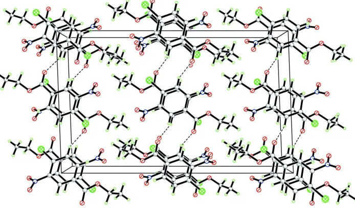
In the crystal structure, intermolecular C—H···O hydrogen bonds (Table 1) link the molecules (Fig. 2), in which they
may be effective in the stabilization of the structure.
S2. Experimental
For the preparation of the title compound, 4-chloro-3-nitrobenzoic acid (35.0 g, 174 mmol) was suspended in ethanol
(150 ml) and cooled to 273 K. Concentrated sulfuric acid (15 ml) was slowly added with stirring, and then the mixture
was heated under reflux for 17 h. Upon cooling to room temperature, a precipitate formed, which was collected by
filtration and washed with cold ethanol (2 × 50 ml) and hexane (2 × 50 ml) to afford the ethyl ester as a white solid
(yield; 29.9 g, 75%) (Daniel et al., 2004). Crystals of (I) suitable for X-ray analysis were obtained by slow evaporation of
a methanol solution.
S3. Refinement
H atoms were positioned geometrically, with C—H = 0.93, 0.97 and 0.96 Å for aromatic, methylene and methyl H,
respectively, and constrained to ride on their parent atoms, with Uiso(H) = xUeq(C), where x = 1.5 for methyl H, and x =
Figure 1
The molecular structure of the title molecule, with the atom-numbering scheme. Displacement ellipsoids are drawn at the
30% probability level.
Figure 2
A packing diagram of (I). Hydrogen bonds are shown as dashed lines.
Ethyl 4-chloro-3-nitrobenzoate
Crystal data
C9H8ClNO4 Mr = 229.61 Monoclinic, C2/c Hall symbol: -C 2yc a = 12.930 (3) Å b = 7.4820 (15) Å c = 20.945 (4) Å β = 92.11 (3)° V = 2024.9 (7) Å3 Z = 8
F(000) = 944 Dx = 1.506 Mg m−3
Mo Kα radiation, λ = 0.71073 Å Cell parameters from 25 reflections θ = 9–14°
[image:3.610.132.479.307.511.2]Data collection
Enraf–Nonius CAD-4 diffractometer
Radiation source: fine-focus sealed tube Graphite monochromator
ω/2θ scans
Absorption correction: ψ scan (North et al., 1968)
Tmin = 0.866, Tmax = 0.964
1984 measured reflections
1984 independent reflections 1449 reflections with I > 2σ(I) Rint = 0.000
θmax = 26.0°, θmin = 2.0° h = −15→15
k = 0→9 l = 0→25
3 standard reflections every 120 min intensity decay: none
Refinement
Refinement on F2
Least-squares matrix: full R[F2 > 2σ(F2)] = 0.045 wR(F2) = 0.130 S = 1.06 1984 reflections 136 parameters 0 restraints
Primary atom site location: structure-invariant direct methods
Secondary atom site location: difference Fourier map
Hydrogen site location: inferred from neighbouring sites
H-atom parameters constrained w = 1/[σ2(F
o2) + (0.06P)2 + 1.5P]
where P = (Fo2 + 2Fc2)/3
(Δ/σ)max < 0.001
Δρmax = 0.16 e Å−3
Δρmin = −0.21 e Å−3
Special details
Geometry. All e.s.d.'s (except the e.s.d. in the dihedral angle between two l.s. planes) are estimated using the full covariance matrix. The cell e.s.d.'s are taken into account individually in the estimation of e.s.d.'s in distances, angles and torsion angles; correlations between e.s.d.'s in cell parameters are only used when they are defined by crystal symmetry. An approximate (isotropic) treatment of cell e.s.d.'s is used for estimating e.s.d.'s involving l.s. planes.
Refinement. Refinement of F2 against ALL reflections. The weighted R-factor wR and goodness of fit S are based on F2,
conventional R-factors R are based on F, with F set to zero for negative F2. The threshold expression of F2 > σ(F2) is used
only for calculating R-factors(gt) etc. and is not relevant to the choice of reflections for refinement. R-factors based on F2
are statistically about twice as large as those based on F, and R- factors based on ALL data will be even larger.
Fractional atomic coordinates and isotropic or equivalent isotropic displacement parameters (Å2)
x y z Uiso*/Ueq
C5 0.90841 (17) 0.1923 (3) 0.48080 (10) 0.0408 (5) H5A 0.8426 0.1492 0.4700 0.049* C6 0.97087 (18) 0.2546 (3) 0.43393 (11) 0.0444 (5) C7 1.06993 (18) 0.3170 (3) 0.44914 (11) 0.0459 (6) C8 1.10485 (18) 0.3167 (3) 0.51210 (12) 0.0489 (6) H8A 1.1711 0.3576 0.5229 0.059* C9 1.04200 (17) 0.2561 (3) 0.55916 (11) 0.0446 (5) H9A 1.0660 0.2569 0.6016 0.054*
Atomic displacement parameters (Å2)
U11 U22 U33 U12 U13 U23
Cl 0.0766 (5) 0.0632 (5) 0.0794 (5) 0.0006 (4) 0.0387 (4) 0.0099 (4) O1 0.0616 (11) 0.0952 (15) 0.0424 (10) −0.0104 (10) 0.0052 (8) 0.0096 (9) O2 0.0497 (10) 0.0774 (13) 0.0612 (11) −0.0127 (9) 0.0065 (8) 0.0051 (9) O3 0.130 (2) 0.116 (2) 0.0628 (13) −0.0096 (17) 0.0003 (14) 0.0378 (14) O4 0.1026 (17) 0.0833 (16) 0.0601 (12) 0.0024 (14) −0.0146 (11) −0.0164 (11) N 0.0721 (15) 0.0704 (16) 0.0461 (12) 0.0122 (13) 0.0057 (11) 0.0025 (12) C1 0.109 (3) 0.109 (3) 0.074 (2) 0.028 (2) 0.037 (2) 0.012 (2) C2 0.088 (2) 0.107 (3) 0.0461 (15) −0.011 (2) 0.0176 (14) 0.0144 (16) C3 0.0456 (13) 0.0368 (12) 0.0455 (13) 0.0046 (10) 0.0015 (10) −0.0005 (10) C4 0.0422 (11) 0.0301 (11) 0.0441 (12) 0.0041 (9) 0.0044 (9) −0.0015 (9) C5 0.0397 (11) 0.0347 (11) 0.0480 (13) 0.0031 (9) 0.0012 (9) −0.0023 (10) C6 0.0526 (13) 0.0385 (12) 0.0424 (12) 0.0085 (10) 0.0053 (10) 0.0005 (10) C7 0.0517 (13) 0.0340 (12) 0.0531 (14) 0.0057 (10) 0.0157 (11) 0.0025 (10) C8 0.0412 (12) 0.0402 (13) 0.0657 (16) −0.0015 (10) 0.0054 (11) −0.0038 (11) C9 0.0424 (12) 0.0443 (13) 0.0472 (13) 0.0021 (10) 0.0009 (10) −0.0032 (10)
Geometric parameters (Å, º)
Cl—C7 1.724 (2) C2—H2B 0.9700 O1—C3 1.319 (3) C3—C4 1.492 (3) O1—C2 1.459 (3) C4—C5 1.378 (3) O2—C3 1.191 (3) C4—C9 1.388 (3) N—O3 1.222 (3) C5—C6 1.375 (3) N—O4 1.220 (3) C5—H5A 0.9300 N—C6 1.466 (3) C6—C7 1.389 (3) C1—C2 1.463 (5) C7—C8 1.378 (4) C1—H1A 0.9600 C8—C9 1.377 (3) C1—H1B 0.9600 C8—H8A 0.9300 C1—H1C 0.9600 C9—H9A 0.9300 C2—H2A 0.9700
C2—C1—H1B 109.5 C4—C5—H5A 119.9 H1A—C1—H1B 109.5 C5—C6—C7 120.7 (2) C2—C1—H1C 109.5 C5—C6—N 116.6 (2) H1A—C1—H1C 109.5 C7—C6—N 122.6 (2) H1B—C1—H1C 109.5 C8—C7—C6 119.1 (2) O1—C2—C1 109.1 (3) C8—C7—Cl 117.85 (19) O1—C2—H2A 109.9 C6—C7—Cl 123.07 (19) C1—C2—H2A 109.9 C9—C8—C7 120.3 (2) O1—C2—H2B 109.9 C9—C8—H8A 119.9 C1—C2—H2B 109.9 C7—C8—H8A 119.9 H2A—C2—H2B 108.3 C8—C9—C4 120.5 (2) O2—C3—O1 125.0 (2) C8—C9—H9A 119.8 O2—C3—C4 123.5 (2) C4—C9—H9A 119.8 O1—C3—C4 111.5 (2)
C3—O1—C2—C1 −109.8 (3) C3—C4—C5—C6 178.6 (2) C2—O1—C3—O2 −3.0 (4) C5—C4—C9—C8 0.3 (3) C2—O1—C3—C4 177.6 (2) C3—C4—C9—C8 −179.2 (2) O4—N—C6—C5 −38.6 (3) C4—C5—C6—C7 1.0 (3) O3—N—C6—C5 138.0 (3) C4—C5—C6—N −179.2 (2) O4—N—C6—C7 141.2 (3) C5—C6—C7—C8 −0.3 (3) O3—N—C6—C7 −42.2 (3) N—C6—C7—C8 179.8 (2) O2—C3—C4—C5 −5.1 (3) C5—C6—C7—Cl 177.82 (17) O1—C3—C4—C5 174.2 (2) N—C6—C7—Cl −2.0 (3) O2—C3—C4—C9 174.4 (2) C6—C7—C8—C9 −0.3 (3) O1—C3—C4—C9 −6.2 (3) Cl—C7—C8—C9 −178.58 (18) C9—C4—C5—C6 −1.0 (3) C7—C8—C9—C4 0.3 (4)
Hydrogen-bond geometry (Å, º)
D—H···A D—H H···A D···A D—H···A
C2—H2B···O2 0.97 2.29 2.706 (3) 104 C8—H8A···O2i 0.93 2.53 3.357 (3) 148