A second polymorph of
b
-arteether
Jerry P. Jasinski,a* Ray J. Butcher,bH. S. Yathirajan,c B. Narayanadand T. V. Sreevidyad
a
Department of Chemistry, Keene State College, 229 Main Street, Keene, NH 03435-2001, USA,bDepartment of Chemistry, Howard University, 525 College Street NW, Washington, DC 20059, USA,cDepartment of Studies in Chemistry,
University of Mysore, Manasagangotri, Mysore 570 006, India, anddDepartment of
Studies in Chemistry, Mangalore University, Mangalagangotri 574 199, India Correspondence e-mail: [email protected]
Received 21 November 2007; accepted 24 November 2007
Key indicators: single-crystal X-ray study;T= 103 K; mean(C–C) = 0.002 A˚; Rfactor = 0.041;wRfactor = 0.101; data-to-parameter ratio = 24.3.
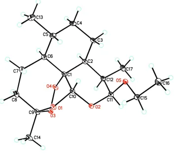
The crystal structure of the title compound, C17H28O5,
reported here is a polymorph of the structure first reported by El-Feraly, Al-Yahya, Orabi, McPhail & McPhail [J. Nat. Prod.(1992).55, 878–883]. It is a derivative of the antimalaria compound artemisinin and consists primarily of three substituted ring systems fused together. A cyclohexane ring (distorted chair conformation) fused to a tetrahydropyran group (distorted chair) is adjacent to an oxacycloheptane unit containing an endo-peroxide bridge, giving the molecule its particular three-dimensional arrangement. The crystal packing is stabilized by intermolecular C—H O interactions between an O atom from the endo-peroxide bridge and H atoms from both the cyclohexane and seven-membered oxacycloheptane fused rings, as well as between an O atom and H atom from adjacent tetrahydropyran rings. The two polymorphs have the same space group and similar cell parameters for the a and b axes, but significantly different values for thecaxis.
Related literature
For the first polymorph of this compound, see: El-Feralyet al.
(1992). For crystal structures of similar compounds, see: Brossi
et al. (1988); Flippen-Anderson et al. (1989); Karle & Lin (1995); Li et al. (2006); Luo et al. (1984); Yue et al. (2006); Butcher et al. (2007); Jasinski et al. (2008). For biological activity of artemisinin derivatives in vitro and in vivo, see: Graceet al.(1998); Liet al.(2001); Maggset al.(2000); Yanget al. (1997). For endo-peroxide sesquiterpene lactone deriva-tives, see: Saxenaet al.(2003); Venugopalanet al.(1995); Wuet al.(2001). For the synthesis of artemisinin and its derivatives, see: Lui et al. (1979); Liu (1980); Robert et al. (2001). For related literature, see: Cremer & Pople (1975); Lisgartenet al.
(1998); Qinghaosu Research Group (1980); Shen & Zhuang (1984); Wu & Li (1995).
Experimental
Crystal data
C17H28O5 Mr= 312.39 Trigonal,P3221 a= 10.0253 (6) A˚
c= 28.628 (3) A˚
V= 2491.8 (3) A˚3
Z= 6
MoKradiation
= 0.09 mm1
T= 103 (2) K 0.420.220.18 mm
Data collection
Bruker APEXII CCD area-detector diffractometer
Absorption correction: multi-scan (SADABS; Sheldrick, 1996)
Tmin= 0.963,Tmax= 0.984
27842 measured reflections 4935 independent reflections 4517 reflections withI> 2(I)
Rint= 0.034
Refinement
R[F2> 2(F2)] = 0.040 wR(F2) = 0.101
S= 1.04 4935 reflections
203 parameters
H-atom parameters constrained
max= 0.38 e A˚
3
min=0.17 e A˚
3
Table 1
Hydrogen-bond geometry (A˚ ,).
D—H A D—H H A D A D—H A
C5—H5A O4i 1.00 2.45 3.3150 (15) 144
C7—H7A O4i
0.99 2.55 3.4704 (16) 155
Symmetry code: (i)yþ1;x;z.
Data collection:APEX2(Bruker, 2006); cell refinement:APEX2; data reduction: SAINT (Bruker, 2006); program(s) used to solve structure:SHELXS90(Sheldrick, 2008); program(s) used to refine structure: SHELXL97 (Sheldrick, 2008); molecular graphics:
SHELXTL(Sheldrick, 2008); software used to prepare material for publication:SHELXTL.
RJB acknowledges the Laboratory for the Structure of Matter at the Naval Research Laboratory, Washington DC, USA, for access to their diffractometers. BN thanks Strides Arco Labs, Mangalore, India, for a gift sample of the title compound.
Supplementary data and figures for this paper are available from the IUCr electronic archives (Reference: AT2510).
organic compounds
Acta Cryst.(2008). E64, o585–o586 doi:10.1107/S1600536807062812 #2008 International Union of Crystallography
o585
Acta Crystallographica Section EStructure Reports
Online
Brossi, A., Venugopalan, B., Dominguez Gerpe, L., Yeh, H. J. C., Flippen-Anderson, J. L., Buchs, P., Luo, X. D., Milhousand, W. & Peters, W. (1988).J. Med. Chem.31, 645–650.
Bruker (2006).APEX2andSAINT. Bruker AXS Inc., Madison, Wisconsin, USA.
Butcher, R. J., Jasinski, J. P., Yathirajan, H. S., Bindya, S. & Narayana, B. (2007).Acta Cryst.E63, o3291–o3292.
Cremer, D. & Pople, J. A. (1975).J. Am. Chem. Soc.97, 1354–1358. El-Feraly, F. S., Al-Yahya, M. A., Orabi, K. Y., McPhail, D. R. & McPhail, A. T.
(1992).J. Nat. Prod.55, 878–883.
Flippen-Anderson, J. L., George, C., Gilardi, R., Yu, Q.-S., Dominguez, L. & Brossi, A. (1989).Acta Cryst.C45, 292–294.
Grace, J. M., Aguilar, A. J., Trotman, K. M. & Brewer, T. G. (1998).Drug Metab. Dispos.26, 313–317.
Jasinski, J. P., Butcher, R. J., Yathirajan, H. S., Narayana, B. & Sreevidya, T. V. (2008).Acta Cryst.E64, o89–o90.
Karle, J. M. & Lin, Ai. J. (1995).Acta Cryst.B51, 1063–1068.
Li, Y., Shan, F., Wu, J. M., Wu, G. S., Ding, J., Xiao, D., Yang, W. Y., Atassi, G., Leonce, S., Caignard, D. H. & Renard, P. (2001).Bioorg. Med. Chem. Lett.
11, 5–8.
Li, S.-H., Yue, Z.-Y., Gao, P. & Yan, P.-F. (2006).Acta Cryst.E62, o1898–o1900. Lisgarten, J., Potter, B. S., Bantuzeko, C. & Palmer, A. (1998). J. Chem.
Crystallogr.28, 539–542.
Liu, X. (1980).Chin. Pharm. Bull.15, 183–183.
(1979).Acta Chim. Sinica,37, 129–141.
Luo, X. D., Yeh, H. J. C., Brossi, A., Flippen-Anderson, J. L. & Gillardi, R. (1984).Helv. Chim. Acta,67, 1515–1522.
Maggs, J. L., Bishop, L. P. D., Edwards, G., O’Neill, P. M., Ward, S. A., Winstanley, P. A. & Park, K. (2000).Drug Metab. Dispos.28, 209–217. Qinghaosu Research Group (1980).Sci. Sin. (Engl. Ed.),23, 380–396. Robert, A., Benoit-Vical, F., Dechy-Cabaret, O. & Meunier, B. (2001).Pure
Appl. Chem.73, 1173–1188.
Saxena, S., Pant, N., Jain, D. C. & Bhakuni, R. S. (2003).Curr. Sci.85, 1314– 1329.
Sheldrick, G. M. (1996).SADABS. University of Go¨ttingen, Germany. Sheldrick, G. M. (2008).Acta Cryst.A64, 112–122.
Shen, C. C. & Zhuang, L. (1984).Med. Res. Rev.4, 57–59.
Venugopalan, B., Karnik, P. J., Bapat, C. J., Chatterjee, D. K., Iyer, N. & Lepcha, D. (1995).Eur. J. Med. Chem.30, 697–706.
Wu, Y.-L. & Li, Y. (1995).Med. Chem. Res.5, 569–586.
Wu, J. M., Shan, F., Wu, G. S., Ying, L., Ding, J., Xiao, D., Han, J.-X., Atassi, G., Leonce, S., Caignard, D. H. & Renard, P. (2001).Eur. J. Med. Chem.36, 469– 479.
Yang, X. P., Pan, Q. C., Liang, Y.-G. & Zikang, Y.-L. (1997).Cancer,16, 186– 187.
supporting information
sup-1
Acta Cryst. (2008). E64, o585–o586supporting information
Acta Cryst. (2008). E64, o585–o586 [doi:10.1107/S1600536807062812]
A second polymorph of
β
-arteether
Jerry P. Jasinski, Ray J. Butcher, H. S. Yathirajan, B. Narayana and T. V. Sreevidya
S1. Comment
Artemisinin and its derivatives, dihydroartemisinin, artemether, arteether and artesunate, are antimalarial drugs which possess bioactivity with reduced toxicity (Wu & Li, 1995). Artemisinin is isolated from the leaves of the plant Artemisia annua (Qinghao). It is a sesquiterpene lactone with an endo-peroxide linkage. Artemisinin derivatives are more potent than artemisinin and are active by virtue of the endo-peroxide. Because of their activity against strains of the parasite that had become resistant to conventional chloroquine therapy and the ability, due to the lipophilic structure, to cross the blood brain barrier, it was particularly effective for the deadly cerebral malaria (Shen & Zhuang, 1984). Because of their shorter lifetime and decreasing activity, they are used in combination with other antimalarial drugs. The notable activity of artemesinin derivatives in vitro and in vivo has been reported in the literature (Li et al. 2001 & Yang et al. 1997). However, some derivatives of artimisinine showed moderate cytotoxicity in vitro. The electronegativity and bulk of the substituents that are attached to the aryl group play an insignificant role in cytotoxicity. The antimalarial activity and cytotoxicity of some sesquiterpenoids has been reported in the literature (Venugopalan et al., 1995; Wu et al., 2001; Saxena et al., 2003). The endo-peroxide group present in these compounds plays an important role in antimalarial activity. Its 1,2,4-trioxane ring is unique in nature. After being opened in the plasmodium it liberates singlet oxygen and forms free radicals, which in turn produce oxidative damage of the parasite's membrane. Artemisinin is hydrophobic in nature and is partitioned into the membrane of the plasmodium. The crystal structure of an ether dimer of deoxydihydro-qinghaosu, a potential metabolite of the antimalarial arteether, has been reported (Flippen-Anderson et al., 1989). The correlation of the crystal structures of diastereomeric artemisinin derivatives with their proton NMR spectra in CDCl3 has
been reported (Karle & Lin, 1995). The crystal structure of artemisinin has been reported (Lisgarten et al., 1998). The crystal structure of a dimer of α- and β-dihydroartemisinin (Yue et al., 2006) and that of 9,10-dehydro-deoxyartemisin has recently been reported (Li et al., 2006). The synthesis of artemisinin and its derivatives have been described (Lui et al., 1979; Liu, 1980; Robert et al., 2001). β-Arteether (AE) is an endo-peroxide sesquiterpene lactone derivative currently being developed for the treatment of severe, complicated malaria caused by multidrug-resistant Plasmodium falciparum (Grace et al., 1998). β-Artemether (AM), the O-methyl ether prodrug of dihydroartemisinin (DHA), is an endo-peroxide antimalarial (Maggs et al., 2000). In view of the importance of the title compound, C17H28O5 (I), β-arteether, as an
antimalarial drug, this paper describes a new polymorphic form of the crystal structure first reported by El-Feraly et al.
(1992), from data measured at 103 (2) K.
A substantial increase in the length of the unit cell c axis from 25.720 to 28.628 Å in the new structure along with a redetermination of the cell constants and the cell volume for (I) at room temperature (296 K) [a = 10.1557 (14), c = 28.714 (4) Å and V = 2564.8 (8) Å3] provides solid support for the recognition of this new polymorphic form for (I). The
hydrodeoxyartemisinin (Shu-Hui Li et al., 2006). The seven-membered ring (C1/C6–C9/O1–C10) contains the important peroxy linkage [O3—O4 = 1.4759 (13) Å]. The six-membered ring C (O1/O3/O4/C1/C9/C10) which contains both an oxygen bridge and a peroxy bridge is best described by a twist-boat conformation with puckering parameters Q, θ and φ
of 0.749 (2) Å, 94.8 (5)° and 276.8 (8)°, respectively. For an ideal twist-boat conformation, θ and φ are 90° and (60n + 30)°, respectively. This conformation is consistent with 9,10-dehydrodeoxyartemisinin (Li et al., 2006), dihydro-artemisinin (Qinghaosu Research Group, 1980; Jasinski et al., 2008) and artemether (Butcher et al., 2007)
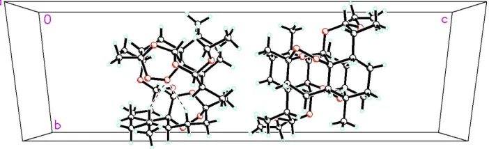
Crystal packing is stabilized by intermolecular C—H···O interactions between hydrogen atoms from the cyclohexane ring (H5A and H7A) and an oxygen atom (O4) from the endo-peroxide bridge (Fig. 2).
S2. Experimental
The title compound (C17H28O5) was obtained in the pure form from Strides Arco Labs, Mangalore, India. X-ray
diffraction quality crystals were grown from acetone [m.p.: 353 K]).
S3. Refinement
All H atoms were initially located in a difference Fourier map. The methyl H atoms were then constrained to an ideal geometry with C—H distances of 0.98Å and Uiso(H) = 1.5Ueq(C), but each group was allowed to rotate freely about its C
[image:4.610.130.485.356.670.2]—C bond. All other H atoms were placed in geometrically idealized positions and constrained to ride on their parent atoms with C—H distances in the range 0.90–1.00 Å and Uiso(H) = 1.17–1.22Ueq(C).
Figure 1
supporting information
[image:5.610.132.483.73.180.2]sup-3
Acta Cryst. (2008). E64, o585–o586Figure 2
The molecular packing for (I) viewed down the c axis. Dashed lines indicate C–H···O intermolecular hydrogen bonds.
β-Arteether
Crystal data
C17H28O5
Mr = 312.39 Trigonal, P3221
Hall symbol: P 32 2"
a = 10.0253 (6) Å
c = 28.628 (3) Å
V = 2491.8 (3) Å3
Z = 6
F(000) = 1020
Dx = 1.249 Mg m−3
Mo Kα radiation, λ = 0.71073 Å Cell parameters from 5075 reflections
θ = 2.4–30.0°
µ = 0.09 mm−1
T = 103 K Chunk, colourless 0.42 × 0.22 × 0.18 mm
Data collection
Bruker APEXII CCD area-detector diffractometer
Radiation source: fine-focus sealed tube Graphite monochromator
φ and ω scans
Absorption correction: multi-scan (SADABS; Sheldrick, 1996)
Tmin = 0.963, Tmax = 0.984
27842 measured reflections 4935 independent reflections 4517 reflections with I > 2σ(I)
Rint = 0.034
θmax = 30.8°, θmin = 2.1°
h = −11→14
k = −14→14
l = −39→39
Refinement
Refinement on F2
Least-squares matrix: full
R[F2 > 2σ(F2)] = 0.040
wR(F2) = 0.101
S = 1.05 4935 reflections 203 parameters 0 restraints
Primary atom site location: structure-invariant direct methods
Secondary atom site location: difference Fourier map
Hydrogen site location: inferred from neighbouring sites
H-atom parameters constrained
w = 1/[σ2(F
o2) + (0.0551P)2 + 0.4841P]
where P = (Fo2 + 2Fc2)/3
(Δ/σ)max = 0.005
Δρmax = 0.38 e Å−3
Δρmin = −0.17 e Å−3
Geometry. All e.s.d.'s (except the e.s.d. in the dihedral angle between two l.s. planes) are estimated using the full covariance matrix. The cell e.s.d.'s are taken into account individually in the estimation of e.s.d.'s in distances, angles and torsion angles; correlations between e.s.d.'s in cell parameters are only used when they are defined by crystal symmetry. An approximate (isotropic) treatment of cell e.s.d.'s is used for estimating e.s.d.'s involving l.s. planes.
Refinement. Refinement of F2 against ALL reflections. The weighted R-factor wR and goodness of fit S are based on F2,
conventional R-factors R are based on F, with F set to zero for negative F2. The threshold expression of F2 > σ(F2) is used
only for calculating R-factors(gt) etc. and is not relevant to the choice of reflections for refinement. R-factors based on F2
are statistically about twice as large as those based on F, and R- factors based on ALL data will be even larger.
Fractional atomic coordinates and isotropic or equivalent isotropic displacement parameters (Å2)
x y z Uiso*/Ueq
O1 0.71973 (10) 0.35233 (10) −0.03898 (3) 0.01837 (18) O2 0.57224 (11) 0.39122 (11) 0.01212 (3) 0.01993 (19) O3 0.47049 (10) 0.15908 (12) −0.05520 (3) 0.0219 (2) O4 0.45576 (11) 0.06611 (11) −0.01350 (3) 0.02042 (19) O5 0.60247 (12) 0.45429 (11) 0.09195 (3) 0.02137 (19) C1 0.58206 (14) 0.15089 (14) 0.01942 (4) 0.0169 (2) C2 0.50124 (14) 0.12064 (15) 0.06716 (4) 0.0182 (2)
H2A 0.4266 0.0075 0.0686 0.022*
C3 0.61370 (16) 0.15728 (16) 0.10811 (4) 0.0222 (3)
H3A 0.6848 0.2699 0.1097 0.027*
H3B 0.5550 0.1246 0.1377 0.027*
C4 0.70709 (16) 0.07586 (16) 0.10279 (4) 0.0232 (3)
H4A 0.7788 0.1028 0.1295 0.028*
H4B 0.6367 −0.0370 0.1031 0.028*
C5 0.79822 (16) 0.12223 (15) 0.05735 (4) 0.0213 (2)
H5A 0.8658 0.2368 0.0574 0.026*
C6 0.68604 (15) 0.07809 (14) 0.01577 (4) 0.0185 (2)
H6A 0.6163 −0.0360 0.0174 0.022*
C7 0.77196 (15) 0.11190 (16) −0.03110 (4) 0.0221 (3)
H7A 0.8677 0.2129 −0.0287 0.027*
H7B 0.8024 0.0330 −0.0361 0.027*
C8 0.68289 (16) 0.11487 (16) −0.07417 (4) 0.0235 (3)
H8A 0.5935 0.0104 −0.0791 0.028*
H8B 0.7505 0.1408 −0.1019 0.028*
C9 0.62470 (15) 0.23007 (15) −0.07075 (4) 0.0204 (2) C10 0.66650 (14) 0.32307 (14) 0.00757 (4) 0.0162 (2)
H10A 0.7569 0.3776 0.0290 0.019*
C11 0.49721 (16) 0.37047 (16) 0.05576 (4) 0.0204 (3)
H11A 0.4239 0.4102 0.0527 0.024*
C12 0.40444 (15) 0.20099 (16) 0.06911 (4) 0.0204 (2)
H12A 0.3232 0.1506 0.0446 0.024*
C13 0.90173 (19) 0.05086 (19) 0.05316 (6) 0.0329 (3)
H13A 0.9584 0.0664 0.0824 0.049*
H13B 0.9748 0.1002 0.0275 0.049*
supporting information
sup-5
Acta Cryst. (2008). E64, o585–o586C14 0.62461 (17) 0.30416 (18) −0.11675 (4) 0.0270 (3)
H14A 0.5651 0.3570 −0.1135 0.040*
H14B 0.5779 0.2247 −0.1409 0.040*
H14C 0.7308 0.3788 −0.1256 0.040*
C15 0.68313 (17) 0.61729 (15) 0.08415 (5) 0.0241 (3)
H15A 0.6102 0.6513 0.0743 0.029*
H15B 0.7612 0.6446 0.0593 0.029*
C16 0.7603 (2) 0.69468 (18) 0.12949 (5) 0.0367 (4)
H16A 0.8216 0.8063 0.1247 0.055*
H16B 0.8277 0.6557 0.1398 0.055*
H16C 0.6817 0.6722 0.1533 0.055*
C17 0.31881 (17) 0.17746 (18) 0.11531 (5) 0.0279 (3)
H17A 0.2512 0.2216 0.1130 0.042*
H17B 0.3935 0.2286 0.1405 0.042*
H17C 0.2569 0.0670 0.1220 0.042*
Atomic displacement parameters (Å2)
U11 U22 U33 U12 U13 U23
O1 0.0161 (4) 0.0163 (4) 0.0141 (4) 0.0017 (3) 0.0008 (3) −0.0006 (3) O2 0.0224 (5) 0.0212 (4) 0.0165 (4) 0.0111 (4) 0.0022 (3) 0.0025 (3) O3 0.0158 (4) 0.0247 (5) 0.0149 (4) 0.0025 (4) −0.0014 (3) 0.0020 (3) O4 0.0157 (4) 0.0187 (4) 0.0149 (4) −0.0004 (3) −0.0015 (3) 0.0003 (3) O5 0.0256 (5) 0.0165 (4) 0.0181 (4) 0.0076 (4) −0.0010 (4) −0.0004 (3) C1 0.0143 (5) 0.0152 (5) 0.0134 (5) 0.0015 (4) −0.0007 (4) −0.0012 (4) C2 0.0159 (5) 0.0161 (5) 0.0150 (5) 0.0024 (5) 0.0003 (4) 0.0006 (4) C3 0.0255 (6) 0.0191 (6) 0.0163 (5) 0.0068 (5) −0.0032 (5) 0.0007 (4) C4 0.0220 (6) 0.0184 (6) 0.0217 (6) 0.0046 (5) −0.0041 (5) 0.0046 (5) C5 0.0181 (6) 0.0150 (5) 0.0260 (6) 0.0045 (5) −0.0020 (5) 0.0039 (5) C6 0.0168 (6) 0.0124 (5) 0.0208 (5) 0.0031 (4) 0.0015 (4) 0.0007 (4) C7 0.0187 (6) 0.0177 (6) 0.0246 (6) 0.0052 (5) 0.0040 (5) −0.0009 (5) C8 0.0209 (6) 0.0209 (6) 0.0200 (6) 0.0040 (5) 0.0019 (5) −0.0044 (5) C9 0.0167 (6) 0.0201 (6) 0.0145 (5) 0.0019 (5) 0.0004 (4) −0.0014 (4) C10 0.0158 (5) 0.0142 (5) 0.0142 (5) 0.0041 (4) −0.0009 (4) 0.0001 (4) C11 0.0202 (6) 0.0210 (6) 0.0171 (5) 0.0083 (5) −0.0007 (4) −0.0009 (4) C12 0.0175 (5) 0.0218 (6) 0.0164 (5) 0.0058 (5) 0.0007 (4) −0.0004 (5) C13 0.0279 (8) 0.0321 (8) 0.0408 (8) 0.0166 (7) 0.0010 (6) 0.0085 (6) C14 0.0254 (7) 0.0309 (7) 0.0147 (5) 0.0066 (6) −0.0010 (5) 0.0006 (5) C15 0.0282 (7) 0.0160 (6) 0.0243 (6) 0.0082 (5) 0.0009 (5) 0.0022 (5) C16 0.0504 (10) 0.0186 (7) 0.0300 (7) 0.0090 (7) −0.0053 (7) −0.0018 (6) C17 0.0240 (7) 0.0294 (7) 0.0229 (6) 0.0079 (6) 0.0055 (5) −0.0007 (5)
Geometric parameters (Å, º)
O1—C10 1.4105 (14) C7—H7A 0.990
O1—C9 1.4386 (15) C7—H7B 0.990
O2—C11 1.4192 (15) C8—C9 1.536 (2)
O3—O4 1.4759 (13) C9—C14 1.5122 (17)
O4—C1 1.4619 (14) C10—H10A 1.000
O5—C11 1.4163 (15) C11—C12 1.5224 (18)
O5—C15 1.4327 (16) C11—H11A 1.000
C1—C10 1.5330 (16) C12—C17 1.5295 (17)
C1—C2 1.5399 (16) C12—H12A 1.000
C1—C6 1.5462 (18) C13—H13A 0.980
C2—C3 1.5382 (17) C13—H13B 0.980
C2—C12 1.5412 (19) C13—H13C 0.980
C2—H2A 1.000 C14—H14A 0.980
C3—C4 1.526 (2) C14—H14B 0.980
C3—H3A 0.990 C14—H14C 0.980
C3—H3B 0.990 C15—C16 1.512 (2)
C4—C5 1.5225 (18) C15—H15A 0.990
C4—H4A 0.990 C15—H15B 0.990
C4—H4B 0.990 C16—H16A 0.980
C5—C13 1.532 (2) C16—H16B 0.980
C5—C6 1.5427 (18) C16—H16C 0.980
C5—H5A 1.000 C17—H17A 0.980
C6—C7 1.5380 (17) C17—H17B 0.980
C6—H6A 1.000 C17—H17C 0.980
C7—C8 1.5315 (19)
C10—O1—C9 113.55 (9) O3—C9—C14 104.63 (11)
C11—O2—C10 116.17 (9) O1—C9—C14 107.15 (11)
C9—O3—O4 108.17 (9) O3—C9—C8 111.90 (11)
C1—O4—O3 111.72 (8) O1—C9—C8 110.02 (10)
C11—O5—C15 113.02 (10) C14—C9—C8 114.05 (11)
O4—C1—C10 110.01 (10) O1—C10—O2 105.06 (9)
O4—C1—C2 103.89 (9) O1—C10—C1 112.44 (9)
C10—C1—C2 110.98 (10) O2—C10—C1 113.28 (10)
O4—C1—C6 105.93 (9) O1—C10—H10A 108.6
C10—C1—C6 113.17 (10) O2—C10—H10A 108.6
C2—C1—C6 112.30 (10) C1—C10—H10A 108.6
C3—C2—C1 112.25 (10) O5—C11—O2 111.96 (11)
C3—C2—C12 115.18 (10) O5—C11—C12 109.68 (10) C1—C2—C12 109.64 (10) O2—C11—C12 111.59 (11)
C3—C2—H2A 106.4 O5—C11—H11A 107.8
C1—C2—H2A 106.4 O2—C11—H11A 107.8
C12—C2—H2A 106.4 C12—C11—H11A 107.8
C4—C3—C2 111.63 (11) C11—C12—C17 111.82 (12)
C4—C3—H3A 109.3 C11—C12—C2 112.41 (10)
C2—C3—H3A 109.3 C17—C12—C2 113.72 (11)
C4—C3—H3B 109.3 C11—C12—H12A 106.1
C2—C3—H3B 109.3 C17—C12—H12A 106.1
H3A—C3—H3B 108.0 C2—C12—H12A 106.1
supporting information
sup-7
Acta Cryst. (2008). E64, o585–o586C5—C4—H4A 109.5 C5—C13—H13B 109.5
C3—C4—H4A 109.5 H13A—C13—H13B 109.5
C5—C4—H4B 109.5 C5—C13—H13C 109.5
C3—C4—H4B 109.5 H13A—C13—H13C 109.5
H4A—C4—H4B 108.0 H13B—C13—H13C 109.5
C4—C5—C13 111.57 (11) C9—C14—H14A 109.5
C4—C5—C6 109.36 (11) C9—C14—H14B 109.5
C13—C5—C6 111.87 (12) H14A—C14—H14B 109.5
C4—C5—H5A 108.0 C9—C14—H14C 109.5
C13—C5—H5A 108.0 H14A—C14—H14C 109.5
C6—C5—H5A 108.0 H14B—C14—H14C 109.5
C7—C6—C5 111.24 (11) O5—C15—C16 107.69 (11)
C7—C6—C1 112.97 (10) O5—C15—H15A 110.2
C5—C6—C1 112.30 (10) C16—C15—H15A 110.2
C7—C6—H6A 106.6 O5—C15—H15B 110.2
C5—C6—H6A 106.6 C16—C15—H15B 110.2
C1—C6—H6A 106.6 H15A—C15—H15B 108.5
C8—C7—C6 116.04 (11) C15—C16—H16A 109.5
C8—C7—H7A 108.3 C15—C16—H16B 109.5
C6—C7—H7A 108.3 H16A—C16—H16B 109.5
C8—C7—H7B 108.3 C15—C16—H16C 109.5
C6—C7—H7B 108.3 H16A—C16—H16C 109.5
H7A—C7—H7B 107.4 H16B—C16—H16C 109.5
C7—C8—C9 114.06 (11) C12—C17—H17A 109.5
C7—C8—H8A 108.7 C12—C17—H17B 109.5
C9—C8—H8A 108.7 H17A—C17—H17B 109.5
C7—C8—H8B 108.7 C12—C17—H17C 109.5
C9—C8—H8B 108.7 H17A—C17—H17C 109.5
H8A—C8—H8B 107.6 H17B—C17—H17C 109.5
O3—C9—O1 108.78 (10)
C13—C5—C6—C1 −179.43 (10) C15—O5—C11—C12 −173.97 (11) O4—C1—C6—C7 69.20 (12) C10—O2—C11—O5 69.63 (13) C10—C1—C6—C7 −51.41 (13) C10—O2—C11—C12 −53.74 (14) C2—C1—C6—C7 −178.04 (10) O5—C11—C12—C17 57.32 (15) O4—C1—C6—C5 −163.99 (9) O2—C11—C12—C17 −178.03 (11) C10—C1—C6—C5 75.39 (12) O5—C11—C12—C2 −72.01 (13) C2—C1—C6—C5 −51.23 (13) O2—C11—C12—C2 52.64 (14) C5—C6—C7—C8 −164.04 (11) C3—C2—C12—C11 75.94 (13) C1—C6—C7—C8 −36.68 (15) C1—C2—C12—C11 −51.80 (13) C6—C7—C8—C9 56.69 (15) C3—C2—C12—C17 −52.41 (15) O4—O3—C9—O1 −73.25 (12) C1—C2—C12—C17 179.85 (10) O4—O3—C9—C14 172.50 (10) C11—O5—C15—C16 166.33 (13)
Hydrogen-bond geometry (Å, º)
D—H···A D—H H···A D···A D—H···A
C5—H5A···O4i 1.00 2.45 3.3150 (15) 144
C7—H7A···O4i 0.99 2.55 3.4704 (16) 155