THE ROLE of MORN2 AND TRAF3IP2 GENETIC VARIANTS IN MODULATING INNATE IMMUNE RESPONSE TO PERIODONTAL PATHOGENS
Lu Sun
A dissertation submitted to the faculty at the University of North Carolina at Chapel Hill in partial fulfillment of the requirements for the degree of Doctor of Philosophy in the
Curriculum in Oral and Craniofacial Biomedicine in the School of Dentistry.
Chapel Hill 2018
Approved by:
Steven Offenbacher Toni Darville
Jennifer Webster-Cyriaque Mitsuo Yamauchi
Sompop Bencharit
ii
© 2018 Lu Sun ALL RIGHTS RESERVED
iii
ABSTRACTLu Sun: The role of MORN2 and TRAF3IP2 genetic variants in modulating innate immune response to periodontal pathogens
(Under the direction of Steven Offenbacher)
Periodontal disease is a polygenic disease that is associated with inflammatory response to the oral biofilm. Although it is believed that microbial pathogens are necessary to the causal pathway, a key factor to determine whether individuals will develop periodontitis is the way how the hosts respond to the microflora resides in their periodontium. Genetic polymorphisms that will cause a change in the encoded protein or its expression, which may alter host innate or adaptive immunity, such as host barrier function and inflammatory responses, to microorganism and determine susceptibility to inflammatory disease. Thus it is necessary to seek candidate genetic function to explain the differences in susceptibility to periodontal disease.
Our recent genome-wide association study (GWAS) identified a missense single nucleotide polymorphism SNP (rs3099950) in the gene MORN2 that codes for a key membrane protein. We have also identified a missense SNP (rs13190932) in the gene TRAF3IP2 locus which is involved IL-17 signaling.
These two genetic variants are associated with a P.gingivalis (P.g) dominant and A.
actinomycetemcomitans (A.a) dominant periodontal disease subtypes. To date, only one study has described the function of MORN2 that serves as a macrophage protein that promotes the phagocytosis and killing of organisms via LC3-asscociated phagocytosis. TRAF3IP2 is involved in IL-17 signaling and mucosal immunity serving as an adaptor protein for the IL-17 receptor. However, the role of MORN2 and TRAF3IP2 in periodontal disease is unknown.
In Chapter 1, the background of periodontal disease and how the SNPs were identified by GWAS were described. In Chapter 2, we investigated the role of MORN2 in modulating innate immune response to periodontal pathogens and our studies suggest that MORN2 plays a critical role in the LC3-associated
iv
phagocytosis – mediated killing of periodontal pathogen (P.g) as well as cytokine/chemokine response through Ca2+ flux and NF-kB activation. In Chapter 3, we explored the role of TRAF3IP2 mediated IL-17 pathway in periodontal disease. Our study suggests that TRAF3IP2 engaged homeostatic IL-17 pathway plays a protective role in a P.g induced alveolar bone loss and colonization through neutrophil recruitment, maintenance epithelial barrier and induction of antimicrobial peptides. Defective TRAF3IP2 shifts the oral commensal communities. Finally in Chapter 4, we discuss the significance and future study directions. In summary, these findings clarify the molecular mechanism of MORN2 and TRAF3IP2 function and provide insight into the genetic basis of MORN2 and TRAF3IP2 in periodontal disease susceptibility.
v
ACKNOWLEDGEMENTS
It is a special and amazing journey of the four years Ph.D. training at School of Dentistry of University of North Carolina at Chapel Hill. When I was in dental school, I realized periodontics is the most active specialty involved in research and is the foundation of dentistry that have a strong interaction with other specialties. No specialty can be compared to periodontics in my mind, which leading me to choose periodontics as my specialty and pursue Ph.D. involved in periodontal research. It has always been a dream of mine to study in the top dental school worldwide.
I was fortunate enough to be accepted into the Oral and Craniofacial Biomedicine program at UNC-CH and had a chance to work with Dr. Offenbacher. The first time I knew him is from the term periodontal medicine, as first suggested by Dr. Offenbacher, when I was a dental student. I would like to give my ardent gratitude to my great mentor – Dr. Steven Offenbacher. I feel extremely lucky to be your Ph.D. student for the past four years. Your enthusiasm for science, foresight of research, great diligence for work never stop to inspire and enlighten me. Your smile, your hello, your email and even your view of back is always great encouragement to me to move forward. It is my cherished experience during discussing our studies and exchanging ideas or thoughts. Thank you Dr. Offenbacher for your guidance, patience and countless support and I really appreciate that you accompany with me during this journey. I am grateful to my committee members, Dr. Toni Darville, Dr. Jennifer Webster-Cyriaque, Dr. Mitsuo Yamauchi, Dr. Sompop Bencharit, who afford constructive comments and encouragement for my dissertation, which always keeping me on the right track.
I am thankful to our program director Dr. Ceib Phillips who recruited me into this fantastic
program and has provided endless support to make sure I am always on the right direction. I also want to give many thanks to Dr. Ricardo Teles – an inspired and committed periodontist. It is my honor to work with you in the same department and thank you so much for your encouragement, resolute support as well as your constructive suggestions to my work. Dr. Flavia Teles, appreciate your guidance for the
vi
progress of the rotation projects and your time for helping me to revise my poster. I will cherish the time I worked with you in my life and it is your support that makes me have a smooth transition from first year to second year. I am thankful to Dr. Stanley Lemon, a featured immunologist. Your positive attitude to experiments and passionate to research ignites my passionate to science and the skills and knowledge I learned in your lab benefits me a lot in my later work. Dr. Roland Arnold, I learned how to critical thinking from you during comprehensive exam. Thank you for training me in bacteria cultivation and answering me questions when I was confused. I am grateful to work with many passionate, diligent colleagues in the Dr.
Offenbacher’s lab and GO Health Center. Thank you for all being my teachers and my friends. Thank you for the people in Koury Oral Health Science Building who raise your hands when I need help. Thank you for all my friends in my life.
At last, I want to thank my parents for their endless love and you are my strength and energy for
trusting my heart, fighting against obstacles, winning the battle and pursuing my dream.
vii
TABLE OF CONTENTS
LIST OF TABLES……….x
LIST OF FIGURES………..xi
LIST OF ABBREVIATIONS………...xii
CHAPTER 1: INTRODUCTION……….1
CHAPTER 2: THE ROLE of MORN2 GENETIC VARIANTS IN MODULATING INNATE IMMUNE RESPONSE TO PERIODONTAL PATHOGENS………8
2.1. Introduction….……….……….…….……...8
2.2. Materials and methods………..……...…………....11
2.2.1. Reagents and Antibodies……….11
2.2.2. Bacterial strain………...11
2.2.3. Cell culture………..11
2.2.4. Bioinformatic approaches……….12
2.2.5. GWAS characterization of the Pg trait………12
2.2.6. MORN2 expression analysis in gingival tissue by RT-qPCR………..12
2.2.7. Immunohistochemistry and immunofluorescence of human gingival biopsies……….13
2.2.8. Lentivirus production and transduction………...14
2.2.9. Phagocytosis and survival assay of Pg ……….14
2.2.10. LC3 puncta formation assays………14
2.2.11. Transmission electronic microscopy……….15
2.2.12. Real-time PCR……….15
2.2.13. ELISA………15
2.2.14. Intracelluar calcium flux assay………..16
2.2.15. Westernblot………..16
2.2.16. Statistical analysis………...16
2.3. Results …...……….………..……..………17
2.3.1. Characterization of the Pg dominant trait, MORN2 SNP identification and in silico analysis……17
2.3.2. MORN2 SNP is associated with periodontal disease severity………17
2.3.3. Identification of MORN2 mRNA expression and localization in human gingival tissue…………...17
viii
2.3.4. MORN2 promotes the LC3-associated phagocytosis in macrophages………18
2.3.5. Role of MORN2 on production of cytokines and chemokines by HGEs and marophages………19
2.3.6. Knockdown MORN2 attenuates calcium flux and NF-κB activation……….19
2.4. Discussion …...…………...……….……….………...…21
2.5. Conclusions …....……….………..25
2.6. Figures and Tables….……….………..26
CHAPTER3: THE ROLE of TRAF3IP2 GENETIC VARIANTS IN MODULATING INNATE IMMUNE RESPONSE TO PERIODONTAL PATHOGENS ...……….41
3.1. Introduction.…..……….……….………41
3.2. Materials and methods……….……….44
3.2.1. Bioinformatic approaches………44
3.2.2. GWAS characterization of the Aa trait………...44
3.2.3. Bacterial strain………...44
3.2.4. Mice……….44
3.2.5. Murine experimental periodontitis model………..45
3.2.6. Samples and DNA isolation……….45
3.2.7. Alveolar bone measurement………...45
3.2.8. Immunohistochemistry……….46
3.2.9. Oral microbial burden………...46
3.2.10. Gene expression analysis by NanoString………...47
3.2.11. Microbiome evaluation via Illumian MiSeq 16S rRNA amplicon sequencing………47
3.3. Results....……….48
3.3.1. TRAF3IP2 SNP is associated with periodontal disease severity………..48
3.3.2. Traf3ip2-/- mice are susceptible to Pg-induced periodontal bone loss………..48
3.3.3. TRAF3IP2 plays a protective role in a Pg colonization partially through neutrophil recruitment.49 3.3.4. Altered cytokines and chemokines expression in the infected Traf3ip2-/- mice………..49
3.3.5. Absence of TRAF3IP2 adversely influences epithelial barrier function………50
3.3.6. TRAF3IP2-mediated signaling promotes osteoblasts formation………...50
3.3.7. Microbiome shift correlates with the absence of TRAF3IP2 ……….51
3.4. Discussion……….………..52
3.5. Conclusions………..…………..………58
3.6. Figures and Tables……….………...59
CHAPTER 4: DISCUSSION AND FUTURE DIRECTIONS………74
ix
REFERENCES………...78
x
LIST OF TABLES
Table 2.1 Principle Component Trait profiles for Socransky complex, Aa trait and Pg trait……….….26 Table 2.2 Subjects' Demographics and Clinical Parameters………..31 Table 3.1 Primer sequence information for 16S rRNA sequencing………...59
xi
LIST OF FIGURES
Figure 1.1 Principal component trait pattern profiles………..6
Figure 1.2 Model of genetic and microbial patterns of periodontal disease………...7
Figure 2.1 rs3099950 causes a missense mutation.………27
Figure 2.2 rs3099950 is associated with microbial burden and periodontal disease severity....…………..29
Figure 2.3 Identification of MORN2 in human gingival tissues ………..32
Figure 2.4 MORN2 is required for LAP………..34
Figure 2.5 MORN2 is required for neutrophil specific chemokines and G-CSF synthesis in HGEs………36
Figure 2.6 Knockdown of MORN2 decreases proinflammatory cytokine production upon P.g infection…37 Figure 2.7 MORN2 is required for chemokines and HGFs production upon P.g infection………38
Figure 2.8 MORN2 knockdown macrophages attenuates calcium flux and activation of NF-κB…………..40
Figure 3.1 TRAF3IP2 SNP is associated with periodontal disease severity………...60
Figure 3.2 Traf3ip2-/- mice are susceptible to P.g-instigated periodontal bone loss………63
Figure 3.3 Higher colonization of P.g in Traf3ip2-/- mice is correlated with defective neutrophils recruitment………..64
Figure 3.4 Cytokines expression in the Traf3ip2-/- mice infected with P.g is altered………...65
Figure 3.5 Absence of TRAF3IP2 exacerbates gingival epithelial barrier dysfunction………...67
Figure 3.6. TRAF3IP2-mediated signaling promotes osteoblasts formation………70
Figure 3.7. Differences of oral microbiota composition between Traf3ip2-/- mice and WT mice..………….71
xii
LIST OF ABBERVIATIONS
A.a Aggregatibacter actinomycetemcomitans
Act1 NF-κB activator 1
AMPs Antimicrobial peptides
AP-1 Activator protein 1
APC Antigen presenting cell
ARIC Atherosclerosis Risk in Communities
BOP Bleeding on probing
BSA Bovine serum albumin
CAL Clinical attachment loss
CCL6 Chemokine (C-C motif) ligand 6
CCR2 C-C chemokine receptor type 2
CD68 Cluster of differentiation 68
CIKS Connection to IKK and SAPK/JNK
CMC Carboxymethyl cellulose
C.r Campylobacter rectus
CXCL1 C-X-C motif ligand 1
CXCL2 C-X-C motif ligand 2, MIP2-alpha
CXCL3 C-X-C motif ligand 3, MIP2-beta
CXCL5 C-X-C motif ligand 5
DAMP Damage associated molecular signal
xiii
DARIC Dental Atherosclerosis Risk in Community StudyDEFB1 Defensin beta 1
DEFB4 Defensin beta 4
DSC-1 Desmocollin-1
DSG-1 Desmoglein-1
FBS Fetus bovine serum
F.n Fusobacterium nucleatum
FILAGGRIN Filament aggregating protein
GAPDH Glyceraldehyde-3 phosphate dehydrogenase
GCF Gingival crevicular fluid
GCP-2 Granulocyte chemotactic protein, CXCL6
G-CSF Granulocyte colony-stimulating factor
GM-CSF Granulocyte macrophage colony-stimulating factor
GWAS Genome-wide association study
HGEs Human gingival epithelial cells
IFN-γ Interferon-γ
IL-1 Interleukin-1
IL-1β Interleukin 1 beta
IL-4 Interleukin 4
IL-6 Interleukin 6
IL-8 Interleukin 8
IL-17 Interleukin 17
xiv
IL-17R Interleukin-17 receptorIL-23 Interleukin 23
IL-36G Interleukin-36 gamma
IP3 Inositol trisphosphate 3
JNK Jun amino-terminal kinases
KLF4 Kruppel-like factor 4
KRT1 Keratin 1
KRT10 Keratin 10
LAP LC3-associated phagocytosis
LCN2 Lipocalin 2
LPS Lipopolysaccride
MAF Minor allele frequency
MAPK Mitogen-activated protein kinases
MCP-1 Monocyte chemoattractant protein 1, CCL2
MIP-1α Macrophage inflammatory protein 1 alpha, CCL3
MMP3 Matrix metallopeptidase 3
MMP9 Matrix metallopeptidase 9
MORN2 Membrane Occupation and Recognition Nexus repeat-containing-2
NF-κB Nuclear factor kappa-light-chain-enhancer of activated B cells
NLRs Nod like receptors
OCLN Occludin
PAMP Pathogen-associated molecular pattern
xv
PCR Polymerase chain reactionPCT Principle component trait
PD Probing depth
P.g Porphyromonas gingivalis
P.i Prevotella intermedia
P.n Prevotella nigrescens
PIP2 Phosphatidylinositol 4,5-bisphosphate
PRR Pattern recognition receptors
qRT-PCR Quantitative reverse transcription PCR
RANKL Receptor activator of nuclear factor kappa-B ligand
RANTES Regulated on activation, normal T cell expressed and secreted, CCL5
RORγ RAR-related orphan receptor gamma
RUNX2 Runt-related transcription factor 2
S100A8 S100 calcium binding protein A8
S100A9 S100 calcium binding protein A9
SEFIR SEF/IL-17R signaling motif
SNP Single nucleotide polymorphism
STAT3 Signal transducer and activator of transcription 3
T.d Treponema denticola
T.f Tannerella forsythia
TLR Toll-like receptor
TNF-α Tumor necrosis factor α
xvi
TRAF3IP2 Tumor necrosis factor-Receptor Associated Factor 3 Interacting Protein 2
Treg Regulatory T cell
Th1 Type 1 T helper cells
Th2 Type 2 T helper cells
Th17 T helper 17 cells
ZNF750 Zinc Finger Protein 750
1
CHAPTER 1: INTRODUCTION
Periodontitis is an inflammatory response to the oral bacterial flora and represents one of the most prevalent infectious disease in human worldwide. Approximately 11% of the world population (Richards 2014) exhibits severe periodontitis and 46% United States adults, representing 64.7 million people, had periodontitis, with 8.9% having severe periodontitis (Eke, Dye et al. 2015). Periodontitis is well characterized by local tissue destruction that involves loss of alveolar bone and supporting ligament around the teeth, and if left untreated, can lead to loss of teeth. Increasing evidence suggests that the chronic inflammation of periodontitis represents potential risk factor for association with various systemic disease including adverse pregnancy, cardiovascular disease, rheumatoid arthritis, stroke as well as diabetes (Offenbacher and Beck 2014, Hasturk and Kantarci 2015, Duda-Sobczak, Zozulinska- Ziolkiewicz et al. 2018, Sen, Giamberardino et al. 2018).
Although pathogenic bacteria are assumed to be essential to the destruction in host tissues, they are not sufficient serve as a strong predictor of periodontal disease, for the reason that specific
microorganisms, in most cases, are not sufficient to cause disease. Thus, periodontitis is considered as a multifactorial oral disease that involves a susceptible host influenced by genetic factors, dysbiotic oral microbial shift and other factors such as smoking(Haffajee and Socransky 2001, Merchant, Pitiphat et al.
2002). One of key factors to determine whether individuals will develop periodontitis is the way how the hosts respond to the microflora resides in their periodontium (Taba, Souza et al. 2012). Genetic polymorphisms that will cause a change in the encoded protein or its expression that may alter host innate or adaptive immunity and/or microbial colonization patterns, which results in the manifestation as the clinical phenotype of disease. For example, genetic mutations within the NLRP6-inflammasome complex results in alteration of the colonic microbial ecology and the inflammatory pathways associated with colitis (Elinav, Strowig et al. 2011). Thus, it is understood that genetic variants in inflammatory pathways can induce dysbiotic shifts in the microbiome that combine to create chronic inflammatory conditions at mucosal surfaces including periodontal disease(Walker, Sanderson et al. 2011, Craven,
2
Egan et al. 2012, Darveau, Hajishengallis et al. 2012, Hajishengallis, Darveau et al. 2012, Hajishengallis and Lamont 2012, Henao-Mejia, Elinav et al. 2012). However, we cannot identify these implicit variants based on clustering of clinical signs (probing depth, attachment loss, bleeding index) alone. There are also several lines gained from family studies to support the genetic role in determining the predisposition of periodontitis. Familial aggregation of aggressive periodontitis is not unusual in clinic and a pilot study of the aggregation within families also suggests a genetic predisposition(Nibali, Donos et al. 2008). Case- control (Schaefer, Jochens et al. 2014, Guedes, Planello et al. 2015) and cross-sectional study (Casado, Aguiar et al. 2015) exploring candidate genes have also demonstrated association of gene polymorphic variants with periodontitis. These candidate genes include Interleukin-1 (Trevilatto, de Souza Pardo et al.
2011, Braosi, de Souza et al. 2012, Armingohar, Jorgensen et al. 2014), Matrix Metalloproteinase (Song, Kim et al. 2013, Emingil, Han et al. 2014), Fc receptor (Hans, Mehta et al. 2015), TNF-α (Ozer Yucel, Berker et al. 2015) and pattern recognition receptor (Han, Ding et al. 2015) in periodontitis could be chosen based on previously association study with other types of chronic inflammatory disease, such as inflammatory bowel disease and rheumatoid arthritis, or their relationship with immune response.
However, these studies explored only one locus or several loci and some candidate gene association studies performed with contradictory results (Laine, Loos et al. 2010, Zhang, Sun et al. 2011, Laine, Crielaard et al. 2012) because of small cohort size of subjects involved, limited number of loci tested and the lack of power and replication. Thus, the identification of considerable genetic risk variants that
contributes to periodontitis needs a large population that can detect small effects given by genetic variant.
Genome wide associate study (GWAS) is a powerful tool to uncover millions of variations in genomic DNA at one time to determine whether any genetic locus is associated with disease phenotype (Loos, Papantonopoulos et al. 2015). Unlike candidate gene association study, GWAS is an open-ended method and not based on a prior knowledge of disease pathogenesis and phenotypes. Single nucleotide polymorphisms (SNPs) are important genetic markers that link DNA variants to phenotype changes. The SNP identified by GWAS covering the entire human genome including coding regions and regulatory regions. By testing all common variants, we could highlight key genes and shed light on the underlying mechanisms. Recently, GWAS of periodontitis have been performed by our group (Divaris, Monda et al.
2012, Divaris, Monda et al. 2013) and other groups (Teumer, Holtfreter et al. 2013, Lee and Kim 2015, Shimizu, Momozawa et al. 2015) and highlighted loci might be associated with this type of prevalent
3
disease. However, these association studies does not meet strict genome-wide significance criteria and could only illustrate a small portion of the total population variance, which indicates the need to explore the gene centric of this condition by using more distinct clinic parameters.
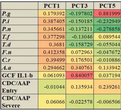
Recently, our group create distinct periodontal complex traits (PCT) by using principle component analysis to interrogate in GWAS analysis to identify novel potential candidate genetic loci related to the biological basis and pathogenesis of chronic periodontitis. Specifically, we have completed a genome- wide association study of 975 European American adult individuals [394 (40%) subjects with periodontal health, which includes subjects with gingivitis, 389 (39.9%) subjects with mild-moderate periodontitis and 192 (19.7%) subjects with severe periodontal disease] selected from Dental Atherosclerosis Risk in Communities (DARIC) cohort (Beck, Elter et al. 2001, Elter, Champagne et al. 2004) including eight periodontal pathogens [P. gingivalis (P.g), Prevotella intermedia (P.i), T. denticola (T.d), T. forsythia (T.f), C. rectus (C.r), Fusobacterium nucleatum (F.n), A. actinomycetemcomitans (A.a), P. nigrescens (P.n)], inflammatory mediator in gingival crevicular fluid (interleukin1-β) and clinical disease classification
(CDC/AAP chronic periodontitis classification) as well as probing depth and interproximal attachment loss.
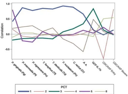
The principle component profile illustrating correlations with the constitutive parameters was shown in Figure 1.1. The first PCT was referred as the Socransky Trait since the microbial community structure in PCT1 has a high correlation and positive loading with all eight periodontal organisms that associated with microbial clusters identified by Socransky et al (Socransky, Haffajee et al. 1998). PCT3 has a high positive loading of A. actinomycetemcomitans and GCF-IL-1β and is designated as A.a trait. PCT5 is characterized by the highest loading of P. gingivalis with an eigenvalue of 0.882 (P.g trait) across all PCTs. Significant gene-centric associations were found in the first six complex traits by using MAGENTA.
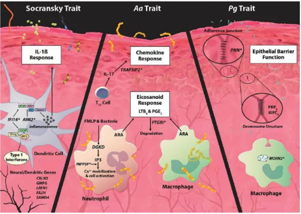
Genes in loci associated with PCT1 (Socransky trait), PCT3 (A.a trait) and PCT5 (P.g trait) suggest a biological basis that controls the epithelial barrier function and innate immune response (Figure 1.2).
Interestingly, there are two genes with nonsynonymous substitutions emerge as potential
candidates for regulating immune response in PCT3 and PCT5. The MORN2 locus within PCT5 contains a missense SNP that is significantly associated with the highest level of P.g counts and severe
periodontal disease clinical scores. MORN2 was identified as a phagosome protein of macrophage that is involved in the recruitment of LC3 in phagocytosis to M. tuberculosis-containing phagosomes and
subsequent maturation to degradative phagolysosomes (Abnave, Mottola et al. 2014). Our structural
4
analysis of MORN2 variant demonstrates that MORN2 variant changes the charge distribution across a PIP3 binding region of MORN2 structure that may result in impaired membrane interactions or
phagolysosome fusion. The A.a trait (PCT3) showed one gene TRAF3IP2 (Act1) contains a non- synonymous SNP that is significantly associated with the higher levels of A.a and severe periodontal disease scores, which point to potential abnormalities in the innate immune response. The paradigm that T helper 1 (Th1) cell and T helper 2 (Th2) cell characterized by the secretion of distinct cytokines was first postulated by Coffman, Mosmann and colleagues in 1986 (Mosmann, Cherwinski et al. 1986). Naïve CD4+ T cells can differentiate into either interferon-γ (IFN-γ)-producing Th1 cells or IL-4-producing Th2 cells when they first encounter foreign antigens presented by antigen-presenting cells (APCs), which is largely controlled by various environmental factors, especially by signals coming directly from APCs (Glimcher and Murphy 2000). Uncontrolled T cell responses can drive the onset of allergy or
autoimmunity such as inflammatory bowel disease (IBD), rheumatoid arthritis (RA) or psoriasis (Bouma and Strober 2003, Lowes, Bowcock et al. 2007). Substantial advances and efforts have been made to resolve some complicated pathological situations that cannot be explained by simple Th1/Th2 paradigm, which resulted in the expanded research and the discovery of a third subset of effector T cells that
produce IL-17 and exhibit distinct functions from Th1 and Th2 cells. It was not until 2005 that investigators proposed that IL-17-producing T cell, a distinct CD4+ T helper cell subset that are critically responsible for the production of IL-17, which was named Th17 cells (Harrington, Hatton et al. 2005, Park, Li et al. 2005).
IL-17 have pro-inflammatory properties (Kolls and Linden 2004) and act on different cell types to induce the gene expression of cytokines, chemokines and antimicrobial peptides (Jovanovic, Di Battista et al.
1998, Kolls and Linden 2004, Abusleme and Moutsopoulos 2017). In 2000, ACT1 was first identified as a protein that activates both NF-κB and JNK constitutively, which indicates that it functions at a point before bifurcation of these two signal pathways (Li, Commane et al. 2000). In the same year, Leonardi’s group identified CIKS (connection to IKK and SAPK/JNK) may represent a point of multiple points convergence in AP-1 and NF-κB pathways (Leonardi, Chariot et al. 2000). Later, Novatchkova’s group identified ACT1 and CIKS were the same protein and a conserved sequence segment in transmembrane receptors and soluble factors (like CIKS/ACT1) in eukaryotes – SEFIR domain (Novatchkova, Leibbrandt et al. 2003).
Bioinformatics analysis identified this conserved motif in the cytoplasmic domains of IL-17R that was homologous to the Toll/IL-1R (TIR) domain and named the conserved region as SEFIR domain (O'Quinn,
5
Palmer et al. 2008). Evidence that IL-17RA and ACT1 interact based on overexpression studies and co- immunoprecipitation studies with endogenous protein (Chang, Park et al. 2006, Maitra, Shen et al. 2007, Qian, Liu et al. 2007). Indeed, ACT1 deficiency by RNAi-mediated knockdown or in knockout mice
impairs IL-17 induced activation of NF-κB (Linden 2007, Dong 2008). It has been shown that homozygous deletion of IL-17RA abrogates the increase in splenic neutrophil progenitors resulting from the
overexpression of IL-17A (Ye, Rodriguez et al. 2001). IL-17RA-/- mouse fibroblasts fail to produce CXCL1 production in response to IL-17A and in human epithelial cells, a monoclonal antibody against IL-17RA effectively blocks both IL-17A and IL-17F induced expression of G-CSF and CXCL1 (McAllister, Henry et al. 2005). IL-17RA deficient mice are susceptible to many pathogens such as Bacteroides fragilis, Candida albicans, Klebsiella pneumoniae and Toxoplasmosis gondii (Chung, Kasper et al. 2003, Huang, Na et al. 2004, Kelly, Kolls et al. 2005, Ouyang, Kolls et al. 2008) and P.g-driven bone destruction is increased in IL-17RA knockout mice (Yu, Ruddy et al. 2007). Collectively, these data indicate that TRAF3IP2 is critical component involved in IL-17 signaling and mucosal immunity.
Thus, we hypothesize that MORN2 and TRAF3IP2 genetic variant may negatively modify the immune response to periodontal pathogen and influences dysbiotic shifts in the oral pathogen, which contributes to creation of chronic inflammatory conditions in periodontal disease. The objective of this study is to clarify the molecular mechanism of MORN2 and TRAF3IP2 function and to provide insight into the molecular and genetic basis of MORN2 and TRAF3IP2 in periodontal disease susceptibility.
.
6
Figure 1.1. Principal component trait pattern profiles.
(Adapted from Offenbacher S, et al. Genome-wide association study of biologically informed periodontal complex traits offers novel insights into the genetic basis of periodontal disease. Hum Mol Genet. 2016 May 15;25(10): 2113-2129. Adapted with permission)
7
Figure 1.2. Model of genetic and microbial patterns of periodontal disease.
(Adapted from Offenbacher S, et al. Genome-wide association study of biologically informed periodontal complex traits offers novel insights into the genetic basis of periodontal disease. Hum Mol Genet. 2016 May 15;25(10): 2113-2129. Adapted with permission)
8
CHAPTER 2: THE ROLE of MORN2 GENETIC VARIANTS IN MODULATING INNATE IMMUNE RESPONSE TO PERIODONTAL PATHOGENS
2.1. Introduction
Periodontitis is a polygenic disease that represents one of the most prevalent infectious diseases worldwide that is associated with an exaggerated inflammatory response to the oral bacterial flora.
Approximately 11% of the world population (Richards 2014) exhibit severe periodontitis and 46% of United States adults have periodontitis, with 8.9% having severe periodontitis (Eke, Dye et al. 2015). The disease is characterized by local tissue destruction that involves loss of alveolar bone and supporting ligament around the teeth, and if left untreated, can lead tooth loss. Although pathogenic bacteria are assumed to be essential to the destruction in host tissues, they are not a strong predictor of periodontal disease severity, suggesting that microbial challenge alone is not sufficient to cause disease. Thus, periodontitis is considered as a multifactorial oral disease with the host response playing a key factor to determine whether individuals will develop periodontitis, as well as the severity of the condition (Taba, Souza et al. 2012).
Genetic variants that will cause a functional change in the encoded protein may alter host barrier function and host innate or adaptive immunity that modifies inflammatory responses to microorganism to determine susceptibility and severity. There are several reported family studies that support the genetic role in determining the predisposition to periodontitis. Familial aggregation studies of aggressive periodontitis suggests a genetic predisposition (Nibali, Donos et al. 2008) and studies by Michalowicz provides the first estimates of heritability that suggested that about half (48%) of variance in disease expression in the population was attributable to genetics (Michalowicz, Diehl et al. 2000). Case-control (Schaefer, Jochens et al. 2014, Guedes, Planello et al. 2015) and cross-sectional (Casado, Aguiar et al.
2015) studies exploring candidate genes have also demonstrated association of gene polymorphic variants with periodontitis. Taken together, this evidence supports the concept that genetic alterations
9
controlling the immune response of the host can lead to alterations of microbial communities and predispose individuals to periodontal disease.
Genome-wide associate study (GWAS) of chronic periodontitis (CP) have been performed in an attempt to identify single nucleotide polymorphisms (SNPs) that either contribute to the pathogenesis and/or risk of developing periodontal disease (Divaris, Monda et al. 2013, Rhodin, Divaris et al. 2014, Schaefer, Jochens et al. 2014). However, no single marker association in these reports met strict genome-wide significance criteria, except four loci (NIN/ABHD12B, WHAMM/AP3B2, KCNK1 and DAB2IP) met gene-centric statistical significance criteria (Divaris, Monda et al. 2013), which underscore the need to continue exploring for the genetic basis of this condition by using larger samples with more distinct clinic parameters and high-quality phenotypes.
Recently, our group created distinct periodontal complex traits by using principle component analysis (PCA) to interrogate in GWAS analysis to identify novel potential candidate genetic loci related to the biological basis and pathogenesis of chronic periodontitis (Offenbacher and Beck 2014). PCA was carried out among 975 participants including eight periodontal pathogens, gingival crevicular fluid IL-1β levels (GCF IL-1β) and clinical disease classification (CDC/AAP chronic periodontitis classification), which created distinct periodontal complex traits (PCTs) to interrogate in GWA. Each PCT defines a specific microbial community structure with varying levels of IL-1β. PCT5 (named P.g trait) is associated with the strongest emergence of P.gingivalis across all PCTs with a correlation coefficient of 0.882 followed by A.
actinomycetemcomitans (0.134) and the correlation with P.g can be seen in (Table 2.1). Strikingly, MORN2 locus contains a missense SNP (rs3099950) (Figure 2.1A), a novel loci significantly associated with disease within PCT5 as assessed by Meta-Analysis Gene-set Enrichment of variaNT Associations (MEGENTA) analysis. To our knowledge, the expression and function of MORN2 has not been reported in any disease. Only one study through RNAi screening of planarians identified MORN2 as being involved with phagocytosis and promoting the recruitment of LC3. LC3 (Microtubule-associated protein light chain 3) is a soluble protein distributed ubiquitously in mammalian tissues and cultured cells that is involved in autophagy, localizes to the phagosomal membrane to facilitate maturation of degradative
phagolysosomes-mediated macrophage restriction of intracellular pathogens including Mycobacterium tuberculosis, L. pneumophila and S. aureus (Abnave, Mottola et al. 2014).
10
In this report we demonstrate the first functional relevance of the identified gene MORN2 associated with Pg dysbiosis and chronic periodontitis. We examine the gene-environment interaction of the MORN2 risk locus with levels of periodontal microorganisms and disease severity. We perform structural analysis of the predicted conformational change induced by rs3099950. We further provide analysis of MORN2 protein expression in gingival samples obtained from healthy individuals and individuals with periodontitis and evaluate the role of MORN2 in macrophage clearance of periodontal micro-organisms and the innate immune response in gingiva epithelial cells and macrophages. Our findings suggest that MORN2 might play a role in the LC3-associated phagocytosis mediated killing of Pg and cytokine/chemokine response by modulating Ca2+ flux and NF-kB activation. The function of MORN2 in mediated phagocytosis, Pg killing and innate inflammatory responses are demonstrated as a potential functional link between the MORN2 genetic variant, Pg dysbiosis and periodontal disease.
11
2.2. Materials and methods2.2.1. Reagents and antibodies
Puromycin was from Invitrogen. 3-methyladenine (3-MA) and Polybrene were from Sigma- Aldrich. Primary human gingival epithelia cells (pooled) were from CELLnTEC. RNA stabilization buffer was from Qiagen. Anti-rabbit HRP-DAB (CTS005, R&D SYSTEMS). ProLong® Diamond Antifade Mountant was from Life Technologies. Lysogeny Broth was from Thermo Fisher Scientific. Wilkins- Chalgren Anaerobe Broth was from Oxoid. Brucella Blood Agar was from Anaerobe Systems. Antibodies used in this study included the following: rabbit anti-MORN2 (ab188524, Abcam), rabbit anti-LC3 (#2775, Cell Signaling Technology), rabbit anti-phospho-IKKα/β (#2697, Cell Signaling Technology), rabbit anti- phospho-IκBα (#2859, Cell Signaling Technology), rabbit anti-phospho-NF-κB p65 (#3033, Cell Signaling Technology), rabbit anti-NF-κB p65 (#8242, Cell Signaling Technology), goat anti-beta Actin (Ab8229, Abcam), rabbit IgG polyclonal isotype control (ab27478, Abcam),
2.2.2. Bacterial strain
The bacteria strain Porphyromonas gingivalis 33277 was obtained from ATCC. Strains were characterized and tested for purity by colony forming on Brucella Agar plate (AS-141, Anaerobic System) and Gram staining kit according to the manufacture’s instruction (212539, BD). All stocks were frozen in 10% skim milk at -80 ℃. Bacteria were grown in Wilkins-Chalgren Anaerobe Broth (CM0643, Oxoid) or Brucella Agar plate in an anaerobic chamber (Thermo Scientific) in an atmosphere of 85% N2, 5% H2, and 10% CO2. An aliquot of the initial stock solution was used for each experiment without sub-culturing.
2.2.3. Cell culture
THP-1 human monocytic cell line were cultivated in suspension in RPMI-1640 medium (11875093, Gibco), supplemented with 10% fetal bovine serum (Corning) at 37 ℃, 5% CO2. Culture medium was changed every 2nd to 3rd. THP-1 cells were differentiated into macrophages by treatment with 100 nM phorbol 12-myristate 13-acetate (PMA). Primary human gingival epithelia cells (pooled) were thawed by gentle swirling in water bath at 37 ℃ and adding necessary amount of CnT-Prime epithelial culture medium to the cell culture dish. CnT medium was changed 12 h after seeding to remove the
12
residual DMSO from the freezing medium. During routine cultivation, medium was changed every 2nd to 3rd day.
2.2.4. Bioinformatic approaches
The primary sequences of the wild type and a SNP variant of MORN2 were obtained from UniProtKB/Swiss-Prot accession number Q502X0 (http://www.uniprot.org/). The sequences were used to create a wild type and a SNP variant homology models using Phyre2
(http://www.sbg.bio.ic.ac.uk/phyre2/html/page.cgi?id=index) (Kelley, Mezulis et al. 2015). Functional significance of the SNP variant compared to the wild type including residue change, physico-chemical properties and variant position assessed using PyMol (1.7.4). The influence of SNP on the functional side chains on the protein surface was analyzed using the surface electrostatic potentials.
2.2.5. GWAS characterization of the P.g trait
Locus zoom plot for the MORN2 regions was created using the GWAS as described elsewhere (Offenbacher, Divaris et al. 2016) and http://locuszoom.org. Previously-reported GWAS data was used to examine the association of the lead MORN2 locus polymorphism with levels of eight periodontal
pathogens, P.g and F.n, extent of probing depth ≥ 4mm (EPD4) and interproximal attachment loss ≥5mm (IAL5).
2.2.6. MORN2 expression analysis in gingival tissue by RT-qPCR
A total of twenty-six healthy subjects and nineteen chronic periodontitis subjects were recruited from the Department of Periodontology, University of North Carolina at Chapel Hill. Besides obtaining demographic information and periodontal clinical parameters, informed consent was obtained from each subject prior to enrollment. All procedures were approved by the Institutional Review Board of the University of North Carolina at Chapel Hill. Medical and dental histories of subjects were obtained
followed by a periodontal examination. The gingival biopsies were obtained that represented interproximal samples that included epithelium and connective tissue, as well as inflammatory infiltrate, if present. The gingival biopsies were immediately placed in the RNA stabilization buffer (RNA later, Qiagen, Germany) for gene expression or fixed in 10% formalin for immunohistochemistry and immunofluorescence assays.
Total RNA was extracted from the gingival tissue samples by using RNeasy Mini Kit (Qiagen, Germany) and cDNA was synthesized using the SuperScript VILO cDNA synthesis Kit (Thermo Fisher, USA)
13
according to the manufacturer’s instructions. The total RNA concentration and purity of each sample were assessed by spectrophotometry using a NanoDrop1000. The MORN2 (Taqman gene expression,
ThermoFisher, USA) mRNA relative expression level was performed by quantitative PCR on
StepOnePlus Real-Time PCR system (Applied Biosystems). The gene expression levels were calculated using ΔΔCT method and the GAPDH (Hs02786624_g1, ThermoFisher, USA) gene was used to
normalize for cell count. Means of 2 technical replicates were generated for each of 2 biological replicates and these values were used for statistical analysis.
2.2.7. Immunohistochemistry and immunofluorescence of human gingival biopsies Human gingival tissues were fixed in 10% neutral buffered formalin overnight at room
temperature, dehydrated in graded alcohol and then embedded in paraffin. The paraffin-embedded tissue blocks were sectioned into 5-um thickness in the sagittal direction including the epithelial layer and connective tissues. The slides were subjected to immunohistochemistry and immunofluorescence to analyze the expression characteristics of MORN2. The slides were stained with anti-MORN2 (rabbit anti- human, ab188524, Abcam) and anti-rabbit HRP-DAB according to instructions provided by the
manufacturer (CTS005, R&D SYSTEMS) and then counterstained with hematoxylin. Rabbit IgG, polyclonal isotype control was used as a negative control (ab27478, Abcam). Photo images for DAB staining were captured using an Olympus microscope. Sequential immunofluorescence stain with CD68 and MORN2 was carried in the Leica Bond-Rx fully automated staining system. Epitope retrieval for CD68 was done for 20 min in Bond-epitope retrieval solution 2 pH 9.0 (AR9640) and for MORN2 for 30 min in solution 1 pH 6.0 (AR9661). The epitope retrieval was followed with 5 min endogenous peroxidase blocking. After pretreatment CD68 was applied for 15 min, then Bond post primary and polymer for 8 min (Bond Refine Detection kit #DS9800); the TSA-Cy5 (#SAT705A001EA, Perkin Elmer) was used to visualize CD68. After completion of CD68 stain the epitope retrieval for the MORN2 was performed as described above, then MORN2 (1:200) was applied for 1h followed with the Bond polymer (#DS9800) and TSA-Cy3 (#SAT704A001EA, Perkin Elmer). Stained slides were counterstained with Hoechst 33258 (#H3569, Life Technologies Carlsbad, CA) and mounted with ProLong® Diamond Antifade Mountant (P36961, Life Technologies Carlsbad, CA). High resolution acquisition of IF slides in the DAPI, Cy3 and Cy5 channels was performed in the Aperio ScanScope FL (Leica) using 20x objective. Nuclei were visualized in DAPI channel (blue), MORN2 in Cy3 (green) and CD68 in Cy5 (red).
14
2.2.8. Lentivirus production and transductionFor MORN2 targeted shRNA bacterial glycerol stocks (Sigma Aldrich) were grown in Lysogeny broth for 16h at 37 ℃ and plasmids DNA were isolated using NucleoBond Xtra Midi Kit (Clonetech) according to manufacturer’s instruction. Plasmids DNA were sent to the lenti-shRNA core facility at UNC- CH to assemble lentiviral particles. Lentivirus containing medium were aliquoted and stored in -
80 ℃.MORN2 is targeted by 2 different shRNAs driven by the U6 promoter on pLKO.1 vector backbone.
Knockdown of human MORN2 was achieved by transduction of specific shRNAs or unspecific negative control shRNA (sh-scramble) lentivirus in pooled human gingival epithelial cells (HGEs) and THP-1 cells according to the manufacturer’s instruction. Transduction was facilitated by the addition of 8 µg/ml polybrene. HGEs and THP-1 cells were subjected to selection with puromycin. The quality of gene down- regulation was evaluated via RT-qPCR. MORN2 lentiviral vector (LV226383, Abm) was packaged into lentiviral particles for MORN2 overexpression in THP-1. Empty version of the vector was used as a control during lentiviral experiments.
2.2.9. Phagocytosis and survival assay of P.g
The MORN2 knockdown and scrambled THP-1 cells were plated into 24-well plates and allowed to differentiate to macrophage in the presence of PMA. Before infection, culture medium were replaced by new medium and cells were exposed to P.g at a multiplicity of infection (MOI) of 100 for 1h, then
incubated with metronidazole (200 µg/ml) and gentamicin (300 µg/ml) for 1h to kill extracelluar bacteria.
Cells were lysed with 1 ml of sterile distilled water and 100 µl of 10x diluent was placed on Brucella Blood Agar, and cultured under Forma Anaerobic System (Thermo Scientific) at 37 ℃. For bacteria survival, cells were further incubated 24 hours after infected by P.g 1 h and extracelluar bacteria were killed by antibiotics. Cells were lysed with 1 ml of sterile distilled water and 100 µl of 10x diluent was placed on Brucella Blood Agar, and cultured under Forma Anaerobic System (Thermo Scientific) at 37 ℃.
2.2.10. LC3 puncta formation assays
Differentiated THP-1 cells seeded in 8-well Chamber Slide (Thermo Fisher Scientific) and pre- incubated with 3-Methyladenine. Cells were challenged by P.g 33277 at MOI 100 for 1 hour, washed twice with PBS, fixed in 4% paraformaldehyde in PBS (pH 7.4), permeabilized by 0.2% Triton X-100, blocked by 10% serum. LC3 puncta were stained by rabbit anti-LC3B antibody kit (L10382, Thermo
15
Fisher Scientific) followed by Alexa Fluor 647 chicken anti-rabbit dye staining (Thermo Fisher Scientific).
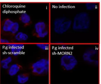
Slides were mounted in anti-fade media after DAPI staining. Cells treated with 60 µM Chloroquine diphosphate for 14 hours were used as positive control. The average number of LC3+ puncta per 50 macrophages was obtained from Zeiss LSM 700 confocal microscope.
2.2.11. Transmission electronic microscopy (TEM)
Cell monolayers grown on plastic tissue culture plates challenged by P.g at MOI of 100 were fixed in 3% paraformaldehyde/0.15M sodium phosphate buffer for one hour at room temperature. Following three rinses with 0.15M sodium phosphate buffer (pH 7.4), the cells were post-fixed with 1% osmium tetroxide/1.25% potassium ferrocyanide/0.15M sodium phosphate buffer for 1 hour at room temperature.
After washes in deionized water, the cells were dehydrated using increasing concentrations of ethanol and embedded in Polybed 812 epoxy resin (Polysciences, Inc. Warrington, PA). The cells were sectioned en face to the substrate at 70nm using a diamond knife and Leica Ultracut UCT ultramicrotome (Leica Microsystems, Inc., Buffalo Grove, IL). Ultrathin sections were collected on 200 mesh copper grids and stained with 4% aqueous uranyl acetate for 15 minutes, followed by Reynolds’ lead citrate for 7 minutes.
Samples were viewed with a LEO EM910 transmission electron microscope (Carl Zeiss Microscopy, LLC, Peabody, MA) with an acceleration voltage of 80 kV. Digital images were taken using a Gatan Orius SC 1000 CCD Camera and DigitalMicrograph 3.11.0 software (Gatan, Inc., Pleasanton, CA).
2.2.12. Real-time PCR
RNA was extracted from HGEs and THP-1 cells infected by P.g at indicated time with the RNeasy Mini kit (Qiagen, Germany) and cDNA was synthesized using the SuperScript VILO cDNA synthesis Kit (Thermo Fisher, USA) according to the manufacturer’s protocol. The expression of human genes encoding IL-1β, IL-6, TNF-α, CXCL1, CXCL2, GCP-2, IL-8, CXCL3, CCL3, RANTES, GM-CSF, G-CSF, MCP-1 and GAPDH was assessed by real-time PCR with human Taqman Gene Expression Assays (Thermo Fisher Scientific). Results were normalized to expression of the gene GAPDH and were quantified by the ∆∆ct method.
2.2.13. ELISA
Cell culture supernatants were collected and assayed for cytokines. Cytokine production was measured by enzyme-linked immunosorbent assay of human IL-1β, IL-6, TNF-α, IL-8 according to the instruction of
16
manufacturer (R&D Systems).2.2.14. Intracelluar calcium flux assay
Differentiated THP-1 cells were plated in 96-well plate and Fluo-8 dye-loading solution was added according to the manufacturer’s protocol (Abcam). The calcium flux assay was performed by monitoring the fluorescence intensity at Ex/Em=490/525 nm after cells were stimulated by P.g at MOI of 100.
2.2.15. Westernblot
THP-1 differentiated macrophages were stimulated with P.g 33277 for the indicated time points at a MOI 100:1. Cells were washed twice and were lysed with RIPA buffer containing phosphatase and proteinase inhibitors. Samples were separated by SDS-PAGE with 4-12% NuPAGE Bis-Tris gels
(Invitrogen), transferred into a 0.2 µm polyvinylidene difluoride (PVDF) membranes, blocked with 5% skim milk and incubated overnight at 4℃ with the respective primary antibodies: rabbit anti-LC3 (#2775, Cell Signaling Technology), rabbit anti-phospho-IKKα/β (#2697, Cell Signaling Technology), rabbit anti- phospho-IκBα (#2859, Cell Signaling Technology), rabbit anti-phospho-NF-κB p65 (#3033, Cell Signaling Technology), rabbit anti-NF-κB p65 (#8242, Cell Signaling Technology), goat anti-beta Actin (Ab8229, Abcam). Secondary antibodies against the corresponding primary antibodies were used. The blots were developed using chemiluminescence (ECL, Thermo Scientific) and visualized using ImageQuant LAS4000 luminescent image reader (GE Healthcare Life Sciences, USA).
2.2.16. Statistical analysis
Results are reported as mean ± SEM. Differences between two groups was analyzed with paired t-test. Statistical analysis of experiments was carried out using one-way ANOVA followed by Dunn’s post hoc test, as appropriate, for the distribution of variance among groups. P-values < 0.05 were considered statistically significant. Statistical analyses were performed using Prism 6.0 software (GraphPad Software).
17
2.3. Results2.3.1. Characterization of the Pg dominant trait, MORN2 SNP identification and in silico analysis
Our analysis of the MORN2 locus, as identified in our previous work (Offenbacher, Divaris et al.
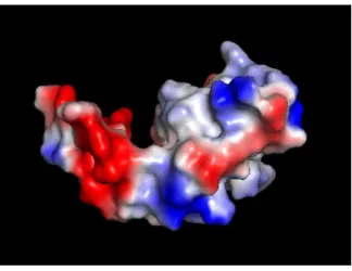
2016) demonstrates that it contains a missense SNP rs3099950 (p=1.65 x 10⁻⁶) that is predicted to be probably damaging with a score of 1.0 by Polypen-2 (Figure 2.1B). Molecular models (PyMol) predict that rs3099950 causes a missense mutation which results in an amino acid change from glutamate, which is a highly negative charged (red area) (Figure 2.1C, D), to lysine, which is a highly positively charged (blue area) (Figure 2.1E, F). This model suggests an alteration in the surface electrostatic potential of the protein, which will cause the change of the charge distribution across a PIP3 binding region of the
MORN2 structure (cleft region) that may result in impaired membrane interactions and/or phagolysosome fusion (Figure 2.1G).
2.3.2. MORN2 SNP is associated with periodontal disease severity
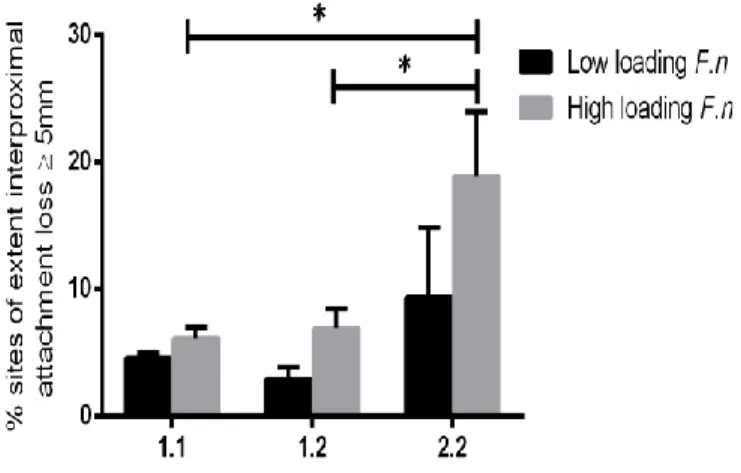
We show that the newly identified SNP rs3099950 has a significant association with the level of total pathogens (ANOVA p=0.007) (Figure 2.2A), the level of Pg (ANOVA p=0.046) (Figure 2.2B), and F.nucleatum (ANOVA p=0.046) (Figure 2.2C) stratified by rs3099950 genotype. For example, the Pg count was 1.4 times as high in 2.2 individuals (homozygous for the minor allele, G/G) as compared to 1.1 individuals (homozygous for the major allele, A/A). This finding further supports the SNP in the MORN2 region potentially affect the biological host response of the individual, resulting in increased numbers of periodontal pathogens present in plaque samples. rs3099950 is also significantly associated with EPD4 and IAL5 in the presence of high Pg or Fn (dichotomized at 75th percentile loading of Pg and Fn) (Figure 2.2D-G). Because the presence of SNP in the MORN2 region is associated with an increased number of total periodontal pathogens, with strong emergence of Pg and Fn. The combination of the 2.2 genotype in the presence of high pathogen load (upper quartile) suggests a strong gene-environment interaction that results in clinical disease. These findings suggest that the presence of the minor allele frequency in MORN2 locus may increase the susceptibility of the individual to periodontal disease by perhaps promoting dysbiosis and when the pathogens emerge, there is an association of more severe disease.
2.3.3. Identification of MORN2 mRNA expression and localization in human gingival tissue
18
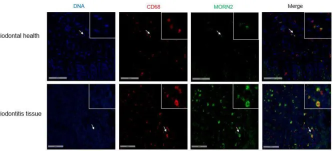
The presence of MORN2 expression has never been evaluated in periodontal tissues. Therefore, we evaluated the MORN2 expression in full-thickness gingival tissue samples from subjects free of periodontitis and from individuals with chronic periodontitis (CP). The demographic information and clinical parameters of the participants are shown in Table 2.2. Our results indicate that, in comparison to periodontitis-free samples, the mRNA level of MORN2 was significantly lower in CP groups (P < 0.01) (Figure 2.3A). To identify the cellular localization of MORN2 protein expression in the cells of the periodontium, immunohistochemistry and immunofluorescence were performed. The major cell types expressing MORN2 were epithelial and infiltrated immune cells. Representative low-resolution (x10) and high-resolution (x40) images of periodontitis-free and periodontitis tissues show a similar cellular pattern of expression among samples (Figure 2.3B). In addition, co-localization of MORN2 and CD68
(macrophage marker) by confocal microscope demonstrated that the infiltrated immune cells expressing MORN2 are mostly identified as macrophages (Figure 2.3C). In periodontitis the macrophage plays a critical role in clearing invasive pathogens and in innate immune response by recruiting other immune cells such as neutrophils and lymphocytes to the lesion. This descriptive localization analysis indicates that MORN2 is expressed predominantly by macrophages within the periodontal inflammatory lesion and the epithelium.
2.3.4. MORN2 promotes the LC3-asscociated phagocytosis in macrophages
Given that MORN2 is expressed in the macrophages of periodontal gingival tissues and MORN2 facilitates phagocytosis-mediated restriction of M. tuberculosis, L. pneumophila, and S. aureus in
macrophages (Abnave, Mottola et al. 2014). We investigated the functional implication of MORN2 in macrophage-mediated uptake and killing of P.g. Our functional analysis revealed that the silencing of MORN2 slightly reduced the phagocytosis of P.g in a log-10 form and markedly resulted in a 1.8 ± 0.6 fold increased intracellular survival of P.g at 24 hours infection(p<0.05) (Figure 2.4A, B). Currently, the most significant ultrastructural difference that distinguishes LAP from canonical autophagy is that bacteria is enclosed within single-membrane phagosomes, instead of double membrane auophagosomes (Lerena and Colombo 2011, Martinez, Almendinger et al. 2011). EM analysis of macrophages infected with P.g.
We observed that single-membrane phagosomes (white arrow in Figure 2.4C). To elucidate the
mechanism of MORN2 in phagocytosis-mediated killing of P.g, we next investigated whether MORN2 has a role in LC3 recruitment. The cytosolic form of LC3 (LC3-I) is conjugated to phosphatidylethanolamine
19
(PE) and in this form (LC3-II) it is recruited to the phagosome or autophagosome membrane. Conjugation was assessed by measuring the levels of LC3-II in the presence of 3-MA. This treatment blocks the fusion of autophagosomes with lysosomes, thereby precluding the effect of autophagy on the accumulation of LC3-II. LC3-II levels showed a decrease after MONR2 knockdown in differentiated THP-1 cells
challenged by P.g (Figure 2.4D). The formation of LC3 puncta viewed by fluorescence microscopy is widely used as a marker for LC3 recruitment. Given that P.g infection of macrophages resulted in decrease in the LC3-II formation, we then assessed the formation of LC3 puncta. The silencing of MORN2 in macrophages decreased the LC3 puncta formation at 1h by immunofluorescence (Figure 2.4E). Collectively, these data demonstrate that MORN2 plays a role in the LC3-associated phagocytosis for the efficient clearance of P.gingivalis.
2.3.5. Role of MORN2 on production of proinflammatory cytokines and chemokines by HGEs and Macrophages
Given that MORN2 expressed both in human gingival epithelial cells and macrophages, we investigated the role of MORN2 on production of inflammatory mediators on these two type of cells. To test this prediction, we first demonstrated the knockdown efficiency (knockdown mRNA expression by 45% in HGEs and >90% in THP-1) and specificity of MORN2 silencing in HGEs and THP-1 cells with one or two corresponding shRNAs. We observed that neutrophil specific chemokines (CXCL1, CXCL2, IL-8) and G-CSF synthesis was downregulated in MORN2 knockdown HGEs at mRNA level (p<0.05) (Figure 2.5A-D). The proinflammatory cytokines (IL-1β, IL-6, TNF-α) and IL-8 production was reduced at both mRNA and protein level (p<0.05) in MORN2 knockdown macrophages (Figure 2.6A-F). In addition, silenced MORN2 in macrophages significantly impaired mRNA levels of neutrophil specific chemokines (CXCL1, CXCL2, GCP-2), monocytic and T cells recruitment chemokines (CXCL3, CCL3, RANTES) and hematopoietic growth factors (GM-CSF, G-CSF) production (p<0.05), except MCP-1 expression was upregulated (p<0.05) (Figure 2.7A-K). Based on these results, MORN2 posit that MORN2 plays an important role in regulating inflammatory response to periodontal pathogens.
2.3.6. Knockdown MORN2 attenuates calcium flux and NF-κB activation
NF-κB is an important transcriptional factor in several types of inflammations, which results in the production of a plethora of pro-inflammatory cytokines and chemokines. Considering that NF-κB can be
20
stimulated through TLR to activate the IKK complex, which leads to the translocation of heterodimers of the NF-κB subunits p65 and p50 to the nucleus (Kawai and Akira 2007). To determine whether MORN2 is involved in TLR-mediated NF-κB activation, we knocked down MORN2 expression by MORN2-specific shRNA in THP-1 cells and stimulated those differentiated cells with P.g 33277. Cell lysates were used to assess the phosphorylation of IKKα/β, IκBα and p65. We found that specific knockdown of MORN2 diminished the phosphorylation of IKKα/β, IκBα and p65, especially at 30 min and 60 min after P.g stimulation, compared THP-1 cells transduced with control scramble shRNA (Figure 2.8A). Calcium signals have been described in various cells of the immune system, including T and B cells, DCs, monocytes and macrophages, in which they contribute to the cell’s activation, differentiation or gene expression (Feske 2007). An influx of Ca2+ into cytosol occurs following the engagement of
immunoreceptors on the cell surface. To further determine the molecular mechanisms by which MORN2 knockdown affects NF-κB signaling, we performed intracellular calcium flux assay. A transient increase in
Ca2+ was evoked at 60s after the start of P.g treatment and totally diminished at 300s (Figure 2.8B).
Lower intracellular calcium flux was observed in MORN2 knockdown group compared to control at 60s and 90s. Therefore, the results indicate that the initial transient intracellular increase in Ca2+ may play a role in NF-κB activation. Taken together, these results suggest that activation of NF-κB depends on the transient increase in Ca2+ in P.g stimulated differentiated macrophages from THP-1 cells and specific knockdown of MORN2 decreases intracellular calcium flux mediated NF-κB activation in response to P.g stimulation.
21
2.4. DiscussionStudying the role of human genes in infection has been termed infectogenomics and has the potential to serve as a powerful tool to identify and understand gene function relevant to host response to pathogens and chronic infection associated diseases. Our group’s previous GWAS has highlighted a potential role for MORN2 in periodontal disease (Offenbacher, Divaris et al. 2016), which was emerged from GWAS gene-centric data relative to Pg dominated dysbiosis and is corroborated by the role of Pg as keystone pathobiont in periodontitis (Hajishengallis, Darveau et al. 2012). In the present study we have identified a potentially functionally important MORN2 SNP. Subjects with the 2.2 MORN2 genotype (homozygous for the minor allele) have more severe disease as reflected by greater extent of probing depth ≥ 4mm and interproximal attachment loss ≥ 5mm, only in the presence of high bacterial loading, as compared to subjects with 1.1 (homozygous for the major allele) or 1.2 (heterozygous). This suggests that compromised function of MORN2 induced by the minor allele of rs3099950 in MORN exon 5 may render patients more susceptible to periodontal disease. Prediction analysis using UniProtKB suggested that the function of MORN2 is altered by the presence of rs3099950 due to an amino acid change from a negatively charged glutamate to a positively charged lysine at position 48. We identified that MORN2 is expressed in epithelial cells and macrophages of gingival tissues, which suggests a potential role of MORN2 in epithelial barrier function and monocytic response to periodontal pathogens. The mRNA expression of MORN2 is suppressed in chronic periodontitis, as compared to health.
The knowledge base regarding MORN2 is scarce and its function in any disease, including periodontal disease, is unclear. To date, only one study that explored whole-transcriptome analysis of the planarian response to bacterial challenge through a functional RNAi-based screening approach has identify identified MORN2 as a critical gene for bacterial resistance in planarians with human orthologs.
Using human macrophages they demonstrated that MORN2 promotes the recruitment of LC3 to microbe- containing phagosomes and phagosome-lysosome fusion (Abnave, Mottola et al. 2014). This suggests that MORN2 function may be important for bacterial clearance and phagocytic cell function. The function of MORN2 in LC3-associated phagocytosis (LAP) of the periodontal keystone pathogen P.g was studied by creating stable MORN2 knockdown human monocytic cell line. LC3 can be recruited into both
phagosomes and autophagosomes, which are both involved in microbial clearance. LAP distinct from the conventional autophagy is that LC3 is recruited into the double-membrane autophagosome and LC3 is
22
recruited directly to the single-membrane phagosome (Sanjuan, Dillon et al. 2007, Martinez, Almendinger et al. 2011). Our study pinpoints a role for MORN2 in LAP of P.g (strain 33277) in THP-1 differentiated macrophage. Analysis by EM indicated most of the Pg are located in single-membrane phagosomes. We found that in vitro MORN2 in human macrophages promotes the phagocytosis and killing of Pg. Similarly, we provide evidence that during phagocytosis MORN2 expression promotes the cytosolic form of LC3-I complexation to membrane associated phosphatidylethanolamine (PE) to form LC3-II and increases the number of LC3-positive puncta formation independently of autophagy. This finding is consistent with a role for MORN2 in LC3 recruitment to M. tuberculosis containing phagosomes as previously report (Abnave, Mottola et al. 2014). Collectively, our findings support a function for MORN2 in the LAP of Pg in macrophages. It is not known whether this loss of MORN2 function induces Pg dysbiosis, as further research is indicated.
Invading pathogens are recognized by mammalian cells through receptors either in the cytoplasm (NLRs) or at the cell surface (TLRs) (Xu, Jagannath et al. 2007, Anand, Malireddi et al. 2012). It has been shown that P.g can interact with host-expressed TLR2 (Burns, Eliyahu et al. 2010, Papadopoulos,
Weinberg et al. 2013, Maekawa, Krauss et al. 2014) and LPS of P.g has also been reported to engage TLR4 (Ogawa, Asai et al. 2002, Darveau, Pham et al. 2004). The TLR2 and TLR4 binding of P.g is strain specific due to the presence of an LPS structure that contains multiple lipid A (Darveau, Pham et al. 2004, Herath, Darveau et al. 2013). Proteomics analysis of purified phagosome membranes in cells that have engulfed different cargoes by phagocytosis revealed that the absence of TLR signaling results in failure of LC3 recruitment to phagosome membranes (Sanjuan, Dillon et al. 2007, Shui, Sheu et al. 2008). The triggering of LAP by TLRs is associated with enhanced phagosomal fusion with lysosomal compartment, results in a more efficient degradation of pathogens to reduced intracellular survival. Also, LAP might serve as another host defense strategy to protect from microorganisms by facilitating peptide presentation on MHC class II molecules to initiate adaptive immunity or delivering phagosomes cargo to other
intracellular pathogen recognition receptors.
The activation of Toll-like receptors that occurs upon P.gingivalis exposure results in activation of the NF-κB intracellular signaling pathway (Lerena and Colombo 2011), which will triggers increased expression of many pro-inflammatory cytokines [IL-1β, IL-6, TNF-α] (Geivelis, Turner et al. 1993,
Yavuzyilmaz, Yamalik et al. 1995, Salvi, Brown et al. 1998) and chemokines [IL-8, MCP-1, RANTES, etc]
23
(Gurkan, Eren et al. 2016, Diomede, Zingariello et al. 2017). Considering the broad implication for
MORN2 in antibacterial immunity and completely unknown the role of MORN2 in modulating inflammatory mediators, it will be of interest to assess whether MORN2 is involved in TLR-induced NF-κB signaling pathway to prevent pathological consequences. To define the biological function of MORN2 in vitro, we generated MORN2 knockdown HGEs and THP-1 cells with shRNA targeting MORN2 expression. Our study using MORN2-knockdown cells provide evidence that MORN2 is a critical regulator of TLR-induced NF-κB signaling pathway. MORN2 knockdown HGEs stimulated by P.g produce lower levels of
chemokines that target neutrophil recruitment and activation. MORN2 knockdown causes macrophages to produce less proinflammatory cytokines and chemokines in response to TLR stimulation, with the exception of MCP-1, accompanied by diminished phosphorylation of IKKα/β, IκBα and p65.
Intracelluar calcium flux is an important second messenger that plays a role in diverse array of cellular processes, including activation of NF-κB (Pahl and Baeuerle 1996, Gewirtz, Rao et al. 2000, Han and Logsdon 2000). Phosphatidylinositol 4,5-bisphosphate (PIP2) is a minor phospholipid component of plasma membrane and serves as a precursor of important second messengers. PIP2 can be
phosphorylated by phosphatidylinositol-3 kinase (PI3K) to generate phosphatidylinositol 3,4,5-
trisphosphate (PIP3) (Carnero and Paramio 2014),