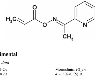
(
E
)-1-(Pyridin-2-yl)ethanone
O
-acryloyl-oxime
Mariusz Mojzych,a* Zbigniew Karczmarzykaand Andrzej Fruzin´skib
aDepartment of Chemistry, University of Podlasie, ul. 3 Maja 54, 08-110 Siedlce, Poland, andbInstitute of General and Ecological Chemistry, Technical University, ul. Z.wirki 36, 90-924 Ło´dz´, Poland
Correspondence e-mail: [email protected]
Received 13 February 2008; accepted 26 February 2008
Key indicators: single-crystal X-ray study;T= 100 K; mean(C–C) = 0.002 A˚; Rfactor = 0.033;wRfactor = 0.098; data-to-parameter ratio = 13.2.
The title compound, C10H10N2O2, was synthesized by the
reaction of the oxime of 2-acetylpyridine and 3-bromo-propanoyl chloride in the presence of triethylamine. The molecule adopts a nearly planar chain-extended conformation with the oxime group in atransand the acryloyl group in ans
-cis conformation. This conformation is stabilized by an intramolecular C—H N hydrogen bond. The screw-related molecules are linked intoC(9) chains by C—H O hydrogen bonds.
Related literature
For general background, see: Robertson, (1995). For the biological activity of oximes, see: Van Heldenet al.(1996). For related structures, see: Mojzychet al.(2007). For the graph-set notation, see: Bernsteinet al.(1995).
Experimental
Crystal data
C10H10N2O2 Mr= 190.20
Monoclinic,P21=n a= 7.0240 (5) A˚
b= 18.4054 (14) A˚
c= 7.8642 (6) A˚
= 114.043 (1)
V= 928.47 (12) A˚3 Z= 4
MoKradiation
= 0.10 mm1 T= 100 (2) K 0.750.130.07 mm
Data collection
Bruker SMART APEXII CCD diffractometer
Absorption correction: multi-scan (SADABS; Sheldrick, 2002)
Tmin= 0.924,Tmax= 0.993
15216 measured reflections 2209 independent reflections 1951 reflections withI> 2(I)
Rint= 0.035
Refinement
R[F2> 2(F2)] = 0.033 wR(F2) = 0.098 S= 1.02 2209 reflections
167 parameters
All H-atom parameters refined
max= 0.40 e A˚
3
min=0.18 e A˚
[image:1.610.71.258.529.682.2]3
Table 1
Hydrogen-bond geometry (A˚ ,).
D—H A D—H H A D A D—H A
C10—H10B N1 0.94 (2) 2.438 (14) 2.8596 (14) 107 (1) C1—H1 O2i
0.96 (2) 2.588 (15) 3.4581 (14) 150 (1)
Symmetry code: (i)xþ1 2;yþ
1 2;zþ
1 2.
Data collection:SMART(Bruker, 2003); cell refinement:SAINT
(Bruker, 2003); data reduction:SAINT; program(s) used to solve structure:SIR92(Altomare et al., 1993); program(s) used to refine structure: SHELXL97 (Sheldrick, 2008); molecular graphics:
ORTEP-3 for Windows(Farrugia, 1997); software used to prepare material for publication:SHELXL97 (Sheldrick, 2008), PLATON
(Spek, 2003) andWinGX(Farrugia, 1999).
Supplementary data and figures for this paper are available from the IUCr electronic archives (Reference: CI2565).
References
Altomare, A., Cascarano, G., Giacovazzo, C. & Guagliardi, A. (1993).J. Appl. Cryst.26, 343–350.
Bernstein, J., Davis, R. E., Shimoni, L. & Chang, N. L. (1995).Angew. Chem. Int. Ed. Engl.34, 1555–1573.
Bruker (2003).SMARTandSAINT. Bruker AXS Inc., Madison, Wisconsin, USA.
Farrugia, L. J. (1997).J. Appl. Cryst.30, 565. Farrugia, L. J. (1999).J. Appl. Cryst.32, 837–838.
Mojzych, M., Karczmarzyk, Z., Fruzin´ski, A. & Rykowski, A. (2007).Anal. Sci. (X-Ray Str. Anal. Online),23, x205–x206.
Robertson, G. M. (1995). Comprehensive Organic Functional Group Transformation, Vol. 3, edited by A. R. Katritzky, O. Meth-Cohn & C. W. Rees, pp. 425–441. Oxford: Elsevier.
Sheldrick, G. M. (2002).SADABS. University of Go¨ttingen, Germany. Sheldrick, G. M. (2008).Acta Cryst.A64, 112–122.
Spek, A. L. (2003).J. Appl. Cryst.36, 7–13.
Van Helden, F. P. M., Busker, R. W., Melchers, B. P. C. & Bruijnzeel, P. L. B. (1996).Arch. Toxicol.70, 779–786.
Acta Crystallographica Section E Structure Reports
Online
supporting information
Acta Cryst. (2008). E64, o653 [doi:10.1107/S1600536808005436]
(
E
)-1-(Pyridin-2-yl)ethanone
O
-acryloyloxime
Mariusz Mojzych, Zbigniew Karczmarzyk and Andrzej Fruzi
ń
ski
S1. Comment
Oximes and their derivatives such as O-ethers and esters are important intermediates in organic chemistry and are well known in both analytical and coordination chemistry (Robertson, 1995). These compounds are also of interest as biologically active compounds (Van Helden et al., 1996). In this in mind we have decided to synthesize and structurally characterize a set of O-acryloyl oximes of 6-membered aza-heterocycles as 1,2,4-triazine, pyridine and pyrazine. These compounds were obtained by reaction of appropriate oximes and 3-bromopropanoyl chloride under Staudinger reaction conditions. As a part of our ongoing studies, we report herein the crystal and molecular structure of the title compound.
The geometric parameters (bond lengths, angles and torsion angles) in the title compound are very similar to those observed in a previously reported structure of (E)-1-(3-methylsulfanyl-1,2,4-triazin-5-yl)-ethanone O-acryloyl oxime (Mojzych et al., 2007). The oxime group is in trans and the acryloyl group in s-cis conformation with the torsion angles O1—N2—C6—C5 and O2—C7—C8—C9 of 179.39 (7) and 2.73 (16)°, respectively. The molecule as a whole adopts a nearly planar chain-extended conformation (Fig. 1). This conformation is stabilized by an intramolecular C10—
H10B···N1 hydrogen bond leading to the formation of a five-membered ring described by the S(5) graph-set symbol (Bernstein et al., 1995).
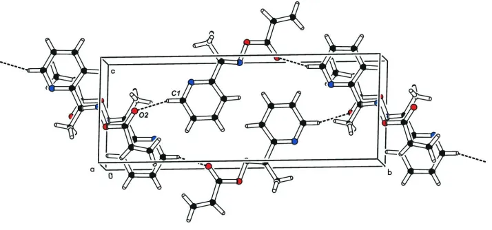
In the crystal structure, the screw-related molecules are linked to form C(9) chains along the [010] direction by C1— H1···O2 intermolecular hydrogen bonds (Fig. 2).
S2. Experimental
To a solution of 2-acetylpyridine (204 mg, 1.5 mmol) and triethylamine (454 mg, 4.5 mmol) in dry CH2Cl2 (5 ml) at 233 K was added 3-bromopropionyl chloride (1.5 mmol) in CH2Cl2 (2 ml) dropwise. The reaction mixture was allowed to warm to room temperature and was stirred for 12 h. It was then washed with water (2 × 10 ml), saturated aqueous sodium bicarbonate (3 × 10 ml), brine (1 × 10 ml) and dried over MgSO4. Removal of the solvent yielded the crude product which was then purified by column chromatography on silica gel using CH2Cl2-hexane mixture (2:1) as eluent to afford the title compound as a colourless solid. Yield: 216 mg (76%) and m.p. 338 K. Single crystals suitable for X-ray diffraction analysis were grown by slow evaporation of an ethanol solution. 1H NMR (CDCl
3) δ: 2.55 (s, 3H), 5.99–6.02 (d, 1H, J = 10.5 Hz), 6.29–6.38 (dd, 1H, J = 10.5 Hz), 6.59–6.65 (d, 1H, J = 17.4 Hz), 7.34–7.38 (t, 1H, J = 6.6 Hz), 7.72– 7.78 (t, 1H, J = 8.1 Hz), 8.12 - 8.15 (d, 1H, J = 8.1 Hz), 8.65–8.67 (d, 1H, J = 6.6 Hz). 13C NMR (CDCl
3) δ: 13.11, 122.28, 125.27, 126.72, 132.59, 136.78, 149.34, 152.89, 163.71, 164.28. HR—MS (m/z) for C10H11N2O2: 191.0822 [M++H]; calcd. 191.0821.
S3. Refinement
Figure 1
The molecular structure of the title compound, showing the atom-numbering scheme. Displacement ellipsoids are drawn at the 50% probability level.
Figure 2
A view of the molecular packing in the title compound. Dashed lines indicate intermolecular hydrogen bonds.
(E)-1-(Pyridin-2-yl)ethanone O-acryloyloxime
Crystal data
C10H10N2O2
Mr = 190.20
Monoclinic, P21/n Hall symbol: -P 2yn
a = 7.0240 (5) Å
b = 18.4054 (14) Å
c = 7.8642 (6) Å
β = 114.043 (1)°
V = 928.47 (12) Å3
Z = 4
F(000) = 400
Dx = 1.361 Mg m−3 Melting point: 338 K
Mo Kα radiation, λ = 0.71073 Å Cell parameters from 166 reflections
θ = 3.8–28.0°
µ = 0.10 mm−1
[image:3.610.129.486.299.466.2]Data collection
Bruker SMART APEXII CCD diffractometer
Radiation source: fine-focus sealed tube
ω scans
Absorption correction: multi-scan (SADABS; Sheldrick, 2002)
Tmin = 0.924, Tmax = 0.993 15216 measured reflections
2209 independent reflections 1951 reflections with I > 2σ(I)
Rint = 0.035
θmax = 28.4°, θmin = 2.2°
h = −9→9
k = −24→24
l = −10→10
Refinement
Refinement on F2 Least-squares matrix: full
R[F2 > 2σ(F2)] = 0.033
wR(F2) = 0.098
S = 1.03 2209 reflections 167 parameters 0 restraints
Primary atom site location: structure-invariant direct methods
Secondary atom site location: difference Fourier map
Hydrogen site location: difference Fourier map All H-atom parameters refined
w = 1/[σ2(F
o2) + (0.0531P)2 + 0.3208P] where P = (Fo2 + 2Fc2)/3
(Δ/σ)max = 0.001 Δρmax = 0.40 e Å−3 Δρmin = −0.19 e Å−3
Special details
Geometry. All e.s.d.'s (except the e.s.d. in the dihedral angle between two l.s. planes) are estimated using the full covariance matrix. The cell e.s.d.'s are taken into account individually in the estimation of e.s.d.'s in distances, angles and torsion angles; correlations between e.s.d.'s in cell parameters are only used when they are defined by crystal symmetry. An approximate (isotropic) treatment of cell e.s.d.'s is used for estimating e.s.d.'s involving l.s. planes.
Refinement. Refinement of F2 against ALL reflections. The weighted R-factor wR and goodness of fit S are based on F2, conventional R-factors R are based on F, with F set to zero for negative F2. The threshold expression of F2 > σ(F2) is used only for calculating R-factors(gt) etc. and is not relevant to the choice of reflections for refinement. R-factors based on F2 are statistically about twice as large as those based on F, and R- factors based on ALL data will be even larger.
Fractional atomic coordinates and isotropic or equivalent isotropic displacement parameters (Å2)
x y z Uiso*/Ueq
H4 0.185 (2) 0.0111 (7) 0.1226 (18) 0.021 (3)* H8 0.363 (2) −0.0726 (8) 0.931 (2) 0.030 (4)* H9A 0.293 (2) −0.1938 (7) 1.002 (2) 0.025 (3)* H9B 0.180 (2) −0.2100 (7) 0.776 (2) 0.025 (3)* H10A 0.389 (2) 0.1079 (8) 0.726 (2) 0.038 (4)* H10B 0.429 (2) 0.1661 (9) 0.605 (2) 0.039 (4)* H10C 0.581 (3) 0.1023 (8) 0.673 (2) 0.039 (4)*
Atomic displacement parameters (Å2)
U11 U22 U33 U12 U13 U23
O1 0.0177 (4) 0.0178 (4) 0.0120 (3) −0.0008 (3) 0.0056 (3) 0.0015 (3) O2 0.0281 (4) 0.0172 (4) 0.0168 (4) −0.0010 (3) 0.0085 (3) −0.0012 (3) N1 0.0150 (4) 0.0154 (4) 0.0200 (4) 0.0006 (3) 0.0072 (3) 0.0004 (3) N2 0.0158 (4) 0.0173 (4) 0.0119 (4) 0.0022 (3) 0.0059 (3) 0.0022 (3) C1 0.0168 (5) 0.0153 (5) 0.0244 (5) 0.0010 (4) 0.0081 (4) 0.0033 (4) C2 0.0154 (5) 0.0227 (5) 0.0181 (5) 0.0023 (4) 0.0064 (4) 0.0067 (4) C3 0.0152 (5) 0.0220 (5) 0.0154 (5) −0.0014 (4) 0.0060 (4) −0.0011 (4) C4 0.0147 (4) 0.0154 (5) 0.0176 (5) 0.0000 (3) 0.0072 (4) 0.0004 (4) C5 0.0104 (4) 0.0162 (5) 0.0155 (5) 0.0013 (3) 0.0058 (3) 0.0005 (4) C6 0.0115 (4) 0.0160 (5) 0.0149 (5) 0.0024 (3) 0.0067 (3) −0.0007 (4) C7 0.0131 (4) 0.0163 (5) 0.0169 (5) 0.0031 (3) 0.0080 (4) 0.0013 (4) C8 0.0167 (5) 0.0237 (5) 0.0156 (5) 0.0008 (4) 0.0073 (4) 0.0013 (4) C9 0.0212 (5) 0.0241 (6) 0.0208 (5) 0.0039 (4) 0.0110 (4) 0.0051 (4) C10 0.0205 (5) 0.0181 (5) 0.0154 (5) −0.0027 (4) 0.0077 (4) −0.0025 (4)
Geometric parameters (Å, º)
O1—C7 1.3698 (12) C4—C5 1.3971 (14) O1—N2 1.4352 (10) C4—H4 0.976 (13) O2—C7 1.2005 (13) C5—C6 1.4954 (13) N1—C1 1.3426 (14) C6—C10 1.4957 (13) N1—C5 1.3442 (13) C7—C8 1.4843 (14) N2—C6 1.2849 (13) C8—C9 1.3224 (15) C1—C2 1.3879 (15) C8—H8 0.989 (15) C1—H1 0.962 (14) C9—H9A 0.972 (15) C2—C3 1.3884 (15) C9—H9B 0.954 (14) C2—H2 0.929 (15) C10—H10A 0.947 (17) C3—C4 1.3868 (14) C10—H10B 0.937 (16) C3—H3 0.954 (15) C10—H10C 0.969 (16)
C3—C2—H2 120.2 (9) C9—C8—H8 124.2 (9) C1—C2—H2 121.0 (9) C7—C8—H8 115.5 (9) C4—C3—C2 118.38 (9) C8—C9—H9A 122.2 (8) C4—C3—H3 121.3 (8) C8—C9—H9B 120.1 (8) C2—C3—H3 120.3 (8) H9A—C9—H9B 117.7 (11) C3—C4—C5 118.96 (9) C6—C10—H10A 111.0 (10) C3—C4—H4 121.0 (8) C6—C10—H10B 110.3 (10) C5—C4—H4 120.0 (8) H10A—C10—H10B 109.1 (13) N1—C5—C4 123.07 (9) C6—C10—H10C 109.4 (9) N1—C5—C6 116.01 (8) H10A—C10—H10C 109.9 (13) C4—C5—C6 120.90 (9) H10B—C10—H10C 107.0 (13)
C7—O1—N2—C6 −176.92 (7) O1—N2—C6—C10 −0.77 (13) C5—N1—C1—C2 0.84 (14) N1—C5—C6—N2 160.49 (8) N1—C1—C2—C3 −2.47 (15) C4—C5—C6—N2 −18.13 (12) C1—C2—C3—C4 1.12 (14) N1—C5—C6—C10 −19.37 (12) C2—C3—C4—C5 1.61 (14) C4—C5—C6—C10 162.02 (9) C1—N1—C5—C4 2.13 (14) N2—O1—C7—O2 0.98 (13) C1—N1—C5—C6 −176.46 (8) N2—O1—C7—C8 −179.69 (7) C3—C4—C5—N1 −3.38 (14) O2—C7—C8—C9 2.73 (16) C3—C4—C5—C6 175.14 (8) O1—C7—C8—C9 −176.58 (9) O1—N2—C6—C5 179.39 (7)
Hydrogen-bond geometry (Å, º)
D—H···A D—H H···A D···A D—H···A
C10—H10B···N1 0.94 (2) 2.438 (14) 2.8596 (14) 107 (1) C1—H1···O2i 0.96 (2) 2.588 (15) 3.4581 (14) 150 (1)