Original Article
Ki-67/MIB-1 immunostaining in a cohort of human
gliomas
Anne J Skjulsvik1,2, Jørgen N Mørk1, Morten O Torp1, Sverre H Torp1,2
1Department of Laboratory Medicine, Children’s and Women’s Health, Faculty of Medicine, Norwegian University of Science and Technology (NTNU), Trondheim, Norway; 2Department of Pathology and Medical Genetics, St. Olavs Hospital, Trondheim University Hospital, Trondheim, Norway
Received October 13, 2014; Accepted December 1, 2014; Epub December 1, 2014; Published December 15, 2014
Abstract: Histopathological malignancy grading of human gliomas is limited by subjective interpretation of the
mor-phological criteria. Assessment of mitotic activity is a cornerstone of grading these tumours, but mitotic figures can
be hard to identify in haematoxylin-eosin stained sections. Thus, determining proliferative activity by means of Ki-67/MIB-1 immunostaining has become a useful supplement. However, this method has drawbacks, so continuous testing and evaluation are required for optimization and standardization. The aim of this study was to analyse and evaluate the Ki-67/MIB-1 proliferative indices (PIs) in a series of gliomas. We found that Ki-67/MIB-1 PIs correlated well with histological malignancy grade in all glioma subtypes, but a considerable overlap of PIs was observed
between the malignancy groups. Consequently, Ki-67/MIB-1 immunostaining alone is not sufficient to adequately
determine the malignancy grade. Therefore, future work is necessary to clarify the role of this immunostaining in the histopathological diagnosis of human gliomas.
Keywords: Astrocytoma, brain tumour, diagnosis, glioblastoma, immunohistochemistry, proliferation
Introduction
Histopathological classification and malignan -cy grading of human gliomas are based on cri-teria issued by the World Health Organization (WHO) [1]. However, these criteria are encum-bered with subjective interpretations, giving rise to inter- and intra-observer variability [2, 3]. Because proliferation is a basic process in glio-magenesis, mitotic counting constitutes a cor-nerstone in the grading of these tumors. Since
identification and counting of mitotic figures in
haematoxylin-eosin stained sections can be
difficult, glioma grading is imprecise and may
unfavorably impact prognosis, treatment, and follow-up.
Immunohistochemical determination of prolif-erative activity is a useful supplement for establishing the histopathological diagnosis of glioma. Ki-67/MIB-1 immunostaining is most commonly used and has been shown to corre-late positively with tumor grade and prognosis [4-6]. Despite its widespread use, the
proce-dure still has many uncertain and limiting fac-tors, including problematic overlap of indices between different glioma grades and inherent problems in the immunohistochemical analysis [5-9]. Thus, publishing data on Ki-67/MIB-1 immunostaining in human gliomas is still worth-while in order to optimize this method, with the superior goal of achieving a standardized proce-dure. The aim of this study was to evaluate the Ki-67/MIB-1 proliferative indices (PIs) in a
series of gliomas and critically evaluate the find -ings and procedure.
Materials and methods
Patients
This study includes a series of gliomas in adults (over 16 years of age) who underwent opera-tions at St. Olavs University Hospital in Trondheim, Norway, during the time period 1998-2013. Both the histopathological
diagno-sis (according to the WHO classification system)
performed in collaboration by AJS and SHT. All patients were found by searching the electronic
patient data files of the pathology department.
Patients were included at primary diagnosis and all cases were diagnosed based solely on
WHO classification system, and in addition the
[image:2.612.92.520.85.299.2]and water bath. The Ki-67/MIB-1 antibody was supplied by Immunotech (Hamburg, Germany) and by DAKO (Glostrup, Denmark). The working dilution was 1:100 or 1:600 depending on the detection system used. The sections were incu-bated for 40 min at room temperature.
Table 1. Ki-67/MIB-1 proliferative indices for glioma subtypes
Diagnosis (abbreviations) grade nWHO years (range)Median age, MIB-1 PIMedian Ki-67/ a (range) Statistical analysisb
Pilocytic astrocytoma (PILOCYT) I 12 38 (17-65) 1.9 (0.7-8.1) vs. diffuse astrocytoma: P = 0.004 Diffuse astrocytoma (A) II 57 44 (18-78) 5.2 (0.5-16.6) vs. oligodendroglioma: P = 0.218
vs. oligoastrocytoma: P = 0.287 vs. anaplastic astrocytoma and glioblastoma: P < 0.001
Oligo-astrocytoma (OA) II 13 42 (26-73) 4.0 (1.0-13.3) vs. anaplastic oligoastrocytoma: P = 0.006 Oligodendroglioma (O) II 27 44 (21-72) 4.4 (1.0-22.0) vs. oligoastrocytoma: P = 0.798
vs. anaplastic oligodendroglioma: P < 0.001 Anaplastic astrocytoma (AA) III 28 52 (19-82) 13.1 (2.1-39.1) vs. anaplastic oligoastrocytoma: P = 0.643
vs. anaplastic oligodendroglioma: P = 0.122 vs. glioblastoma: P = 0.002
Anaplastic oligoastrocytoma (AOA) III 7 53 (33-71) 12.7 (6.3-39.0) vs. anaplastic oligodendroglioma: P = 0.006 Anaplastic oligodendroglioma (AO) III 12 49 (31-78) 21.7 (2.4-41.2)
Glioblastoma (GBM) IV 89 65 (30-89) 19.4 (2.2-80.0) vs. anaplastic oligodendroglioma: P = 0.867 vs. anaplastic oligoastrocytoma: P = 0.035 Sub-ependymoma (SUBEP) I 9 38 (22-62) 1.0 (0.1-8,5) vs. ependymoma: P = 0.126
Ependymoma (EP) II 13 55 (27-74) 2.0 (0.6-25.3) Anaplastic ependymoma III 0
aPI = proliferation index; bSignificance was determined using the Mann-Whitney U test.
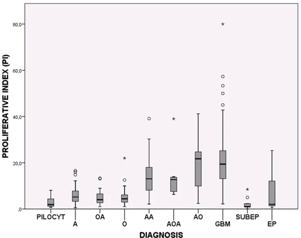
Figure 1. Box plots showing the distribution of Ki-67/MIB-1 PIs among the glioma subtypes. PILOCYT: Pilocytic astrocytoma, A: Diffuse astrocytoma, OA: Oligoas-trocytoma, O: Oligodendroglioma, AA: Anaplastic asOligoas-trocytoma, AOA: Anaplastic oligoastrocytoma, AO: Anaplastic oligodendroglioma, GBM: Glioblastoma, SUBEP: Subependymoma and EP: Ependymoma.
Ki-67/MIB-1 PIs were con-tinuously registered in a spreadsheet. Diagnosis were made independent of Ki-67/MIB-1 PIs, but in cases were the PI was unusually high, a comment was made in the diagno-sis. However, this did not change the WHO grade.
Immunohistochemistry
All tumor samples were
fixed in buffered formalin,
usually for not more than 24 hours, and then
embed-ded in paraffin. Paraffin sections (3-μm-thick) were
cut and mounted on Superfrost glass slides,
[image:2.612.91.396.329.570.2]Automatized immunohistostainers and detec-tion systems were purveyed by DAKO (TechMate 500, Autostainer Plus, Autostainer Link 48). The staining procedures were performed according to the manufacturer’s recommenda-tions. Positive controls were used in each stain-ing run (“sausage block” with tonsil, appendix, pancreas, and liver). First, a standard streptav-idin-biotin-peroxidase technique was used, and later the DAKO EnVision Flex+ System. Diaminobenzidine was used as the chromo-gene and haematoxylin as the counterstain.
Proliferation index evaluation
The immunostained sections were scanned using a 40× objective with an eye grid for the areas with the highest density of labeled tumor cells (hot spots). At least 1000 tumor cells, or
alternatively three high power fields (HPF) were
examined. Only immunoreactive tumor cell nuclei were counted. Necrotic areas and vascu-lar endothelium were excluded. The Ki-67/
MIB-1 PI was defined as the percentage of
immunoreactive tumor cell nuclei among the total number of cells.
Statistical analyses
Statistical analyses were performed using IBM SPSS Statistics version 21. The Mann-Whitney U test was applied to estimate differences in the PIs between groups of tumors. P < 0.05
was considered significant.
Results
A total of 267 glioma subtypes were examined: 186 astrocytomas, 39 oligodendrogliomas, 20
mixed gliomas, and 22 ependymal tumors.
Table 1 shows all data and statistical
correla-tions for the tumor subtypes. The Ki-67/MIB-1 PIs are graphically illustrated in Figure 1. In general, the quality of the Ki-67/MIB-1 immu-nostaining was good (Figure 2). Some variation in staining intensity was observed, however, only distinctly labeled tumor cell nuclei were counted. Normal brain tissue did not show any immunoreactivity. Various distribution patterns were observed for the labeled tumor cells, both homogenous dispersion throughout the tumor tissue and hot spots, with the latter being more frequent in high grade tumors.
The Ki-67/MIB-1 PI correlated significantly with
tumor grade for each glioma type. However, considerable overlap was observed between the malignancy groups (Figure 2). No significant
difference was found between glioma types of the same tumor grade. Anaplastic oligodendro-gliomas and anaplastic oligoastrocytomas had indices comparable to glioblastomas.
Discussion
In our material we found that the Ki-67/MIB-1
PIs correlated significantly with increasing
[image:3.612.93.524.73.233.2]tumor grade in all types of gliomas but an over-lap occurred between the malignancy groups. The positive correlations between Ki-67/MIB-1 PI and tumor grade in our series of gliomas are in agreement with the literature [10-14]. We found that indices were comparable between gliomas of similar malignancy grade, and indi-ces for high-grade gliomas (grade III/IV) were Figure 2. Ki-67/MIB-1 immunostaining showing low PI (~4%) in a grade II astrocytoma (left) and high PI (~30%) in a
significantly higher than in low-grade (grade I/II)
tumors. Thus, Ki-67/MIB-1 is useful for differ-entiating between high and low-grade gliomas, but differentiating between grade I and grade II or grade III and grade IV is more problematic due to the overlap of values between the differ-ent tumor grades. This overlap is a main limita-tion of this immunostaining. For this reason, Ki-67/MIB-1 should not be used alone as a marker of tumor grade but in conjunction with histological features [15, 16].
Histological grading and estimation of Ki-67/ MIB-1 PI are subjected to heterogeneity-induced sampling errors, limiting their diagnos-tic accuracy, especially in small specimens such as stereotactical biopsies [17]. Tumor his-tology can appear discordant with the observed Ki-67/MIB-1 PI. In cases with histologically
ana-plastic glioma tissue in which mitotic figures can be difficult to find, a high index may support
the high grade diagnosis. On the other hand, a low index in a cellular lesion may indicate a reactive condition (e.g., gliosis, microglial response) rather than a neoplasm [5, 16]. If the index is elevated for a glioma with an otherwise benign histology, a more aggressive tumor may be indicated. Such a setting should not lead to a change in tumor grade but a remark in the biopsy report saying “with elevated Ki-67/ MIB-1 PI, see comment” [5, 16]. In these cases one should consider step sections as well as to correlate to radiological images and clinical his-tory [5, 16].
Ki-67/MIB-1 immunostaining to distinguish gli-osis and low-grade gliomas should be interpret-ed with caution [5]. Normally, reactive astro-cytes do not exhibit proliferative activity, but in some non-neoplastic conditions reactive astro-cytes may have a proliferation rate of 1-5% [18]. In such cases, immunohistochemical analyses for mutated p53 and isocitrate dehy-drogenase (IDH) proteins can be useful, though p53 immunoreactivity may occur in both set-tings, and there are gliomas without IDH muta-tion [19-21].
The procedure for Ki-67/MIB-1 immunostaining is not standardized and has various analytical and clinical elements of uncertainty [7]. Nevertheless, the method is regarded as being robust [8, 9], which is also in accordance with our experience during several years with both clinical and experimental use [14, 22, 23]. The
recommended fixative is buffered formalin, and storage time, delay in fixation and fixation time
does not seem to substantially affect the stain-ing results [8, 24, 25]. Loss of immunoreactivity has been described if cut sections are exposed to room air for some months [8]. A prerequisite for satisfactory immunostaining is adequate antigen retrieval [24-26]. Various antibodies against the Ki-67 antigen are commercially available, but MIB-1 is the predominant anti-body [22, 27]. Counting procedures vary across studies. Usually counting is performed in areas with the highest immunoreactivity (“hot spots”), and approximately 1000 cells are counted
using the 40× objective. The PI is calculated as
the percentage of labeled tumor cell nuclei to the total number of tumor cells [5, 9]. As the expression of the Ki-67 antigen changes during the cell cycle [28], the intensity of nuclear stain-ing will vary; principally, all types of stainstain-ing should be regarded as positive [8, 9]. Counting can be done manually or by digitalized image analysis systems, but manual counting has turned out to be applicable for most diagnostic
purposes [5]. Defining a cut-off value is also a
topic of interest due to its impact on the
deter-mination of patients classified as “high Ki-67”,
which is indicative of a poorer outcome. Generally, these patients will receive more
aggressive treatment. However, the definition
of threshold value is not straightforward mostly due to inter-/intra-observer variability and counting procedures. Accordingly, extrapolating values from other laboratories can be decep-tive; thus, Ki-67/MIB-1 immunostaining should be interpreted in the context of one’s own prac-tice [5]. Each pathology department should regularly adjust its Ki-67/MIB-1 PIs by tumor grade and survival and develop its own in-house policy. Such a work-up will constitute an important part of a department’s quality assur-ance and accreditation programs [29]. For astrocytomas, a cut-off of approximately 10% has appeared clinically feasible [6, 16]. However, the predictive value of Ki-67/MIB-1 is ambiguous [7, 30].
This study also has limitations inherent to the Ki-67/MIB-1 immunohistochemistry, including
definition of immunoreactive tumor cell nuclei,
sampling error and counting procedures. In addition, no statistical analysis of intra- or inter-observer variability was done. The statistics
may also be influenced by the fact that not all
Overall, Ki-67/MIB-1 immunostaining is a use-ful supplement to the histopathological diagno-sis of human gliomas. However, the procedure cannot be used alone, but should be used in combination with established histopathological features of malignancy. The analytical and clini-cal performance of Ki-67/MIB-1
immunostain-ing in glioma diagnosis is not sufficiently deter -mined. This limits its clinical utility and under-lines the need for further research and stan-dardization of procedures between laboratories [7]. To improve the diagnostics for human glio-mas, a battery of proliferation markers might be considered [23]. Progress has been made in the recent years towards introducing molecular genetics in glioma diagnosis [31]. This has the potential to move us towards a more personal-ized medicine in the care of glioma patients.
Acknowledgements
We are grateful to Prof. Johannes Attems, Dr. David Scheie, Prof. Ivar Skjåk Nordrum, Prof. Christina Vogt and Dr. Erlend Hassel for their critical reading of the manuscript. Anne Jarstein Skjulsvik, MD, was funded by St. Olavs Hospital, Trondheim University Hospital, Trondheim, Norway. Jørgen Negård Mørk, MD, and Morten Overrein Torp, MD, were funded by the Norwegian State Educational Loan Fund, Norway. Sverre Helge Torp, Prof, PhD, was fund-ed by Norwegian University of Science and Technology (NTNU), Trondheim, Norway.
Disclosure of conflict of interest
None.
Address correspondence to: Dr. Anne J Skjulsvik, Department of Pathology and Medical Genetics, St. Olavs Hospital, Erling Skjalgssonsgate 1. N-7006 Trondheim, Norway. Tel: +47 72 57 32 20; Fax: +47 72 57 64 28; E-mail: [email protected]
References
[1] Louis DN, Ohgaki H, Wiestler OD, Cavenee WK, Burger PC, Jouvet A, Scheithauer BW, Kleihues
P. WHO Classification of Tumours of the Central
Nervous System. Acta Neuropathol 2007; 114: 97-109.
[2] Coons SW, Johnson PC, Scheithauer BW, Yates AJ and Pearl DK. Improving diagnostic accura-cy and interobserver concordance in the
clas-sification and grading of primary gliomas.
Cancer 1997; 79: 1381-1393.
[3] van den Bent MJ. Interobserver variation of the histopathological diagnosis in clinical trials on glioma: a clinician’s perspective. Acta Neuro- pathol 2010; 120: 297-304.
[4] Takei H and Powell SZ. Novel immunohisto-chemical markers in the diagnosis of nonglial tumors of nervous system. Adv Anat Pathol 2010; 17: 150-153.
[5] Prayson RA. The utility of MIB-1/Ki-67 immu-nostaining in the evaluation of central nervous system neoplasms. Adv Anat Pathol 2005; 12: 144-148.
[6] Johannessen AL and Torp SH. The clinical val-ue of Ki-67/MIB-1 labeling index in human as-trocytomas. Pathol Oncol Res 2006; 12: 143-147.
[7] Berghoff AS, Stefanits H, Woehrer A, Heinzl H, Preusser M and Hainfellner JA. Clinical neuro-pathology practice guide 3-2013: levels of evi-dence and clinical utility of prognostic and pre-dictive candidate brain tumor biomarkers. Clin Neuropathol 2013; 32: 148-158.
[8] Dowsett M, Nielsen TO, A’Hern R, Bartlett J, Coombes RC, Cuzick J, Ellis M, Henry NL, Hugh JC, Lively T, McShane L, Paik S, Penault-Llorca F, Prudkin L, Regan M, Salter J, Sotiriou C, Smith IE, Viale G, Zujewski JA and Hayes DF. Assessment of Ki67 in breast cancer: recom-mendations from the International Ki67 in Breast Cancer working group. J Natl Cancer Inst 2011; 103: 1656-1664.
[9] Pathmanathan N and Balleine RL. Ki67 and proliferation in breast cancer. J Clin Pathol 2013; 66: 512-516.
[10] Schiffer D, Cavalla P, Migheli A, Chio A, Giordana MT, Marino S and Attanasio A. Apoptosis and cell proliferation in human neu-roepithelial tumors. Neurosci Lett 1995; 195: 81-84.
[11] Karamitopoulou E, Perentes E, Diamantis I and Maraziotis T. Ki-67 immunoreactivity in human central nervous system tumors: a study with MIB 1 monoclonal antibody on archival mate-rial. Acta Neuropathol 1994; 87: 47-54. [12] Coons SW, Johnson PC and Pearl DK. The
prog-nostic significance of Ki-67 labeling indices for
oligodendrogliomas. Neurosurgery 1997; 41: 878-84
[13] Suzuki S, Oka H, Kawano N, Tanaka S, Utsuki S and Fujii K. Prognostic value of Ki-67 (MIB-1) and p53 in ependymomas. Brain Tumor Pathol 2001; 18: 151-154.
[14] Torp SH. Diagnostic and prognostic role of Ki67 immunostaining in human astrocytomas using four different antibodies. Clin Neuro- pathol 2002; 21: 252-257.
[16] Trembath D, Miller CR and Perry A. Gray zones
in brain tumor classification: evolving con -cepts. Adv Anat Pathol 2008; 15: 287-297. [17] Coons SW and Johnson PC. Regional
heteroge-neity in the proliferative activity of human glio-mas as measured by the Ki-67 labeling index. J Neuropathol Exp Neurol 1993; 52: 609-618. [18] Colodner KJ, Montana RA, Anthony DC,
Folkerth RD, De Girolami U and Feany MB. Proliferative potential of human astrocytes. J Neuropathol Exp Neurol 2005; 64: 163-169. [19] Kurtkaya-Yapicier O, Scheithauer BW, Hebrink
D and James CD. p53 in nonneoplastic central nervous system lesions: an immunohisto-chemical and genetic sequencing study. Neurosurgery 2002; 51: 1246-1254; discus-sion 1254-1245.
[20] Capper D, Sahm F, Hartmann C, Meyermann R, von Deimling A and Schittenhelm J. Application of mutant IDH1 antibody to differentiate dif-fuse glioma from nonneoplastic central ner-vous system lesions and therapy-induced changes. Am J Surg Pathol 2010; 34: 1199-1204.
[21] Kloosterhof NK, Bralten LB, Dubbink HJ, French PJ and van den Bent MJ. Isocitrate de-hydrogenase-1 mutations: a fundamentally new understanding of diffuse glioma? Lancet Oncol 2011; 12: 83-91.
[22] Torp SH. Proliferative activity in human glio-blastomas: evaluation of different Ki67 equiv-alent antibodies. Mol Pathol 1997; 50: 198-200.
[23] Habberstad AH, Gulati S and Torp SH. Evaluation of the proliferation markers Ki-67/ MIB-1, mitosin, survivin, pHH3, and DNA topoi-somerase IIalpha in human anaplastic astrocy-tomas--an immunohistochemical study. Diagn Pathol 2011; 6: 43.
[24] Cattoretti G, Becker MH, Key G, Duchrow M, Schluter C, Galle J and Gerdes J. Monoclonal antibodies against recombinant parts of the Ki-67 antigen (MIB 1 and MIB 3) detect prolif-erating cells in microwave-processed
formalin-fixed paraffin sections. J Pathol 1992; 168:
357-363.
[25] Wester K, Wahlund E, Sundstrom C, Ranefall P, Bengtsson E, Russell PJ, Ow KT, Malmstrom PU
and Busch C. Paraffin section storage and im -munohistochemistry. Effects of time,
tempera-ture, fixation, and retrieval protocol with em -phasis on p53 protein and MIB1 antigen. Appl Immunohistochem Mol Morphol 2000; 8: 61-70.
[26] Taylor CR, Shi SR, Chaiwun B, Young L, Imam SA and Cote RJ. Strategies for improving the immunohistochemical staining of various in-tranuclear prognostic markers in
formalin-par-affin sections: androgen receptor, estrogen re -ceptor, progesterone re-ceptor, p53 protein, proliferating cell nuclear antigen, and Ki-67 antigen revealed by antigen retrieval tech-niques. Hum Pathol 1994; 25: 263-270. [27] Zabaglo L, Salter J, Anderson H, Quinn E, Hills
M, Detre S, A’Hern R and Dowsett M. Comparative validation of the SP6 antibody to Ki67 in breast cancer. J Clin Pathol 2010; 63: 800-804.
[28] Scholzen T and Gerdes J. The Ki-67 protein: from the known and the unknown. J Cell Physiol 2000; 182: 311-322.
[29] Luporsi E, Andre F, Spyratos F, Martin PM, Jacquemier J, Penault-Llorca F, Tubiana-Mathieu N, Sigal-Zafrani B, Arnould L, Gompel A, Egele C, Poulet B, Clough KB, Crouet H, Fourquet A, Lefranc JP, Mathelin C, Rouyer N, Serin D, Spielmann M, Haugh M, Chenard MP, Brain E, de Cremoux P and Bellocq JP. Ki-67: level of evidence and methodological consider-ations for its role in the clinical management of breast cancer: analytical and critical review. Breast Cancer Res Treat 2012; 132: 895-915. [30] Moskowitz SI, Jin T and Prayson RA. Role of
MIB1 in predicting survival in patients with glioblastomas. J Neurooncol 2006; 76: 193-200.
[31] Theeler BJ, Yung WK, Fuller GN and De Groot
JF. Moving toward molecular classification of