Original Article
Expression profiling of long noncoding RNAs
and the dynamic changes of lncRNA-NR024118
and Cdkn1c in angiotensin II-treated cardiac fibroblasts
Xiao-Ying Jiang, Qi-Lan Ning
Department of Genetics and Molecular Biology, School of Medicine, Xi’an Jiaotong University, Xi’an, 710061, Shaanxi, China
Received January 31, 2014; Accepted February 15, 2014; Epub March 15, 2014; Published April 1, 2014
Abstract: A growing body of evidence shows that long non-coding RNAs (lncRNAs) are involved in multiple human dis-eases than previously realized. However, no information is available now about lncRNAs in cardiac fibroblasts. The expression profile of lncRNAs was analyzed in Ang II-treated cardiac fibroblasts using lncRNAs arrays. The analysis showed that 282 of 4376 detected lncRNAs demonstrated >2-fold differential expression in response to the treat-ment with Ang II (100 nm) for 24 h. Among of them, 22 lncRNAs showed a greater than 4-fold changes. Meanwhile, Ang II also induced a widely expression changes in protein-coding genes in cardiac fibroblasts. Quantitative real time PCR confirmed the changes of six lncRNAs (AF159100, BC086588, MRNR026574, MRAK134679, NR024118, AX765700) and mRNAs (IL6, RGS2, PRG4, TIMP1, Cdkn1c, TIMP3, Col I, Col III and Fibronectin) in cardiac fibro-blasts. Bioinformatic analysis indicated the process of cell proliferation. Further studies revealed that the down-regulating of Ang II on the expression of lncRNA-NR024118 was time-dependent, that the level of NR024118 was lowest at 24 h and back at 48 h. Ang II also dynamically down regulated the expression of Cdkn1c in cardiac fibro-blasts. Ang II at a range from 10-9 M to 10-6 M induced a decrease of NR024118 and Cdkn1c in cardiac fibroblasts.
In conclusion, the expression profile of lncRNAs was significantly altered in the Ang II-treated cardiac fibroblasts and Ang II dynamically regulated the expression of lncRNA-NR024118 and Cdkn1c in cardiac fibroblasts, indicating the potential role of NR024118 in cardiac fibroblasts.
Keywords: Angiotensin Π, cardiac fibroblasts, long non-coding RNA
Introduction
Cardiac fibrosis is the excess accumulation of extracellular matrix in the heart, which is close-ly associated with numerous cardiovascular diseases [1, 2]. Cardiac fibroblasts play a piv-otal role in the development of cardiac fibrosis through the synthesis of extracellular matrix (ECM) proteins, the degradation of ECM by pro-ducing matrix metalloproteinases (MMPs) and their endogenous inhibitors (TIMPs), and the secretion of cytokines including interleukin (IL)-6 [3, 4]. Angiotensin II (Ang II) is considered to be a major factor in the pathogenesis of cardi-ac remodeling [5, 6]. Ang II has been shown to induce cardiac fibrosis by stimulation of cell proliferation, ECM synthesis and cytokines secretion in cardiac fibroblasts [7, 8]. At the present time, the molecular mechanisms under-
lying the effects of Ang II on cardiac fibroblasts are still not completely understood.
80% but is the least well-understood ncRNAs now [10]. Although initially thought to be tran-scriptional noise, recent evidence suggests that the expression of lncRNAs is cell- and developmental stage-specific and regulated by common transcription factors [11-13].
Although lncRNAs have been studied in differ-ent types of human cancer and neural diseas-es, the research of lncRNAs in cardiovascular disease is clearly in its infancy [14]. Only sever-al lncRNAs were reported in cardiovascular sys-tem. LncRNA-MIAT has been identified to con-fer risk of myocardial infarction and
lncRNA-ANRIL was considered to be a risk factor of coronary artery diseases [14, 15]. LncRNA-AK143260 (Braveheart) was reported to be necessary for cardiac development [16]. Recently, lncRNAs were reported to be able to be regulated by Ang II in vascular smooth cells [17]. However, no information is avail-able now about lncRNAs in cardiac fibro-blasts. In this study, we found that Ang II (100 nm) for 24 h simultaneously induced widely changes of lncRNAs and protein-coding RNAs in adult rat cardiac fibroblasts. Bioinformatic analysis indicated the pro-cess of cell proliferation. Further studies revealed that Ang II dynamically downregu-lated the expression of lncRNA-NR024118, companying the decrease of Cdkn1c in car-diac fibroblasts. Ang II at a range from 10-9 M to 10-6 M induced a decrease of NR024118 and Cdkn1c in cardiac fibro-blasts. Our current studies indicated the potential role of NR024118 in cardiac fibroblasts.
Methods
Materials and animals
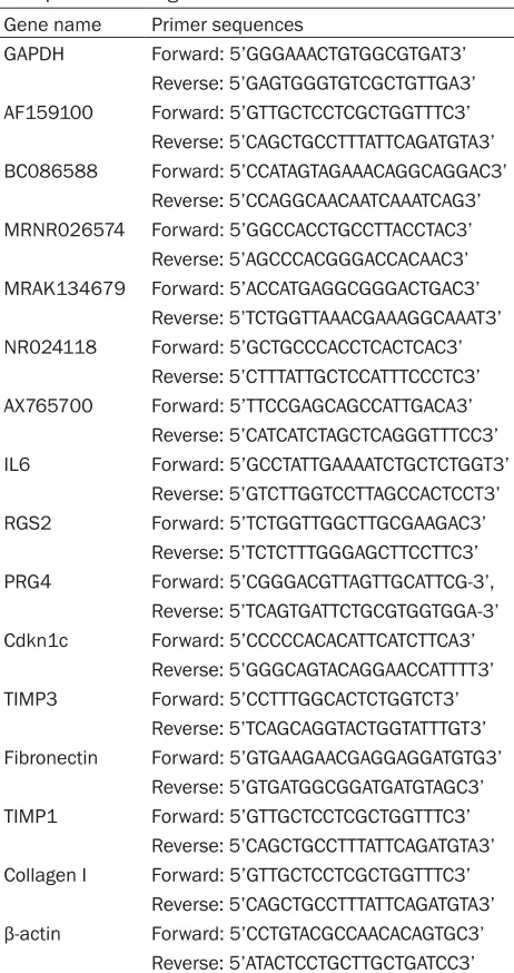
[image:2.612.91.322.93.530.2]Collagenase, trypsin and Ang II were obt- ained from Sigma Chemical (St Louis, MO, USA); Dulbecco’s modified Eagle’s medium (DMEM) and TRIzol were obtained from Life Technologies (Invitrogen, Carlsbad, CA, USA). Rat 4x44K LncRNA expression arrays were purchased from Arraystar (Rockville, USA). Sprague-Dawley (SD) rats were sup-plied from the Experimental Animal Center of Xian Jiaotong University (Xian, China). The animal experiments were approved by the University Committee of Laboratory Animal Care and Use and followed the Table 1. Primers sequences for long non-coding RNAs
and protein-coding RNAs Gene name Primer sequences
GAPDH Forward: 5’GGGAAACTGTGGCGTGAT3’ Reverse: 5’GAGTGGGTGTCGCTGTTGA3’
AF159100 Forward: 5’GTTGCTCCTCGCTGGTTTC3’ Reverse: 5’CAGCTGCCTTTATTCAGATGTA3’ BC086588 Forward: 5’CCATAGTAGAAACAGGCAGGAC3’
Reverse: 5’CCAGGCAACAATCAAATCAG3’
MRNR026574 Forward: 5’GGCCACCTGCCTTACCTAC3’
Reverse: 5’AGCCCACGGGACCACAAC3’
MRAK134679 Forward: 5’ACCATGAGGCGGGACTGAC3’ Reverse: 5’TCTGGTTAAACGAAAGGCAAAT3’
NR024118 Forward: 5’GCTGCCCACCTCACTCAC3’ Reverse: 5’CTTTATTGCTCCATTTCCCTC3’
AX765700 Forward: 5’TTCCGAGCAGCCATTGACA3’ Reverse: 5’CATCATCTAGCTCAGGGTTTCC3’
IL6 Forward: 5’GCCTATTGAAAATCTGCTCTGGT3’ Reverse: 5’GTCTTGGTCCTTAGCCACTCCT3’
RGS2 Forward: 5’TCTGGTTGGCTTGCGAAGAC3’ Reverse: 5’TCTCTTTGGGAGCTTCCTTC3’
PRG4 Forward: 5’CGGGACGTTAGTTGCATTCG-3’, Reverse: 5’TCAGTGATTCTGCGTGGTGGA-3’ Cdkn1c Forward: 5’CCCCCACACATTCATCTTCA3’
Reverse: 5’GGGCAGTACAGGAACCATTTT3’ TIMP3 Forward: 5’CCTTTGGCACTCTGGTCT3’
Reverse: 5’TCAGCAGGTACTGGTATTTGT3’
Fibronectin Forward: 5’GTGAAGAACGAGGAGGATGTG3’ Reverse: 5’GTGATGGCGGATGATGTAGC3’ TIMP1 Forward: 5’GTTGCTCCTCGCTGGTTTC3’
Reverse: 5’CAGCTGCCTTTATTCAGATGTA3’
Collagen I Forward: 5’GTTGCTCCTCGCTGGTTTC3’ Reverse: 5’CAGCTGCCTTTATTCAGATGTA3’ β-actin Forward: 5’CCTGTACGCCAACACAGTGC3’
Reverse: 5’ATACTCCTGCTTGCTGATCC3’
guidelines of the National Animal Research Center.
Isolation and culture cardiac fibroblasts
(mostly fibroblasts) were washed and grown in DMEM with 10% fetal bovine serum. The cardi-ac fibroblasts (passages 3~5) were grown to 80-90% confluence and serum starved for 24 h before treatment.
Preparation of RNA
Following 24 h serum starvation, cardiac fibro-blasts were treated with Ang II (100 nM) for 24 h. Total RNAs were extracted using the TRIZOL
[image:3.612.99.513.69.522.2]reagent as previously described and RNAs were dissolved in RNase-free water [18]. The RNA quantity was determined spectrophotometri-cally as A260 and A260/A280 ratio using NanoDrop spectrophotometer (Thermo Fisher Scientific, Wilmington, DE, USA) and RNA quali-ty were checked by electrophoresis on a 1.2% agarose/formaldehyde gel. Isolated RNAs were stored at -70°C prior to lncRNAs arrays analysis and real time-PCR.
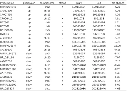
Table 2. Differentially expressed long non-coding RNAs in cardiac fibroblasts (fold change >4)
Probe Name Expression chromosome strand Start End Fold change
MRAK034346 up chr2 + 120115104 120115164 4.25
AF167308 up chr16 - 73031874 73031931 4.26
XR007499 up chr13 - 39625623 39625683 4.26
XR006412 up chr12 + 1021078 1021138 4.61
U57362 up chr8 - 84641434 84641494 4.71
U57361 up chr8 - 84643465 84643525 4.74
BC158638 up chr3 - 113780927 113801831 5.15
U78517 up chr3 + 54716706 54716766 5.40
AF239157 up chr10 - 46291442 46291502 5.92
BC086588 up chr2 - 186049351 186049411 8.92
MRNR026574 up chr1 + 159013775 159013835 12.20
AF159100 up chr16 - 70640308 70640368 47.16
MRAK134679 down chr3 - 62648034 62648094 -8.34
NR_024118 down chr20 - 4128070 4128130 -7.97
AX765700 down chr9 + 60980297 60980357 -7.17
MRAK053938 down chr15 + 109052626 109052699 -6.42
MRAK031289 down chr16 + 64128370 64128430 -6.26
AY973245 down chr16 + 64124051 64124111 -5.46
AJ005396 down chr2 + 210193318 210193378 -5.33
MRAK162711 down chr5 + 28340911 28340971 -5.15
MRAK132609 down chr2 + 210192978 210193038 -5.09
NR_027324 down chr1 - 202822980 202823040 -4.63
Microarray analysis of long ncRNAs and mRNAs expression
The rat LncRNA 4x44K Arrays from Arraystar (Rockville, USA) were used to analyze the expression profile of long non-coding RNAs and mRNAs in adult rat cardiac fibroblasts. The array contains probes of lncRNAs (~9300) and protein-coding genes (~15,200). The microar-ray hybridization was performed based on the manufacturer’s standard protocols [19]. Briefly, total RNAs from three pairs of control cardiac fibroblasts and Ang II-treated cardiac fibro-blasts were extracted and pooled. Next, 1 μg of total RNAs were amplified and transcribed into fluorescent cRNA using Agilent’s Quick Amp Labeling protocol (version 5.7, Agilent Techno- logies). The labeled cRNAs were hybridized onto the Rat LncRNA 4x44K Array (Arraystar, Rockville, USA), washed and the microarrays scanned using an Agilent Scanner G2505B. Agilent Feature Extraction software (version 10.7.3.1) were used to analyze acquired array images. Median normalization and subsequent data processing were performed using the
GeneSpring GX v11.5.1 software package (Agi- lent Technologies).
Quantitative real time-PCR
Quantitative real time-PCR (qPCR) was per-formed to quantify the levels of lncRNAs and mRNAs as previously described [18]. Briefly, total RNAs of cardiac fibroblasts were extracted using TRIzol Reagent. cDNAs were synthesized using the First Strand cDNA Synthesis kit (Fermentas Life Science, Burling, ON, Canada). Reactions were incubated for 60 mins at 42°C, 5 mins at 70°C, and then stored at -20°C. Quantitative PCR was then performed by using SYBR Premix Ex TaqTM II (TaKaRa, Ohtsu, Shiga,
Table 3. Differentially expressed protein-coding RNAs in cardiac fibroblasts (fold change >10)
Probe Name Gene Symbol Description Fold change
CUST3948 Rgs2 Rattus norvegicus regulator of G-protein signaling 2 10.16
CUST10456 Nefh Rattus norvegicus neurofilament, heavy polypeptide 10.38
CUST8969 Spp1 Rattus norvegicus secreted phosphoprotein 1 10.48
CUST12405 Gch1 Rattus norvegicus GTP cyclohydrolase 1 11.92
CUST4425 Cd55 Rattus norvegicus decay accelerating factor 1 (Daf1) 12.69
CUST12794 Ccdc19 Rattus norvegicus coiled-coil domain containing 19 13.079
CUST14423 Pde2a phosphodiesterase 2A isoform 1 13.219
CUST15179 Pde2a phosphodiesterase 2A, cGMP-stimulated 13.28
CUST4963 Gjb2 gap junction membrane channel protein beta 2 13.46
CUST2183 Elf5 E74-like factor 5 13.51
CUST11179 Rnase1 Rattus norvegicus ribonuclease, RNase A family, 1 13.73
CUST1306 Ptprn Rattus norvegicus protein tyrosine phosphatase, receptor type, N 14.33
CUST11361 Hspb7 cardiovascular heat shock protein 14.65
CUST1958 Esm1 Rattus norvegicus endothelial cell-specific molecule 1 15.10
CUST10736 Cldn3 Rattus norvegicus claudin 3 17.50
CUST11657 Prg4 proteoglycan 4 18.11
CUST14921 Lcn2 Rattus norvegicus lipocalin 2 19.78
CUST10982 Hp Rattus norvegicus haptoglobin 20.20
CUST6098 Ccl11 Rattus norvegicus chemokine (C-C motif) ligand 11 21.15
CUST5899 Gja5 gap junction membrane channel protein alpha 5 22.59
CUST6613 RGD1562551 hypothetical protein LOC311760 24.421
CUST10878 Pnoc Rattus norvegicus prepronociceptin 25.39
CUST3944 Cldn11 Rattus norvegicus claudin 11 25.89
CUST11937 Star Rattus norvegicus steroidogenic acute regulatory protein 31.11
CUST5850 Slco4a1 Rattus norvegicus solute carrier organic anion transporter family, member 4a1 47.80
CUST10972 Il6 Rattus norvegicus interleukin 6 49.94
CUST13799 Slc16a3 Rattus norvegicus solute carrier family 16, member 3 55.36
CUST7261 Cilp cartilage intermediate layer protein, nucleotide -30.32
CUST7758 Ces1d Rattus norvegicus carboxylesterase 3 -20.97
CUST4333 Adh7 Rattus norvegicus alcohol dehydrogenase 7 (class IV) -15.41
CUST7819 Cdkn1c Rattus norvegicus cyclin-dependent kinase inhibitor 1C -13.86
CUST8965 Timp3 Rattus norvegicus tissue inhibitor of metalloproteinase 3 -13.85
CUST8211 Arhgap20 Rattus norvegicus Rho GTPase activating protein 20 -13.49
CUST10750 Olfml2a olfactomedin-like 2A -10.41
CUST9855 Flrt3 fibronectin leucine rich transmembrane protein -10.29
CUST4116 Mtss1 metastasis suppressor 1 -10.15
expression was evaluated by the 2(−ΔΔCt) meth-
ods.
Bioinformatic analysis
The Gene Ontology project provides a con-trolled vocabulary to describe gene and gene product attributes in any organism (http://www. geneontology.org). The ontology covers three domains: Biological Process, Cellular Com- ponent and Molecular Function. Fisher’s exact
P-value (Fisher-P value) denotes the signifi-cance of the Pathway correlated to the condi-tions. Lower the P-value, more significant is the Pathway (The recommend P-value cut-off is 0.05).
Statistical analysis
Data were presented as the means ±SEM. The Student’s t-test was used to compare data between the two groups and one-way ANOVA for more than three groups. P<0.05 was con-sidered to indicate a statistically significant dif-ference. *p<0.05, **p<0.01 and ***p<0.001. Results
Arrays analysis of lncRNAs and mRNAs expres-sion in cardiac fibroblasts
Initial studies determined the overall numbers and quantity of lncRNAs and mRNAs that could be detected using an Arraystar microarray (Rockville, USA). This showed that 4376 (~47%) of the 9300 lncRNAs could be detected in untreated cells which is lower fraction than the
protein coding mRNAs, for which 9553 (~63%) of the 15200 could be detected. The average intensity of 4376 lncRNAs was 2841 while the average intensity of 9553 protein-coding genes was 5467. These results are consistent with other studies showing that lncRNAs were gen-erally expressed at lower levels than protein-coding genes (Figure 1) [20].
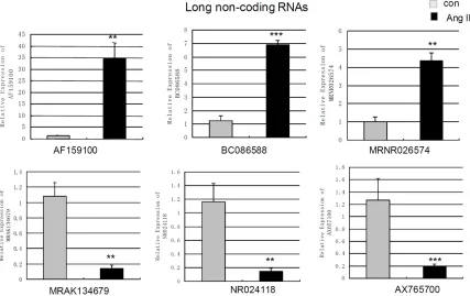
To gain further insights into the putative biologi-cal relevance of lncRNAs in cardiac fibroblasts, we compared the levels of lncRNAs and mRNAs in cardiac fibroblasts with/without Ang II-treat- ment for 24 h. We found that 282 of 4376 detected lncRNAs demonstrated >2-fold differ-ential expression with 178 lncRNAs showing up-regulated and 106 lncRNAs showing down-regulated. When the cut-off was set at 4-fold, 12 lncRNAs were up-regulated while 10 lnc- RNAs were down-regulated (Table 2). Mean- while, 882 mRNAs showed beyond a 2-fold dif-ferential expression in cardiac fibroblasts when compared to control cells. 521 mRNAs were up-regulated while 361 mRNAs were down-reg-ulated. When the cut-off was set at 10-fold, 27 Figure 2. Measurement of changes in long non-coding RNAs using qPCR. The expression levels of AF159100,
BC086588, MRNR026574, MRAK134679, NR024118, AX765700in cardiac fibroblasts treated by angiotensin
[image:6.612.93.520.73.342.2]mRNAs were up-regulated while 9 mRNAs down-regulated (Table 3).
Quantitative real time-PCR analysis of lncRNAs and mRNAs expression
Quantitative real time PCR was used to re-mea-sure the abundance of six lncRNAs (AF159100, BC086588, MRNR026574, MRAK134679, NR- 024118 and AX765700) and 9 mRNAs associ-ated with fibrosis (IL6, RGS2, PRG4, TIMP1, Cdkn1c, TIMP3, Col I, Col III and Fibronectin). qPCR analysis revealed that the levels of AF159100, BC086588 and MRNR026574 in
Ang II-treated cells were up-regulated to 27.42 fold (p=0.0041), 5.50 fold (p<0.001) and 4.37 fold (p=0.0058) compared to control cells (Figure 2). qPCR showed the levels of MR- AK134679, NR024118 and AX765700 were decreased to 7.59 fold (p=0.0057), 8.05 fold (p=0.004) and 6.36-fold (p=0.001) compared to control cells (Figure 2).
[image:7.612.86.522.72.471.2]The levels of 8 mRNAs were also verified by qPCR. It was revealed that the levels of IL6, regulator of G-protein signaling 2 (RGS2) and proteoglycan 4 (PRG4) were increased to 46.93 fold (p=0.0057), 19.28 fold (p=0.001) and Figure 3. Measurement of changes in protein coding mRNAs using qPCR. The expression levels of IL6, RGS2, PRG4,
TIMP1, Cdkn1c, TIMP3, Col I, Col III and Fibronectin in cardiac fibroblasts treated by angiotensin II-treatment (100
nm 24 h) and control cells were measured by qPCR. Expression of mRNAs was normalized to GAPDH expression.
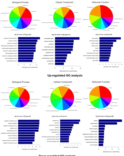
Figure 4.Bioinformatic analysis of the differentially expressed genes. The Gene Ontology (GO) analysis provides a controlled vocabulary to describe differentially expressed transcript attributes in all organisms. The ontology covers three domains: Biological Process, Cellular Component and Molecular Function. The p-value denotes the signifi
-cance of GO terms enrichment in the DE genes. The lower the p-value, the more significant the GO Term (p-value
≤0.05 is recommended).
4.69-fold (p=0.0044) in cardiac fibroblasts
fold (p<0.001) and 8.16-fold (p=0.001) in car-diac fibroblasts compared to control (Figure 3). qPCR also revealed that Ang II up-regulated the level of TIMP1 to 2.41 fold (p=0.0048) (Figure 3). However, the changes of collagen I, collagen III and Fibronectin did not show statistical sig-nificance using qPCR (Figure 3).
Go analysis and pathway analysis
The number (Top ten) of genes associated with GO term and the significance of GO term (Top ten) were shown (Figure 4). The upregulated genes were involved in 843 biological process, 110 cellular components and 165 molecular functions. In the biological process category, the most significant term was the response to external stimulus (p=1.33509E-18). In the cel-lular component category, the most represent-ed GO term was the extracellular region (p=4.20796E-15). Within the molecular compo-nent category, protein binding (p=3.38671E-14) as the most highly represented term. The down-regulated genes were involved in 501 biological process, 40 cellular components and 85 molecular functions. In the biological process category, the regulation of cellular process was enriched most. Within the cellular component category, intracellular part was the most repre-sented GO terms. Among the various molecular functions, binding were most highly represent-ed term.
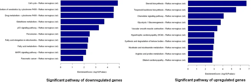
Following Go analysis, KEGG was used to do a pathway enrichment analysis. The downregu-lated genes were involved in 18 pathways while upregulated genes were involved in 30
path-ways. The top 10 pathways of downregulated and upregulated genes were shown (Figure 5). The pathways of downregulated genes include cell cycle, P53 signaling pathway, MAPK signal-ing pathway, indicatsignal-ing the activation of cell proliferation. Among the ten pathways, the most significant pathway was the Cell cycle pathway (p=0.000013705).
The regulation of Ang II on the expression of NR024118 and Cdkn1c
In order to investigate how Ang II regulate the expression of lncRNA-NR024118 and Cdkn1c, we determined the levels of NR024118 and Cdkn1c in cardiac fibroblasts when treated by Ang II at different time and different concentra-tion using quantitative real time PCR. We found that the level of NR024118 was gradually decreasing with the exposure time to Ang II in cardiac fibroblasts. The level of NR024118 reached to the lowest at 24 h but back at 48 h in cardiac fibroblasts (Figure 6A). Meanwhile, the level of Cdkn1c in cardiac fibroblasts was gradually decreasing as the extension of time of treatment by Ang II with the lowest level at 48 h (Figure 6B). All of treatment by Ang II at a range from 10-9 M to 10-6 M induced a decrease of both NR024118 and Cdkn1c in cardiac fibro-blasts (Figure 6C and 6D).
Discussion
[image:9.612.99.523.73.212.2]The current literature on lncRNAs mainly focused on cancer and neural diseases [21-25]. Up to now, only a few lncRNAs were report-ed in cardiovascular system. In our study, the
Figure 5. Pathway analysis of the differentially expressed genes. Pathway analysis is a functional analysis that maps
genes to KEGG (Kyoto Encyclopedia of Genes and Genomes) pathways (http://www.genome.jp/kegg/). The P-value (Fisher-P value) denotes the significance of the Pathway correlated to the conditions. Lower the P-value, more
clear changes of lncRNAs (AF159100, BC08- 6588, MRNR026574, MRAK134679, NR024- 118, AX765700) in adult cardiac fibroblasts in response to Ang II were associated with the changes of several protein-coding RNAs. We suspected that the decrease of Cdkn1c indi-cated that cell proliferation. The upregulated PRG4 induced by Ang II, an extracellular matrix molecule, indicated the enhancing of the syn-thesis of extracellular matrix. The changes of TIMP1 and TIMP3 showed that the degradation of ECM might be affected the activity of MMPs. The obvious increase of IL 6 indicated the inflammation mechanism of cytokines in the developing of cardiac fibrosis. It was reported that production of IL6 in cardiac fibroblasts will lead to TGF-β1 production and stimulates car-diac fibrosis that induced by Ang II [26]. RGS2 has been showed to be a regulator of Ang II-effects in cardiac fibroblast that might have a role in Ang II-induced fibrosis [8].
Go analysis indicated cell response to external stimulus and cell division. Pathways analysis showed several significant pathways related to cell proliferation including cell cycle, P53 sig-naling pathway and MAPK sigsig-naling pathway.
Further studies revealed that Ang II at a range from 10-9 M to 10-6 M decreased the expres-sion of NR024118 and Cdkn1c in cardiac fibro-blasts. Moreover, the regulation of Ang II on the expression of NR024118 and Cdkn1c was in a time-dependent pattern. Moreover, Cdkn1c (p57/KIP2) was common down-regulated in dif-ferent human cancers, indicating the role of Cdkn1c in cell proliferation [32]. Ang II dynami-cally down regulated the level of NR024118 and Cdkn1c in cardiac fibroblasts, strongly sug-gesting the potential role of NR024118 in car-diac fibroblasts.
[image:10.612.91.518.71.312.2]Bioinformatic analysis reveal that LncRNA-NR024118 (793 bp), located in chromosome 20, is defined as Rattus norvegicus tenascin XA, pseudogene 1 (Tnxa-ps1), non-coding RNA. The protein-coding RNA of tenascin X has been reported to facilitate myocardial fibrosis through transforming growth factor-β1 and per-oxisome proliferator-activated receptor γ [27]. Pseudogenes have long been neglected because of being considering nonfunctional. However, recent advances have established that the RNA transcribed from a pseudogene can have diverse functions not only to their Figure 6. The regulation of angiotensin II on the expression of NR024118 and Cdkn1c in cardiac fibroblasts. The ex-pression of NR024118 and Cdkn1cin cardiac fibroblasts treated by angiotensin II (Ang II 100 nm) at different time
parental genes but also to unrelated genes [28]. Pseudogenes are considered not to be pseudo any more now [29]. The pseudogene (PTENP1) of the tumor suppressor PTEN has been reported to regulate the expression of PTEN in mRNA and protein levels [30]. Recently, it was reported that a pseudogene lncRNAs network (PTENpg1 α and β) regulates PTEN transcription and translation in human cells [31]. We anticipate that the next functional studies of ps1 will reveal the roles of Tnxa-ps1 in cardiac fibroblasts.
In conclusion, our current studies showed that the expression profile of lncRNAs was signifi-cantly altered in the Ang II-induced cardiac fibroblasts and Ang II dynamically regulated the expression of lncRNA-NR024118 and Cdkn1c in adult cardiac fibroblasts, strongly indicating the potential role of NR024118 in adult cardiac fibroblasts.
Acknowledgements
This study was supported by National Science Foundation of China (31100834) and the International Cooperation Founds of Shaanxi Province (2012KW-32-02).
Disclosure of conflict of interest None.
Address correspondence to: Xiaoying Jiang, Depart- ment of Genetics and Molecular Biology, School of Medicine, Xi’an Jiaotong University, 76 Yanta West Road, Xi’an, 710061, Shaanxi, China. Tel: (+86)-2982657013; E-mail: [email protected]
References
[1] Takeda N, Manabe I, Uchino Y, Eguchi K, Mat-sumoto S, Nishimura S, Shindo T, Sano M, Otsu K, Snider P, Conway SJ and Nagai R. Cardiac fi-broblasts are essential for the adaptive re-sponse of the murine heart to pressure over-load. J Clin Invest 2010; 120: 254-265. [2] Porter KE and Turner NA. Cardiac fibroblasts:
at the heart of myocardial remodeling. Phar-macol Ther 2009; 123: 255-278.
[3] Díez J. Do microRNAs regulate myocardial fi-brosis? Nat Clin Pract Cardiovasc Med 2009; 6: 88-89.
[4] Jiang X, Tsitsiou E, Herrick SE and Lindsay MA. MicroRNAs and the regulation of fibrosis. FEBS J 2010; 277: 2015-21.
[5] Iwata M, Cowling RT, Yeo SJ and Greenberg B. Targeting the ACE2-Ang-(1-7) pathway in
cardi-ac fibroblasts to treat cardicardi-ac remodeling and heart failure. J Mol Cell Cardiol 2011; 51: 542-547.
[6] Ren J, Yang M, Qi G, Zheng J, Jia L, Cheng J, Tian C, Li H, Lin X and Du J. Proinflammatory protein CARD9 is essential for infiltration of monocyticfibroblast precursors and cardiac fi-brosis caused by Angiotensin II infusion. Am J Hypertens 2011; 24: 701-707.
[7] Lijnen PJ, Van Pelt JF and Fagard RH. Stimula-tion of Reactive Oxygen Species and Collagen Synthesis by Angiotensin II in Cardiac Fibro-blasts. Cardiovasc Ther 2012; 30: e1-e8. [8] Zhang P, Su J, King ME, Maldonado AE, Park C
and Mende U. Regulator of G protein signaling 2 is a functionally important negative regulator of angiotensin II-induced cardiac fibroblast re-sponses. Am J Physiol Heart Circ Physiol 2011; 301: H147-H156.
[9] Taft RJ, Pang KC, Mercer TR, Dinger M and Mattick JS. Non-coding RNAS: regulator of dis-eases. J Pathol 2010; 220: 126-139.
[10] Gibb EA, Brown CJ and Lam WL. The functional role of long non-coding RNA in human carcino-mas. Mol Cancer 2011; 10: 38.
[11] Mercer TR, Dinger ME and Mattick JS. Long non-coding RNAs: insights into functions. Nat Rev Genet 2009; 10: 155-159.
[12] Mercer TR, Dinger ME, Sunkin SM, Mehler MF and Mattick JS. Specific expression of long noncoding RNAs in the mouse brain. Proc Natl Acad Sci U S A 2008; 105: 716-721.
[13] Tsai MC, Manor O, Wan Y, Mosammaparast N, Wang JK, Lan F, Shi Y, Segal E and Chang HY. Long noncoding RNA as modular scaffold of histone modification complexes. Science 2010; 329: 689-693.
[14] Ishii N, Ozaki K, Sato H, Mizuno H, Saito S, Takahashi A, Miyamoto Y, Ikegawa S, Kamatani N, Hori M, Saito S, Nakamura Y and Tanaka T. Identification of a novel non-coding RNA, MIAT, that confers risk of myocardial infarction. J Hum Genet 2006; 51: 1087-1099.
[15] Zhuang J, Peng W, Li H, Wang W, Wei Y, Li W and Xu Y. Methylation of p15INK4b and ex-pression of ANRIL on chromosome 9p21 are associated with coronary artery disease. PLoS One 2012; 7: e47193.
[16] Klattenhoff CA, Scheuermann JC, Surface LE, Bradley RK, Fields PA, Steinhauser ML, Ding H, Butty VL, Torrey L, Haas S, Abo R, Tabebordbar M, Lee RT, Burge CB and Boyer LA. Braveheart, a long noncoding RNA required for cardiovas-cular lineage commitment. Cell 2013; 152: 570-583.
[18] Jiang X, Ning Q and Wang J. Angiotensin II in-duced differentially expressed microRNAs in adult rat cardiac fibroblasts. J Physiol Sci 2013; 63: 31-38.
[19] Yang F, Zhang L, Huo XS, Yuan JH, Xu D, Yuan SX, Zhu N, Zhou WP, Yang GS, Wang YZ, Shang JL, Gao CF, Zhang FR, Wang F and Sun SH. Long noncoding RNA high expression in hepa-tocellular carcinoma facilitates tumor growth through enhancer of zeste homolog 2 in hu-mans. Hepatology 2011; 54: 1679-1689. [20] Guttman M, Garber M, Levin JZ, Donaghey J,
Robinson J, Adiconis X, Fan L, Koziol MJ, Gnirke A, Nusbaum C, Rinn JL, Lander ES and Regev A. Ab initio reconstruction of cell type specific transcriptomes in mouse reveals the con-served multi-exonic structure of lincRNAs. Nat Biotechnol 2010; 28: 503-510.
[21] Prensner JR and Chinnaiyan AM. The emer-gence of lncRNAs in cancer biology. Cancer Discov 2011; 1: 391-407.
[22] Bian S and Sun T. Functions of noncoding RNAs in neural development and neurological diseases. Mol Neurobiol 2011; 44: 359-373. [23] Cui Z, Ren S, Lu J, Wang F, Xu W, Sun Y, Wei M,
Chen J, Gao X, Xu C, Mao JH and Sun Y. The prostate cancer-up-regulated long noncoding RNA PlncRNA-1 modulates apoptosis and pro-liferation through reciprocal regulation of an-drogen receptor. Urol Oncol 2013; 31: 1117-1123.
[24] Silva JM, Boczek NJ, Berres MW, Ma X and Smith DI. LSINCT5 is over expressed in breast and ovarian cancer and affects cellular prolif-eration. RNA Biol 2011; 8: 496-505.
[25] Meola N, Pizzo M, Alfano G, Surace EM and Banfi S. The long noncoding RNA Vax2os1 con-trols the cell cycle progression of photorecep-tor progeniphotorecep-tors in the mouse retina. RNA 2012; 18: 111-123.
[26] Ma F, Li Y, Jia L, Han Y, Cheng J, Li H, Qi Y and Du J. Macrophage-stimulated cardiac fibro-blast production of IL-6 is essential for TGFβ/ Smad activation and cardiac fibrosis induced by angiotensin II. PLoS One 2012; 7: e35144. [27] Jing L, Zhou LJ, Zhang FM, Li WM and Sang Y.
Tenascin-x facilitates myocardial fibrosis and cardiac remodeling through transforming growth factor-β1 and peroxisome proliferator-activated receptor γ in alcoholic cardiomyopa-thy. Chin Med J (Engl) 2011; 124: 390-395. [28] Polisenol L. Pseudogenes: newly discoved
play-ers in human cancer. Sci Signal 2012; 5: re5. [29] Wen YZ, Zheng LL, Qu LH, Ayala FJ and Lun ZR.
Pseudogenes are not pseudo any more. RNA Biol 2012; 9: 27-32.
[30] Poliseno L, Salmena L, Zhang J, Carver B, Have-man WJ and Pandolfi PP. A coding-independent function of gene and pseudogene mRNAs reg-ulates tumour biology. Nature 2010; 465: 1033-1038.
[31] Johnsson P, Ackley A, Vidarsdottir L, Lui WO, Corcoran M, Grandér D and Morris KV. A pseu-dogene long-noncoding-RNA network regu-lates PTEN transcription and translation in hu-man cells. Nat Struct Mol Biol 2013; 20: 440-446.