EVALUATION OF CLINICO
INTERTROCHANTERIC FRACTURES USING DYNAMIC HIP SCREW
STABILISATION PLATE (TSP)
*Jagan Mohan, Reddy Karnati, Monappa A Naik and
Department of Orthopaedics, Kasturba Medical College, Manipal University
ARTICLE INFO ABSTRACT
Back ground:
buttress, or both, after stabilization with a dynamic hip screw (DHS), these fractures tend to have excessive fracture collapse, with significant limb shortening and occasionally fixation failu general for treatment of unstable intertrochanteric fractures, two options exist: extra medullary or intramedullary stabilization. Currently controversies still exist about a suitable device for an unstable intertrochanteric fracture.
Materials & Methods:
between August 2012 and July 2014. A total of 70 cases of unstable intertrochanteric fractures 31 2.2 and above
and Trochanteric stabilization plate (DHS+TSP) and other with Proximal Femoral Nailing per computer generated randomised table at the time of first pres
both clinically and Radiologically. Clinical variables assessed were limp, limb shortening, range of motion, Salvati wilson hip score, Hip abductor power. Radiologically variables like progress of union, varus collapse
Results:
females with mean age of 68.3 years. All fractures united at mean 16.3 weeks in T
weeks for PFN group. There was 1 lag screw back out in TSP group & 2 in PFN group at 6 month follow up. None of the patient required revision surgery. At the end of one
(Limp power
Statistical data was evaluated with the help of SPSS 21 software using Independent T test. Conclusions:
clinic-sample size and comparable groups.
Copyright©2016, Jagan Mohan et al. This is an open access article distributed under the Creative Commons Att distribution, and reproduction in any medium, provided the original work is properly cited.
INTRODUCTION
The incidence of intertrochanteric fractures has increased considerably during recent years because of increasing number of senior citizens with osteoporosis (Kulkarni
Poor bone quality and associated co-morbidity often make the treatment more challenging in these patients.
which are stable and minimally displaced, the dyna screw (DHS) device produces reproducibly reliable results Unstable intertrochanteric fractures are defined as three fractures with an additional posteromedial fragment including
*Corresponding author: Jagan Mohan,
Department of Orthopaedics, Kasturba Medical College, Manipal University
ISSN: 0975-833X
Article History:
Received 22nd May, 2016
Received in revised form 09th June, 2016
Accepted 10th July, 2016
Published online 31st August,2016
Citation: Jagan Mohan, Reddy Karnati, Monappa A Naik and Sharath K Rao
Intertrochanteric fractures using Dynamic Hip Screw (DHS) with Trochanteric Stabilisation Plate (TSP) versus Proximal Femoral
International Journal of Current Research, 8, (08), 37209 Key words: Intertrochanteric fractures, DHS+TSP, PFN, Unstable.
RESEARCH ARTICLE
EVALUATION OF CLINICO- RADIOLOGICAL OUTCOMES OF TREATING UNSTABLE
INTERTROCHANTERIC FRACTURES USING DYNAMIC HIP SCREW (DHS) WITH
STABILISATION PLATE (TSP) VERSUS PROXIMAL FEMORAL NAILING (PFN)
, Reddy Karnati, Monappa A Naik and Sharath K Rao
Department of Orthopaedics, Kasturba Medical College, Manipal University
ABSTRACT
Back ground: Unstable intertrochanteric fractures which lack either
buttress, or both, after stabilization with a dynamic hip screw (DHS), these fractures tend to have excessive fracture collapse, with significant limb shortening and occasionally fixation failu general for treatment of unstable intertrochanteric fractures, two options exist: extra medullary or intramedullary stabilization. Currently controversies still exist about a suitable device for an unstable intertrochanteric fracture.
Materials & Methods: Study was undertaken in Department of Orthopaedics, KMC, Manipal between August 2012 and July 2014. A total of 70 cases of unstable intertrochanteric fractures
2.2 and above) were randomized into two groups (one group treated with
and Trochanteric stabilization plate (DHS+TSP) and other with Proximal Femoral Nailing per computer generated randomised table at the time of first pres
both clinically and Radiologically. Clinical variables assessed were limp, limb shortening, range of motion, Salvati wilson hip score, Hip abductor power. Radiologically variables like progress of union, varus collapse, and screw cut out were analysed at 6 weeks, 3 months, 6 months and at 1 year Results: 20 cases were lost to follow-up. Remainder study group comprised of 32 males and 18 females with mean age of 68.3 years. All fractures united at mean 16.3 weeks in T
weeks for PFN group. There was 1 lag screw back out in TSP group & 2 in PFN group at 6 month follow up. None of the patient required revision surgery. At the end of one
Limp (P value<0.08), Hip Movements, Hip Score (0.087), limb shortening (P value<0.11), Abductor power) and radiological variables (Varus collapse (P value<0.8)
Statistical data was evaluated with the help of SPSS 21 software using Independent T test.
Conclusions: In treatment of unstable trochanteric fractures, DHS+TSP and PFN have comparable -radiological outcomes at the end of one year. However, further studies are required with large sample size and comparable groups.
is an open access article distributed under the Creative Commons Attribution License, which distribution, and reproduction in any medium, provided the original work is properly cited.
The incidence of intertrochanteric fractures has increased increasing number Kulkarni et al., 2006). morbidity often make the treatment more challenging in these patients. In most fractures, which are stable and minimally displaced, the dynamic hip screw (DHS) device produces reproducibly reliable results Unstable intertrochanteric fractures are defined as three-part fractures with an additional posteromedial fragment including
Orthopaedics, Kasturba Medical College, Manipal
the lesser trochanter (31-A2.2 or 31
OTA and AO classifications, respectively
fractures with an additional fragment including the greater trochanter (31-A3.3). These fractures lack the posteromedial buttress, the lateral buttress, or both. Therefore, after stabilization with a dynamic hip screw (DHS), these fractures tend to have a controlled but important telescoping, with lateral dislocation of the greater trochanter fragment, and sometimes an additional derotation of the rotationally unstable head fragment occurs. This instability results in excessive fracture collapse, with significant limb shortening and occasionally fixation failure. Failure rates of
reported for unstable fractures. In general for treatment of unstable intertrochanteric fractures, two options exist:
International Journal of Current Research Vol. 8, Issue, 08, pp.37209-37215, August, 2016
Mohan, Reddy Karnati, Monappa A Naik and Sharath K Rao, 2016. “Evaluation of clinico- radiological outcomes of treating unstable Intertrochanteric fractures using Dynamic Hip Screw (DHS) with Trochanteric Stabilisation Plate (TSP) versus Proximal Femoral
37209-37215.
RADIOLOGICAL OUTCOMES OF TREATING UNSTABLE
(DHS) WITH TROCHANTERIC
VERSUS PROXIMAL FEMORAL NAILING (PFN)
Sharath K Rao
Department of Orthopaedics, Kasturba Medical College, Manipal University
fractures which lack either posteromedial buttress, lateral buttress, or both, after stabilization with a dynamic hip screw (DHS), these fractures tend to have excessive fracture collapse, with significant limb shortening and occasionally fixation failure. In general for treatment of unstable intertrochanteric fractures, two options exist: extra medullary or intramedullary stabilization. Currently controversies still exist about a suitable device for an unstable
Study was undertaken in Department of Orthopaedics, KMC, Manipal between August 2012 and July 2014. A total of 70 cases of unstable intertrochanteric fractures (AO were randomized into two groups (one group treated with (Dynamic Hip Screw and Trochanteric stabilization plate (DHS+TSP) and other with Proximal Femoral Nailing (PFN) ) as per computer generated randomised table at the time of first presentation to hospital, followed up both clinically and Radiologically. Clinical variables assessed were limp, limb shortening, range of motion, Salvati wilson hip score, Hip abductor power. Radiologically variables like progress of , and screw cut out were analysed at 6 weeks, 3 months, 6 months and at 1 year up. Remainder study group comprised of 32 males and 18 females with mean age of 68.3 years. All fractures united at mean 16.3 weeks in TSP group and 15.4 weeks for PFN group. There was 1 lag screw back out in TSP group & 2 in PFN group at 6 month follow up. None of the patient required revision surgery. At the end of one year, clinical variables core (0.087), limb shortening (P value<0.11), Abductor Varus collapse (P value<0.8)) showed no statistical significance. Statistical data was evaluated with the help of SPSS 21 software using Independent T test.
In treatment of unstable trochanteric fractures, DHS+TSP and PFN have comparable radiological outcomes at the end of one year. However, further studies are required with large
ribution License, which permits unrestricted use,
A2.2 or 31-A2.3 according to the OTA and AO classifications, respectively) or four-part fractures with an additional fragment including the greater . These fractures lack the posteromedial buttress, the lateral buttress, or both. Therefore, after stabilization with a dynamic hip screw (DHS), these fractures tend to have a controlled but important telescoping, with lateral ochanter fragment, and sometimes an additional derotation of the rotationally unstable head-neck fragment occurs. This instability results in excessive fracture collapse, with significant limb shortening and occasionally fixation failure. Failure rates of 5 to 12 percent have been reported for unstable fractures. In general for treatment of unstable intertrochanteric fractures, two options exist:
OF CURRENT RESEARCH
extramedullary or intramedullary stabilization. The extramedullary option comprises dynamic hip screw along with various methods, such as medial displacement osteotomy, valgus reduction and additional tension band wiring, valgus osteotomy and trochanter stabilization plate applied to the lateral cortex. The intramedullary methods are basically intramedullary nail devices (Gamma nail, Intramedullary Hip screw, Proximal Femoral Nail) with one or more neck screw options (Parker and Handoll, 2008) which provide more load sharing and allow less collapse (Pajarinen et al., 2005). Currently controversies still exist about a suitable device for an unstable intertrochanteric fracture. Results of randomised clinical studies comparing the results of intramedullary and extramedullary fixation techniques are inconsistent and rare (Simmermacher et al., 1999). The present study is designed accordingly to evaluate and compare the outcomes of unstable intertrochanteric fracture treated with DHS + TSP or PFN.
MATERIALS AND METHODS
A prospective randomised controlled trial was undertaken in Department of Orthopaedics, Kasturba hospital, Manipal from August 2012-July 2014. A total of 70 patients were taken into the study after institutional ethical committee clearance and written consent from subjects.
Inclusion criteria
• 3.1. A2.2, 3.1.A2.3, 3.1.A3.1, 3.1.A3.2, 3.1.A3.3 fractures as per AO classification.
• Presentation within one week of injury
• Should be ambulatory independently before fracture.
Exclusion criteria
• Pathological fractures (exception: osteoporotic or osteopenic fractures)
• Other associated ipsilateral limb fractures and poly trauma patients.
• Neuropathic conditions interfering with rehabilitation.
• Previously operated inter trochanteric fractures.
• Patient not giving consent for surgery
Methodology
Seventy patients with unstable intertrochanteric fractures were randomized with the help of computer generated random table. One group with 35 members was treated with Trochanteric Stabilizing Plate (TSP) superimposed on the regular Dynamic Hip Screw (DHS) with standard lateral approach and other group with Proximal Femoral Nailing (PFN) with a minimal incision for the entry point at our institution between August 2012 and July 2013. Three patients died before the first follow-up, seven patients died between first and second follow up due to comorbidities, seven patients were lost to follow-up, and three patients were bed ridden. Thus, fifty patients were followed for at least twelve months (mean 14 months, range 12 to 20 months). Data collected from patients by interview at the time of admission, follow up at intervals of 6 weeks, 3months, 6 months and 1 year. Pelvis with both hips AP view with both lower limbs parallel and neutral in rotation and trans-lateral
view were taken for each patient and classified according to AO classification system. Clinically limp, shortening, Hip-range of movements, Hip abductor power (MRC grading) and Salvati Wilson hip score were assessed and Confirmed by two Orthopaedicians. Radiological parameters like Varus collapse, Screw cut out, Progress of union (Absence of fracture line, callus formation, at least two cortices union) were assessed.
Post-operative protocol
Active SLR and static quadriceps strengthening exercises were initiated on day1. Patients were allowed partial weight bearing walking with walker from 2nd postoperative day. Full weight bearing started after clinical and radiological signs of union (at least 2 cortices union radio logically).
RESULTS
[image:2.595.307.557.356.398.2]Results were analyzed using SPSS 21 software. The mean age of the participants was 68.5 years. The largest number of patients fell in the age group of 66 to 70 years. In the AO classification system majority of patients, 30(60%) fell in AO type A2.2
Table 1. Distribution of fractures as per AO classification
AO classification No. of patients Percentage
A 2.2 30 60
A 2.3 13 26
A3.1 7 14
Shortening
Patients with <0.5cm shortening is not taken into consideration. At the end of each follow up, P-value was analyzed (Table 2). It showed no much difference between both implant groups at three intervals.
Presence of Limp
At the end of 3 months, 3 out of 30 DHS+TSP patients and 3 out of 20 PFN patients had limp. At the end of 6 months, 3 out of 30 DHS+TSP patients and 3 out of 20 PFN patients had limp. Finally, at the end of 1 year, we noticed that 5(17%) patients had limp in DHS+TSP group (Table 3). 2(10%) patients had limp in PFN group. There is no statistical significance (P<0.08)
Range of movements
Range of movements was measured in both the groups. Results showed that at the end of 3 months, there is statistically significant difference only in internal and external rotation movements (Figure 1). But at the end of 6 months and 1 year (Figure 2, 3), there is no significant clinical or statistical difference. Fx-Flexion, Ex-Extension, Ir-Internal rotation, Er-External rotation, Ab-Abduction, Ad-Adduction, ROM- Range of movements.
Table 2. Shortening of the limb in DHS+TSP and PFN groups
Implant Shortening (3months) Shortening (6months) Shortening (1 year)
<0.5 0.6-1 >1 P value <0.5 0.6-1 >1 P value <0.5 0.6-1 >1 Pvalue
DHS+TSP 28 2 0 0.05 28 2 0 0.09 27 3 0 0.11
[image:3.595.75.525.162.205.2]PFN 18 1 1 17 1 2 17 1 2
Table 3. Distribution of patients with limp and without in DHS+TSP and PFN groups
Follow up DHS+TSP (Limp) PFN (Limp) DHS+TSP (No limp) PFN (No limp)
3 months 3(10%) 3(15%) 27(90%) 17(85%)
6 months 3(10%) 3(15%) 27(90%) 17(85%)
[image:3.595.167.435.235.523.2]1 year 5(17%) 2(10%) 25(83%) 18(90%)
Table 4. Distribution of Range of movements in DHS+TSP and PFN groups
[image:3.595.118.484.545.745.2]Fx-Flexion, Ex-Extension, Ir-Internal rotation, Er-External rotation, Ab-Abduction, Ad-Adduction, ROM- Range of movements.
Figure 2. Range of movements at the end of 6 months
Figure 3. Range of movements at the end of 6 months
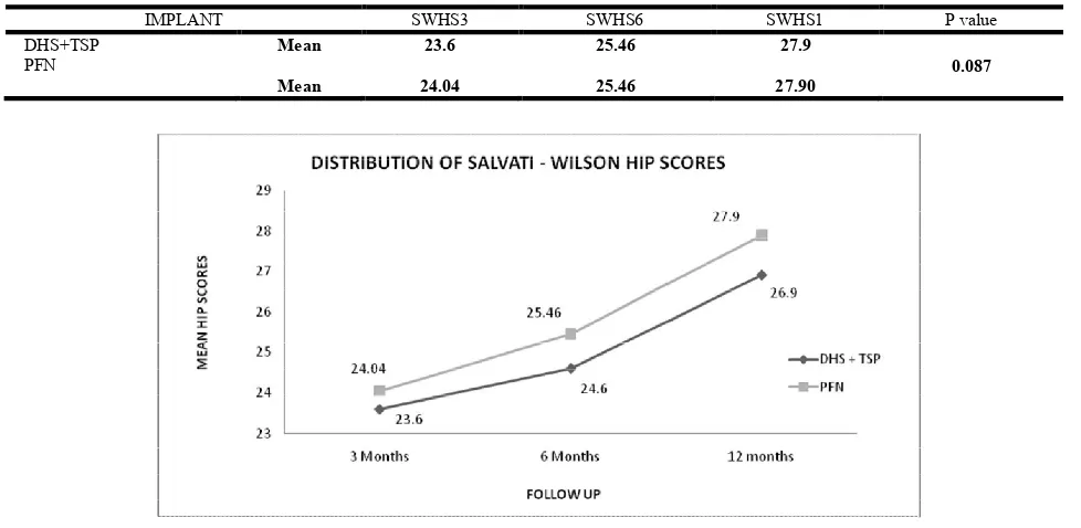
Table 5. Distribution of patients as per to Salvati Wilson Hip score (SWHS) in DHS+TSP and PFN group
IMPLANT SWHS3 SWHS6 SWHS1 P value
DHS+TSP Mean 23.6 25.46 27.9
PFN 0.087
[image:4.595.124.470.281.468.2]Mean 24.04 25.46 27.90
[image:4.595.53.542.527.766.2]Varus collapse: In our study, we noticed that the degree of varus collapse in both the implant groups was almost the same and it is proved statistically. Collapse is negligible in both groups (Table 6).
Hip abductor power
At the end of 1 year, majority of patients in DHS+TSP group(23 out of 30) were found to have grade 4 abductor power,1 out of 30 patients were found to have grade 3 abductor powder and 6 out of 30 patients were found to have grade 5 abductor power. Majority of patients in PFN group (15 out f 20 patients) were found to have grade 4 abductor power, 2 out of 20 were found to have grade 5 abductor power, 3 out of 20 patients were found to have grade 3 abductor power. This shows that though initially abductor power was less in DHS+TSP group, gradually it improved and by the end of 1 year almost both group patients had similar abductor power. All fractures were united by 3-6 months of follow up in both groups. There were 2 lag screw back outs in PFN group and 1 lag screw back out in DHS+TSP group at 6 months follow up, where the patients were asymptomatic. No anti rotation screw cut out in either group or No plate pull out was noticed in DHS+TSP group. No Surgical Site Infection (SSI) was noted in either group. 2 patients in DHS+TSP group developed trochanteric bursitis (confirmed by USG) that was managed conservatively. None of the patient required revision surgery. Clinical variables like Limp (P value<0.08), Hip Movements, Hip Score, Limb shortening (P value<0.08), Abductor power are comparable in both groups. Assessment of both the groups radiologically in Varuscollapse (P value<0.8) showed no statistical significance.
DISCUSSION
The optimal fixation device for the treatment of unstable intertrochanteric fractures is still controversial. Gadegone and Salphale (Koval and Zuckerman, 2001) after reviewing outcomes of 100 asian patients who underwent short proximal femoral nailing for stable and unstable intertrochanteric fractures concluded that short proximal femoral nail is a superior implant for stable and unstable intertrochanteric fractures in terms of operating time, surgical exposure, blood loss and complications, especially for patients with relatively small femora. Simmermacher et al. (1999) reviewed 191patients having proximal femoral fractures treated with proximal femoral nail in one year. After a follow up period of 4 months, technical failures were seen in just 4.6% of the cases. They concluded that the result of this new implant compare favourably to the currently available implants for the treatment of the unstable pertrochanteric femoral fractures. Xiao Huang
et al. (2013) performed meta-analysis included eight RCTs
which showed that PFN fixation and DHS fixation had similar effectiveness in terms of operative time, blood loss and blood
transfusions in the treatment of trochanteric fractures. Parker and Handoll (2010) also compared gamma nail and other intramedullary nails with extra medullary implants for extra capsular hip fractures in adults. In their systematic review the authors enrolled four studies which included PFN and Targon PF nail compared with SHS. The authors concluded that there was no significant difference between the groups in outcomes of blood loss and transfusion, fixation complications, and post-operation complications and hospital stay. Our study has a limitation that operative time, blood loss and blood transfusions were not taken into account. We compared limp and hip movements in both the groups. At the end of 1 year, 5(17%) in DHS+TSP group and 2(10%) in PFN group had limp. This has been found to be statistically not significant (p<0.08). Range of motion assessment showed mean of internal rotation was more in DHS+TSP group and mean of external rotation was more in PFN group at the end of 3 months. Though it was statistically significant, clinically it didn’t show much difference. But by the end of 1 year, there was no much difference in the movements of hip in both implant groups. In our study, functional assessment was evaluated with the help of salvati Wilson score which showed no statistical difference and majority of the patients had good comparable score in both the groups at the end of 3months, 6months and 1 year. Our result contradicts with the study made by Al-Yassari et al. (2002) and Simmermacher et al. (1999). They observed restoration of pre-operative functional status which showed that approximately 40% to 50% of the patients treated with a PFN had better results when compared to DHS group. This probably may be due to good lateral wall reconstruction and preventing varus collapse in DHS +TSP group which maintained good range of movements in our patients. Comparing the amount of shortening, by the end of 1 year, 3 patients had 0.6-1 cm in DHS+TSP group. In PFN group we had 3 patients one with 0.6-1.0cm and two with>1cm shortening respectively. But this did not show any statistical significance. This proves that addition of TSP to DHS minimises the amount of shortening due to collapse at the fracture site and showed comparable results with PFN implant. McGrory et al. (1995) described a positive correlation between abduction strength and the lengths of the lever arm of the abductor muscles after total hip replacement. Collapse at the fracture site will reduce the lever arm and may explain muscle weakness as shown by a positive Trendelenburg sign. Abductor weakness could be a important factor explaining alterations in postoperative versus pre trauma mobility levels. We assessed hip abductor power in our study in both the groups. We noticed that initially at the end of 3 months, in DHS+TSP group, 11(36%) patients had grade 3 power and 15(50%) had grade 4 power which gradually improved probably attributable to the amount of dissection at the time of surgery thereby leading to pain and decreased abductor power in DHS+TSP group. At the end of 1 year of follow up, only 1(3%) patient had grade 3 power and 23(79%) of the patients had grade 4 power. Where as in PFN group,
Table 6. Distribution of patients as per varus collapse (in degrees) in DHS+TSP and PFN groups
Implant Collapse at 3months (In degrees) Collapse at 6months (In degrees) Collapse at 1year (in degrees)
0 1-5 6-10 P-value 0 1-5 6-10 P-value 0 1-5 6-10 P-value
DHS 28 1 1 0.09 26 4 0 0.9 26 4 0 0.06
14(70%) patients had grade 4 power at 3months of follow up and 15(75%) patients had grade 4 power at the end of 1year. This proves that even though initially abductor power is low in DHS+TSP group, at the end of 1 year there was no significant difference in the results in both the groups. Comparing the mean values of SWHS at 3 intervals showed that the difference is negligible as shown in table 4(P-0.087). In several series of unstable fractures operated on with sliding screw plate systems, the incidences of lag screw cut out, varus angulation, limb shortening, and femoral shaft medialization were generally high (Jensen, 1980; Rao et al., 1983; Wolfgang et al., 1982) Babst et al. (1998). Found that the lateralization of the greater trochanter was avoided in all cases by using a Trochanteric stabilisation plate (TSP) with the DHS in thirty patients. The TSP added to DHS reduced the problem with excessive secondary fracture impaction leading to leg shortening and medialization of the femoral shaft previously described when using the sliding screw plate systems (SHS) (Davis et al., 1990; Ekeland et al., 1990; Jensen et al., 1978; Rha et al., 1993). In our study, at all the 3 intervals (as shown in Table 4), varus collapse is comparable in both the groups. This proves that addition of TSP to DHS minimises the varus angulation and showed comparable results with PFN implant. To date, no consistent difference in implant-cutout rates has been found between intramedullary nails and sliding hip screws in randomized trials. It is well known that poor reduction and implant position result in a poor prognosis in hip fracture treatment (Ehrich et al., 2000; Jensen et al., 1980; Parker, 1992). In a randomized controlled study it was proved that implant cutout and other surgical complications were associated with a higher TAD, poor reduction, or reduction more into varus but were independent on the type of implant. Therefore, an increased focus on surgical perfection, rather than implant selection, is probably the best way to address this problem.
Limitations: There were some limitations in this study. Sample size is less to come to a conclusion. Randomised groups were not identical in all aspects. We did not assess the incision size, the amount of blood loss, radiation exposure, postoperative pain, Tip apex distance and correlation of hip abductor power with other variables in this study.
In conclusion, PFN and DHS+TSP are equally effective in the treatment of unstable trochanteric fractures in both clinical and radiological aspects. However further studies are required with a good sample size and comparable groups.
Conflict of interest: Nil.
Acknowledgements: Nil.
REFERENCES
Al-yassari G, Langstaff RJ, Jones JW, Al Lami M. 2002. The AO/ASIF proximal femoral nail (PFN) for the treatment of unstable trochanteric femoral fracture, Injury, 33,395-9. Babst R, Renner N, Biedermann M, Rosso R, Heberer M,
Harder F. 1998. Clinical results using the trochanter stabilizing plate (TSP): The modular extension of the dynamic hip screw (DHS) for internal fixation of selected
unstable intertrochanteric fractures, J Orthop Trauma, 12:392-9.
Davis TR, Sher JL, Horsman A, Simpson M, Porter BB, and Checketts RG. 1990. Intertrochanteric femoral fractures. Mechanical failure after internal fixation. J Bone
Joint Surg Br, 72: 26‐31.
Ehrich EW, Davies GM, Watson DJ, Bolognese JA, Seidenberg BC, Bellamy N. 2000. Minimal perceptible clinical improvement with the Western Ontario and McMaster Universities osteoarthritis index questionnaire and global assessments in patients with osteoarthritis, J
Rheumatol, 27(11), 2635-41.
Ekeland A, Benterud J, K Strømsøe, Alho A. 1990. Telescoping of intertrochanteric and subtrochanteric fractures treated with compression hip screw,
ActaOrthopScand., 61: suppl 235, 10-12.
Jensen JS, Sonne-Holm S, Tøndevold E. 1980. Unstable trochanteric fractures. A comparative analysis of four methods of internal fixation, ActaOrthop Scand, 51(6):949-62.
Jensen JS, Tøndevold E, Mossing N. 1978. Unstable trochanteric fractures treated with the sliding screw-plate system. ActaOrthopScand, 49,392-397.
Jensen SJ 1980. Classification of trochanteric fractures,
ActaOrthopScand, 51:803-810.
Koval KJ, Zuckerman JD. 2001. Intertrochanteric Fractures,In: Rockwood and green’s fractures in adults, Bucholz RW, Heckman JD, Lippincott Williams & Wilkins, Philadelphia.
Kulkarni GS, Limaye R, Kulkarni M, Kulkarni S, 2006. Intertrochanteric fractures, Indian J Orthop., 40,16-23. McGrory BJ, Morrey BF, Cahalan TD, An KN, Cabanela ME
1995. Effect of femoral offset on range of motion and abductor muscle strength after total hip arthroplasty, J Bone
Joint Surg Br, 77,865–869.
Pajarinen J, Lindahl J, Michelsson O, 2005. Pertrochanteric femoral fractures treated with a dynamic hip screw or a proximal femoral nail: a randomised study comparing postoperative rehabilitation. J Bone Joint Surg Br, 87, 76–81.
Parker MJ, Handoll HH. 2008. Gamma and other cephalocondylic intramedullary nails versus extramedullary implants for extracapsular hip fractures in adults, Cochrane
Database Syst rev, 3:CD000093.
Parker MJ. 1992. Cutting-out of the dynamic hip screw related to its position, J Bone Joint Surg Br,74(4):625.
Parker MJ. and Handoll HH. 2010. Gamma and other cephalocondylic intramedullary nails versus extramedullary implants for extracapsular hip fractures in adults, Cochrane
Database Syst Rev, 9:CD000093.
Rao JP, Banzon MT, Weiss AB, Rayhack J. 1983. Treatment of unstable intertrochanteric fractures with anatomic reduction and compression hip screw fixation,
ClinOrthopRelat Res.,175:65-71.
Rha JD, Kim YH, Yoon SI, Park TS, Lee MH. 1993. Factors affecting sliding of the lag screw in intertrochanteric fractures, IntOrthop.,17:320-324.
Wolfgang GL, Bryant MH, O'Neill JP. 1982. Treatment of intertrochanteric fracture of the femur using sliding screw plate fixation, Clin Orthop Relat Res., 163,148-158.
Xiao Huang, Frankie Leung, Zhou Xiang, Pei-Yong Tan, Jing Yang, Dai-Qing Wei, and Xi Yu, 2013. Proximal Femoral Nail versus Dynamic Hip Screw Fixation for Trochanteric Fractures: A Meta-Analysis of Randomized Controlled Trials, The Scientific world Journal, Volume, 805805.