Original Article
The expression and role of LC3II and inflammatory
cytokines in in-stent restenosis after percutaneous
coronary intervention in patients with
coronary heart disease
Chengbao Yu1,2, Qi Tang1,2, Guangyu Xie1,2, Ben Zhang1,2, Na Ma1,2
1Department of Cardiology, The First Hospital of Qiqihar, Qiqihar, Heilongjiang Province, China; 2Department of
Cardiology, Affiliated Qiqihar Hospital, Southern Medical University, Qiqihar, Heilongjiang Province, China
Received March 25, 2019; Accepted June 11, 2019; Epub July 15, 2019; Published July 30, 2019
Abstract: Objective: To investigate the expression and role of microtubule-associated protein 1A/1B-light chain 3-II
(LC3II) and inflammatory cytokines in in-stent restenosis after percutaneous coronary intervention (PCI) in patients
with coronary heart disease (CHD). Methods: We recruited 64 CHD patients with in-stent restenosis in the obser-vation group and an additional 64 CHD patients without in-stent restenosis as the control group. The expression
levels of LC3II in the two groups were measured and compared. Changes in the serum levels of interleukin-6 (IL-6) and interleukin-8 (IL-8) were evaluated. The levels of nitric oxide (NO) and vascular endothelial growth factor (VEGF) were also compared between the two groups. Results: The serum levels of IL-8, IL-6, NO and VEGF in the observa -tion group were higher than those in the control group (all P < 0.05). The expression level of LC3II in the observa-tion group was higher than that in the control group (P < 0.05). The correlations were positive between LC3II levels and
the levels of IL-8, IL-6, NO, and VEGF (all P < 0.001). Conclusion: The serum levels of IL-8, IL-6, NO, and VEGF in CHD patients with in-stent restenosis after PCI were significantly different from those in CHD patients without in-stent
restenosis. These changes are involved in the pathophysiological process of in-stent restenosis and may be associ-ated with autophagy mediassoci-ated by the expression of LC3II.
Keywords: Coronary heart disease, in-stent restenosis, inflammation, autophagy
Introduction
Studies have confirmed that coronary heart dis -ease (CHD) is the leading cause of human death. Although it is decreasing in developed countries, the occurrence of CHD shows an upward trend in developing countries on an annual basis [1]. In China, the incidence of CHD is rising as the issue of population aging increases. Studies have shown that CHD caused approximately 100 deaths in every 100,000 Chinese people in the past ten years and was the major cause of death among Chinese residents. In addition, CHD can result in disability, posing a heavy economic burden on the society [2].
Percutaneous coronary intervention (PCI) is the major treatment for CHD. PCI can treat CHD by restoring the blood supply from coronary
arter-ies, thereby relieving clinical symptoms or pre-venting the occurrence of adverse events [3, 4]. However, a previous study showed that reste- nosis or even occlusion would occur in one quarter to half of the patients within 5-10 years after PCI, degrading their prognosis and quality
of life [5]. Currently, research has confirmed that diabetes, endothelial cell injury, and inflam -matory response are the major reasons for the occurrence of stenosis in PCI patients, but the
specific mechanisms have not been described
[6].
Recent studies have shown that autophagy plays an important role in the in-stent resteno-sis caused by vascular endothelial injury after
PCI treatment, and have confirmed that there is
instead aggravates vascular endothelial injury [7, 8]. Moreover, autophagy can affect vascular endothelial injury through its effect on the
inflammatory response. Therefore, this study
explored the role of LC3II-mediated autophagy
and inflammatory cytokines in in-stent resteno -sis of PCI patients, providing new theoretical basis for the long-term prevention of in-stent restenosis in PCI patients and identifying potential therapeutic targets.
Materials and methods
General information
We recruited 64 patients with in-stent resteno-sis (6 months after PCI) who underwent PCI from January 2016 to December 2017, in the
Department of Cardiology at The First Hospital
of Qiqihar as the observation group, and an additional 64 patients without in-stent resteno-sis who underwent surgery in the same period as the control group. Inclusion criteria: Patients whose follow-up angiography showed that the
in-stent restenosis was ≥ 50% stenosis (< 50%
stenosis was not considered as in-stent
reste-nosis, and one or more coronary arteries with ≥ 50% stenosis was diagnosed as in-stent reste -nosis); patients who had never received PCI before the study; patients had no history of myocardial infarction; patients whose left
ven-tricular ejection fraction ≥ 55%; patients aged <
80 years old. Exclusion criteria: Patients com-plicated with valvular disease; patients who failed to regulate blood glucose levels within a normal range; patients complicated with other organ dysfunction; patients with poor treat-ment compliance; patients complicated with
immune system diseases; patients who took
steroid hormones; patients who had infections
in the two weeks before the study. This study
was approved by the Ethics Committee of The
The specimens were then centrifuged at 3,000 r/min to collect the supernatant as serum. Protein extraction solution (Protein Extraction Kit, BestBio, Shanghai, China) was added to the serum samples. After mixing evenly, the sam-ples were placed at 4°C for 5 min, then centri-fuged at 1,400 r/min for 10 min. The superna-tant, which was serum protein, was collected into pre-cooled sterilized centrifuge tubes and
stored at -80°C. The enzyme-linked immuno
-sorbent assay kit (Santa, USA) and Infinite F50
Absorbance Microplate Reader (Tecan, Switzer- land) were used in the two groups to detect the
serum levels of interleukin-8 (IL-8), interleu
-kin-6 (IL-6), nitric oxide (NO) and vascular endo
-thelial growth factor (VEGF) according to the manufacturers’ instructions. The reagent kit
(Jiangsu Jianglai Biotechnology Co., Ltd., China) was used to detect the serum levels of LC3-II in the two groups.
Statistical analysis
All the data were statistically processed using
SPSS 22.0 software package. Measurement
data are expressed as mean ± standard devia-tion (_x ± sd). Comparison of measurement data between the two groups was based on the t-test. Comparison of rates between the two groups was based on the chi-square test. Pearson correlation analysis was applied to evaluate the correlations between the level of
LC3II and the levels of IL-6, NO, VEGF and IL-8.
P < 0.05 was considered statistically signi-
ficant.
Results
Comparison of baseline characteristics
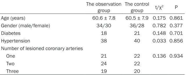
The results of this study showed that there
[image:2.612.91.411.85.215.2]were no significant differences between the Table 1. Comparison of baseline characteristics
The observation
group The control group t/χ2 P Age (years) 60.6 ± 7.8 60.5 ± 7.9 0.175 0.861 Gender (male/female) 34/30 36/28 0.782 0.377
Diabetes 18 21 0.148 0.701
Hypertension 38 40 0.033 0.856 Number of lesioned coronary arteries
One 21 22 0.136 0.934
Two 24 22
Three 19 20
First Hospital of
Qi-qihar and all partici-pants signed informed consent before partic-ipating in the study.
Measurements
From each patient,
two groups in age, gender, cases of diabetes, the number of lesioned coronary arteries and cases of hypertension (all P > 0.05). Therefore, the two groups were comparable. See Table 1.
Comparison of serum levels of LC3II protein
The serum level of LC3II in the observation
group was significantly higher than that in the
[image:3.612.91.286.71.257.2]control group (P < 0.05), indicating that the level of autophagy in the observation group was higher than that of the control group. See Figure 1.
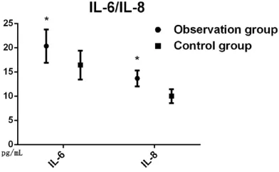
Comparison of IL-6 and IL-8 levels
The serum levels of IL-6 and IL-8 in the
observa-tion group were significantly higher than those
in the control group (both P < 0.05), indicating
that the inflammatory response was possibly
[image:3.612.325.522.74.274.2]involved in the development of in-stent reste-nosis in PCI patients after the surgery. See Figure 2.
Comparison of serum levels of NO and VEGF
The results of this study showed that the se-
rum levels of NO and VEGF in the observation
group were higher than those in the control group (both P < 0.05), indicating that intra- vascular injury in CHD patients complica- ted with diabetes was more severe than that in the control group, as shown in Figures 3 and 4.
Figure 1. Comparison of serum levels of LC3II pro-tein. The serum level of LC3II protein in the observa-tion group was higher than that in the control group after surgery, *P < 0.05. LC3II:
[image:3.612.91.287.342.463.2]microtubule-associat-ed protein 1A/1B-light chain 3-II.
Figure 2. Comparison of IL-6 and IL-8 levels. The se-rum levels of IL-6 and IL-8 in the observation group was higher than those in the control group aftersur-gery, *P < 0.05. IL-6: interleukin-6; IL-8: interleukin-8.
Figure 3. Comparison of serum levels of NO. The se-rum level of NO in the observation group was higher than that in the control group after surgery, *P <
0.05. NO: nitric oxide.
Figure 4. Comparison of serum levels of VEGF. VEGF:
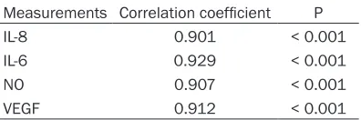
[image:3.612.325.522.355.493.2]Correlation analysis between LC3II level and levels of IL-8, IL-6, NO and VEGF
The results of this study showed that there
were significant positive correlations between
the LC3II level and the levels of IL-8, IL-6, NO
and VEGF (all P < 0.001). See Table 2. Discussion
Prevention of in-stent restenosis after PCI is a common challenge facing both physicians and patients. Although PCI patients currently receive aspirin and antiplatelet drugs and other such measures as lowering cholesterol and controlling blood pressure and glucose levels to prevent in-stent restenosis, a number of CHD patients still suffer restenosis after PCI [9, 10]. Consequently, the chest tightness, chest pain and other symptoms occur again due to isch-emia, affecting the patient’s health while reduc-ing the effect of PCI. Moreover, these clinical symptoms pose greater psychophysiological and economic burdens on the patients. To this
end, it is of great significance to study the
occurrence of in-stent restenosis in CHD patients after PCI [11-14].
Studies have shown that autophagy plays an important role in the development of cardiovas-cular disease. Autophagy, the process by which cells recycle cytoplasm, dispose of excess or defective organelles, and convert them into energy supply, helps relieve myocardial isch-emic and hypoxic injury. However, excessive levels of autophagy can lead to cell damage,
indicating that there are both benefits and risks
to the process of autophagy [15, 16]. Recent studies have shown that autophagy is impor-tant in the development of coronary artery ste-nosis and resteste-nosis after PCI. It may be involved in the above pathophysiological
pro-cesses via the aggravation of local inflamma
-tory response and vascular injuries [17]. Nevertheless, autophagy is associated with the
inflammatory response and vascular injury to a
certain extent. Therefore, this study explored the role of autophagy in restenosis in CHD patients after PCI.
LC3II is one of the proteins representative of autophagy in the body. The results of this study showed that the LC3II levels in the observation group were higher than that in the control group, indicating that the level of autophagy was higher in PCI patients with in-stent reste-nosis than that in PCI patients without in-stent restenosis, which is consistent with the conclu-sions of previous studies [18].
Furthermore, autophagy inhibits the function -ing of neutrophils. At the early stage of
revascu-larization, the inflammatory response height
-ens, resulting in the increase of inflammatory cytokines, IL-6 and IL-8. Autophagy can effec
-tively reduce the levels of inflammatory cyto
-kines, but over-elelvated autophagy worsens the inflammatory response as a result of dam -age to endothelial cells. The results of this study showed that the levels of IL-6 and IL-8 in
the observation group were significantly higher
than those in the control group, indicating that over-elevated autophagy increases the levels
of inflammatory cytokines and thus partici -pates in the pathophysiological process of in-stent restenosis after PCI, which is consiin-stent with the results of previous studies [19].
Studies have confirmed that functional damage
of the coronary endothelium is also an impor-tant factor for the formation of in-stent resteno-sis, and the related literature has also
con-firmed that autophagy can engage in vascular
endothelial injury by affecting the levels of NO
and VEGF [20]. Our study showed that the lev
-els of NO and VEGF in the observation group were significantly higher than those in the con -trol group, indicating that autophagy in CHD patients engages in the process of coronary restenosis after PCI, which corroborates the results of previous studies [21].
By further analyzing the correlations between
[image:4.612.91.289.97.163.2]levels of autophagy and inflammatory response and VEGF, our study found that there are posi -tive correlations between LC3II and the above-mentioned indicators. Autophagy could be con-sidered as a potential therapeutic target for the Table 2. Correlation analysis between LC3II
level and other related factors
Measurements Correlation coefficient P IL-8 0.901 < 0.001 IL-6 0.929 < 0.001 NO 0.907 < 0.001
VEGF 0.912 < 0.001
Note: LC3II, microtubule-associated protein 1A/1B-light
prevention of in-stent restenosis. However, this study is a single-center study with a small
sam-ple size. Therefore, the verification is to be per -formed in the future by conducting multi-center studies with enlarged sample size. The moder-ate levels of autophagy also warrant assess-ments by more accurate indicators.
Disclosure of conflict of interest
None.
Address correspondence to: Qi Tang, Department
of Cardiology, The First Hospital of Qiqihar, No.30
Gongyuan Road, Longsha District, Qiqihar 161000, Heilongjiang Province, China. Tel: +86-0452-245-
9227; E-mail: tangqi153k@163.com
References
[1] Zhu KF, Wang YM, Zhu JZ, Zhou QY and Wang NF. National prevalence of coronary heart dis -ease and its relationship with human develop-ment index: a systematic review. Eur J Prev Cardiol 2016; 23: 530-543.
[2] Belialov FI. Depression, anxiety, and stress in patients with coronary heart disease. Ter Arkh
2017; 89: 104-109.
[3] Lu Y, Zhang H, Wang Y, Zhou T, Welsh J, Liu J, Guan W, Li J, Li X, Zheng X, Spertus JA, Masoudi
FA, Krumholz HM and Jiang L. Percutaneous
Coronary intervention in patients without acute myocardial infarction in China: results from the China peace prospective study of percutane-ous coronary intervention. JAMA Netw Open 2018; 1: e185446.
[4] Zhao XY, Li JX, Tang XF, Xu JJ, Song Y, Jiang L,
Chen J, Song L, Gao LJ, Gao Z, Qiao SB, Yang YJ, Gao RL, Xu B and Yuan JQ. Prognostic value of NT-proBNP in stable coronary artery disease in Chinese patients after percutaneous coro-nary intervention in the drug-eluting stent era. Biomed Environ Sci 2018; 31: 859-866. [5] Xiu WJ, Yang HT, Zheng YY, Ma YT and Xie X.
Drug-eluting balloons versus second-genera-tion drug-eluting stents for treating in-stent re-stenosis in coronary heart disease after pci: a meta-analysis. Cardiol Res Pract 2018; 2018: 7658145.
[6] Noman A, Balasubramaniam K, Alhous MHA, Lee K, Jesudason P, Rashid M, Mamas MA, Za-man AG. Mortality after percutaneous coro-nary revascularization: prior cardiovascular
risk factor control and improved outcomes in
patients with diabetes mellitus. Catheter Car-diovasc Interv 2017; 89: 1195-1204.
[7] Ma X, Liu H, Foyil SR, Godar RJ, Weinheimer CJ,
Hill JA and Diwan A. Impaired autophagosome clearance contributes to cardiomyocyte death
in ischemia/reperfusion injury. Circulation 2012; 125: 3170-3181.
[8] Guan Y, Li X, Umetani M, Boini KM, Li PL and
Zhang Y. Tricyclic antidepressant amitriptyline
inhibits autophagic flux and prevents tube for -mation in vascular endothelial cells. Basic Clin Pharmacol Toxicol 2019; 124: 370-384. [9] Safian RD. Restenosis after pci: battered but
not beaten. Catheter Cardiovasc Interv 2016; 87: 209-10.
[10] Prasad K. Do statins have a role in reduction/ prevention of post-PCI restenosis. Cardiovasc Ther 2013; 31: 12-26.
[11] Luca SR, Koh M, Qiu F, Alter DA, Bagai A, Bha
-tia RS, Czarnecki A, Goodman SG, Lau C, Wijey -sundera HC and Ko DT. Stress testing after percutaneous coronary interventions: a popu-lation-based study. CMAJ Open 2017; 5: E417-E423.
[12] Cheng G, Chang FJ, Wang Y, You PH, Chen HC, Han WQ, Wang JW, Zhong NE and Min ZQ. Fac
-tors influencing stent restenosis after percuta -neous coronary intervention in patients with coronary heart disease: a clinical trial based on 1-year follow-up. Med Sci Monit 2019; 25: 240-247.
[13] Salisbury AC, Sapontis J, Grantham JA, Qintar M, Gosch KL, Lombardi W, Karmpaliotis D, Mo-ses J, Cohen DJ, Spertus JA and Kosiborod M. Outcomes of chronic total occlusion percuta-neous coronary intervention in patients with diabetes: insights from the open CTO registry. JACC Cardiovasc Interv 2017; 10: 2174-2181. [14] Wang Q, Liu H and Ding J. Outcomes of
percu-taneous coronary intervention in patients with coronary chronic total occlusions with versus without type 2 diabetes mellitus: a systematic review and meta-analysis. Medicine (Balti-more) 2017; 96: e8499.
[15] Sasaki Y, Ikeda Y, Iwabayashi M, Akasaki Y and
Ohishi M. The impact of autophagy on cardio-vascular senescence and diseases. Int Heart J 2017; 58: 666-673.
[16] Han Z, Cao J, Song D, Lei T, Kan C, Yue W, Lin
G, Yin Z, Fan Y and Wang C. Autophagy is in -volved in the cardioprotection effect of remote limb ischemic postconditioning on myocardial ischemia/reperfusion injury in normal mice, but not diabetic mice. PLoS One 2014; 9: e86838.
[17] Zhang R, Li T, Guo J, Zhao Y, Liu Y, Yao Y and
Zeng Z. Fufang-zhenzhu-tiaozhi capsule reduc
-es r-estenosis via the downregulation of NF-kappaB and inflammatory factors in rabbits.
Lipids Health Dis 2018; 17: 272.
[18] Zhu H and Zhang Y. Life and death partners in post-pCI restenosis: apoptosis, autophagy, and
[19] Qing Y, Dong X, Hongli L and Yanhui L. Berber-ine promoted myocardial protection of postop-erative patients through regulating myocardial autophagy. Biomed Pharmacother 2018; 105: 1050-1053.
[20] Bharath LP, Cho JM, Park SK, Ruan T, Li Y, Mueller R, Bean T, Reese V, Richardson RS, Cai J, Sargsyan A, Pires K, Anandh Babu PV, Boudi -na S, Graham TE, Symons JD. Endothelial cell autophagy maintains shear stress-induced ni-tric oxide generation via glycolysis-dependent purinergic signaling to endothelial nitric oxide
synthase. Arterioscler Thromb Vasc Biol 2017;
37: 1646-1656.